. 2002 Nov;283(5):R1079-86.
doi: 10.1152/ajpregu.00207.2002.

Hypocretin release in normal and narcoleptic dogs after food and sleep deprivation, eating, and movement

Affiliations

Hypocretin release in normal and narcoleptic dogs after food and sleep deprivation, eating, and movement

M-F Wu et al. Am J Physiol Regul Integr Comp Physiol. 2002 Nov.

Abstract

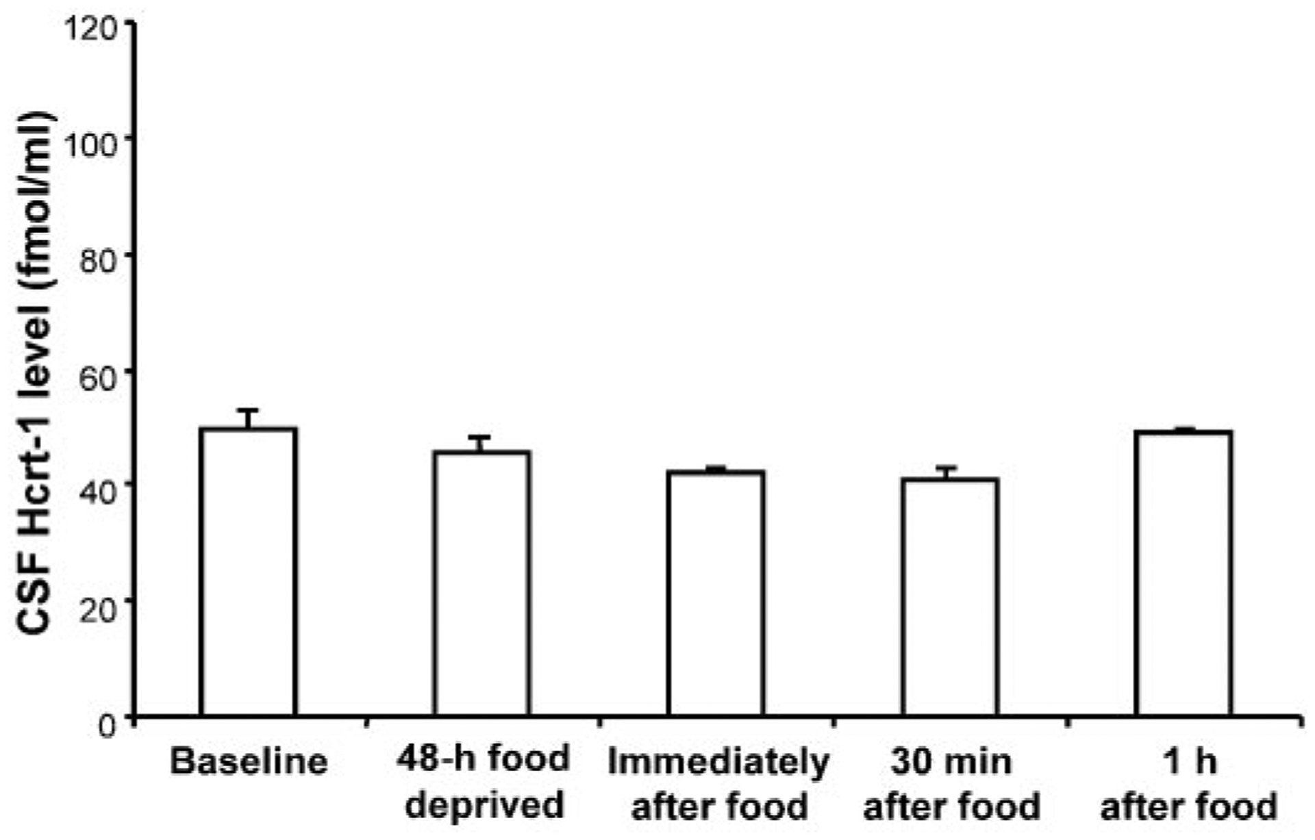
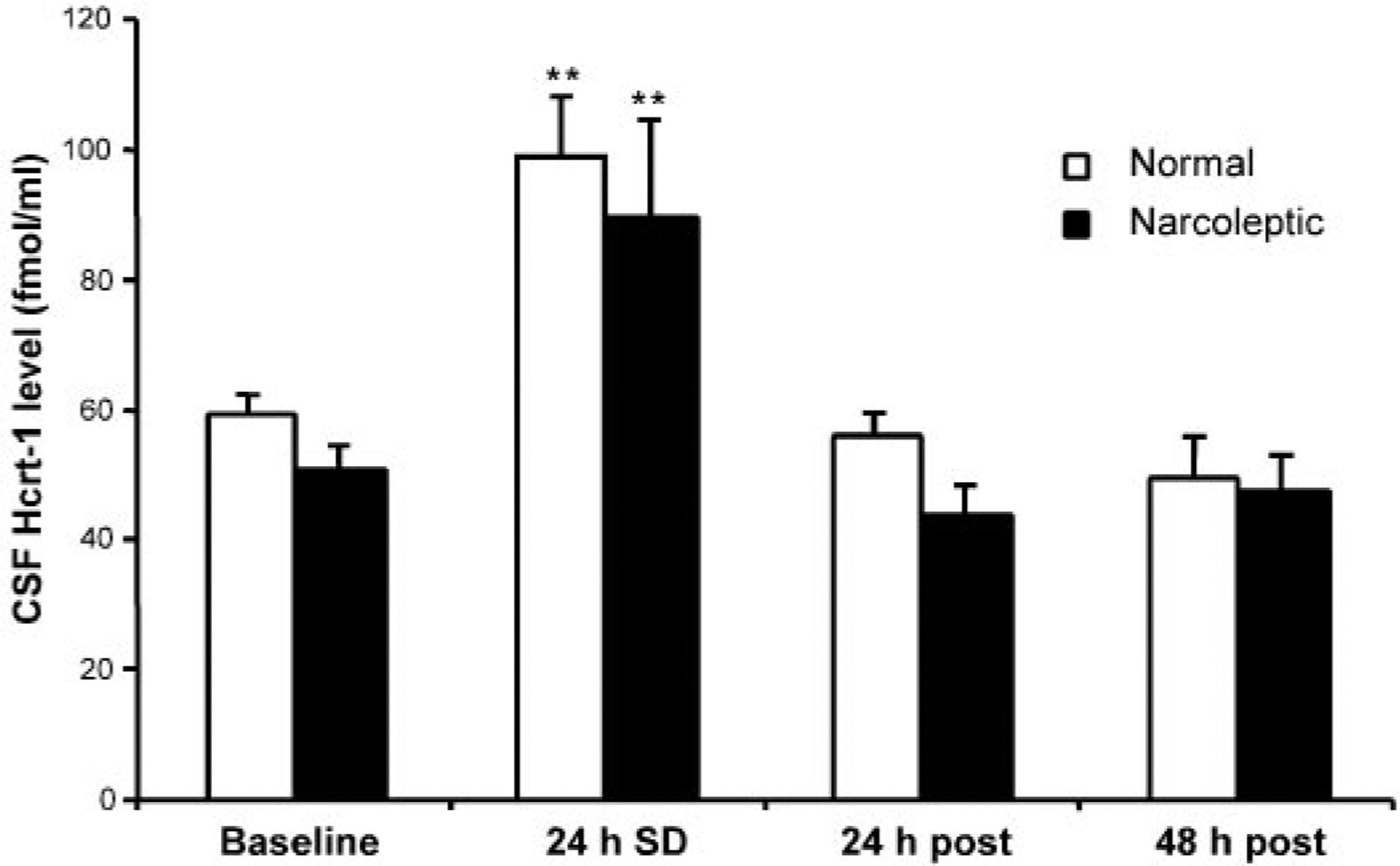
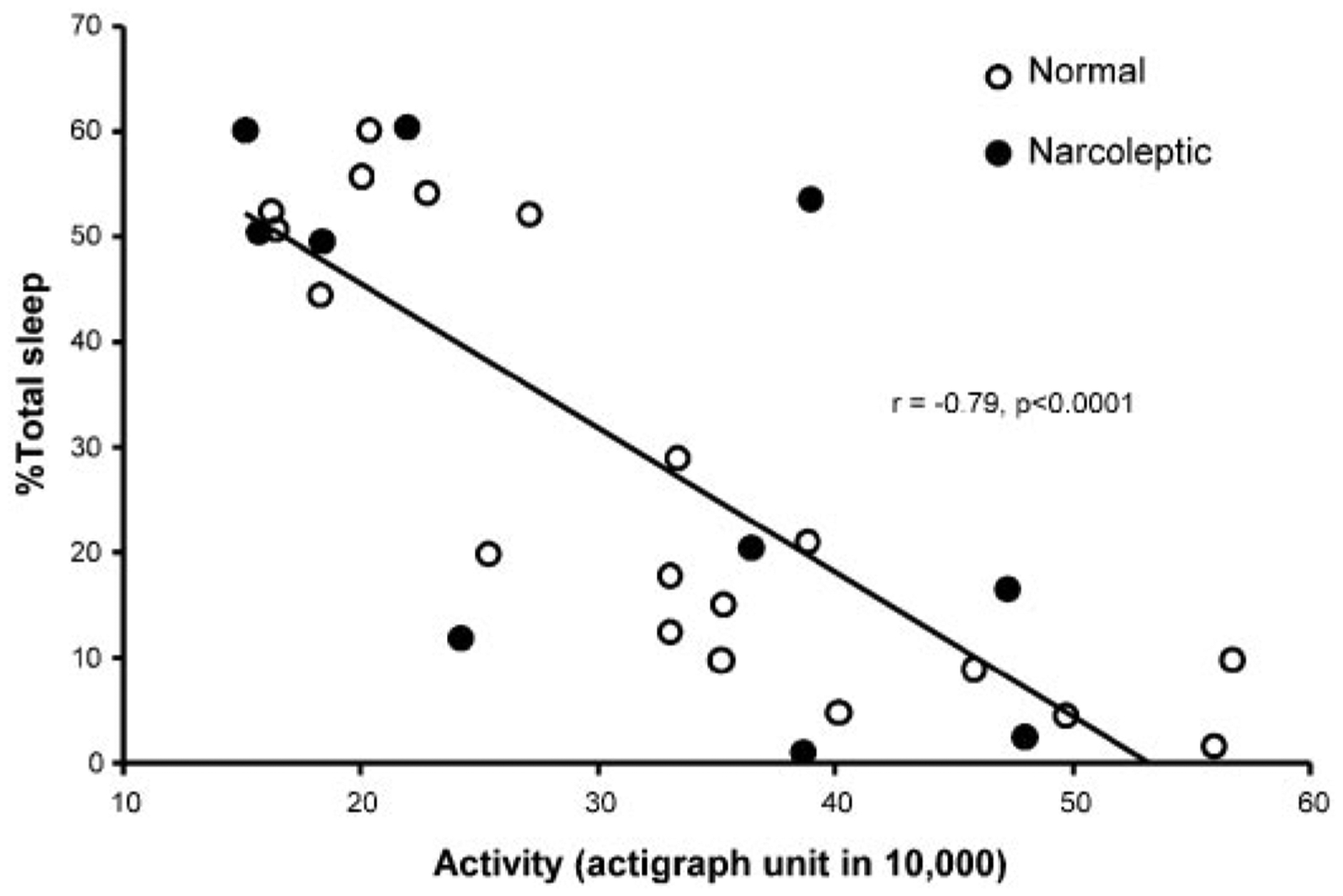
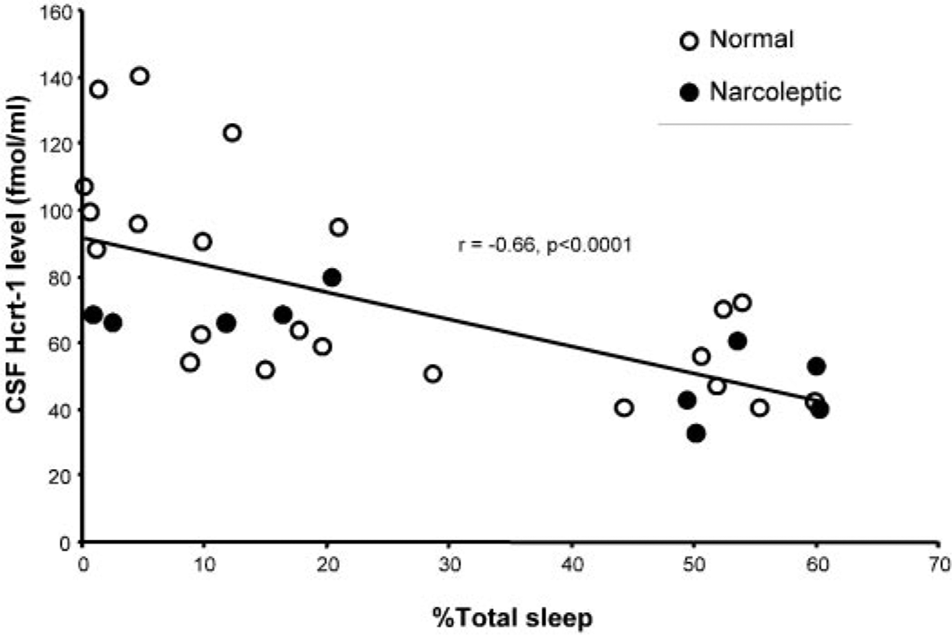
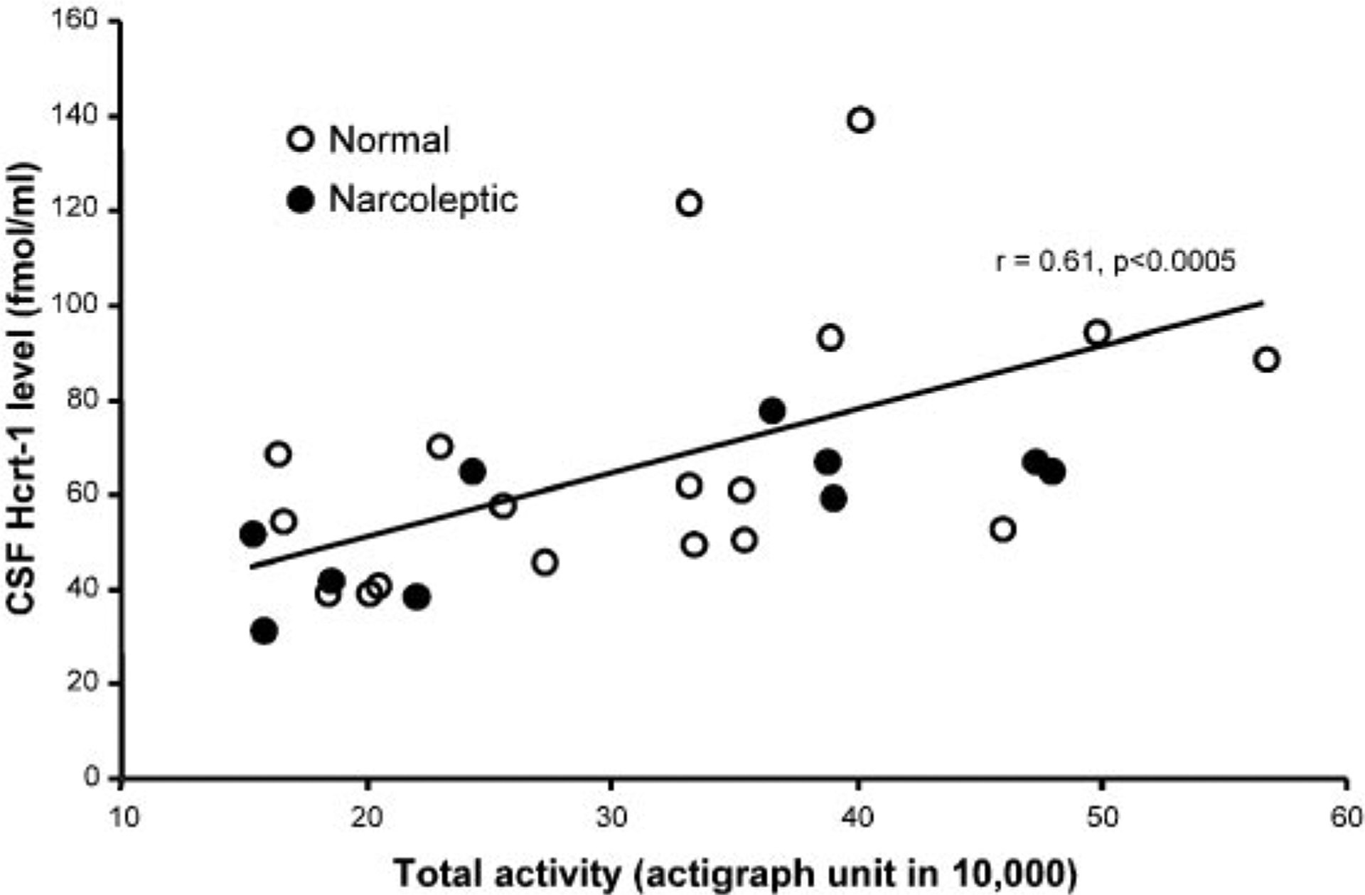
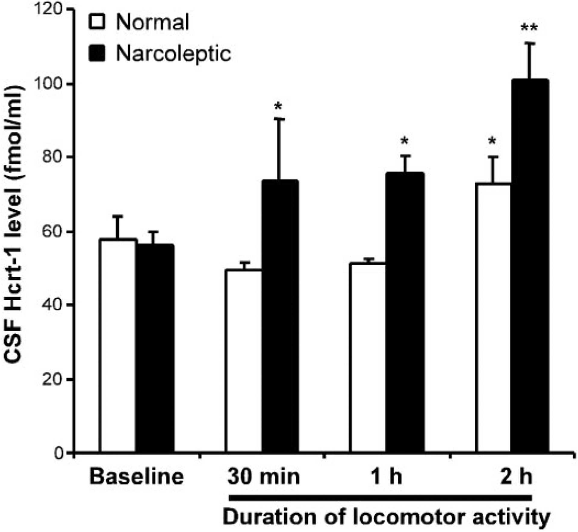
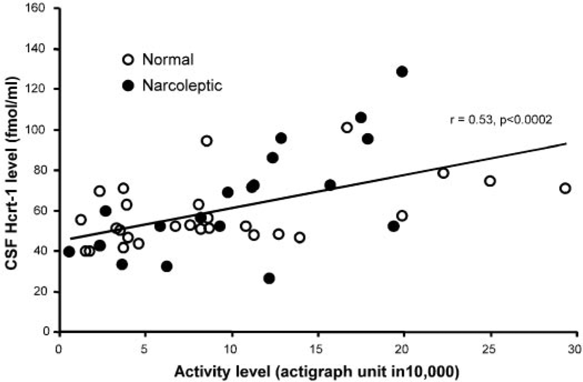
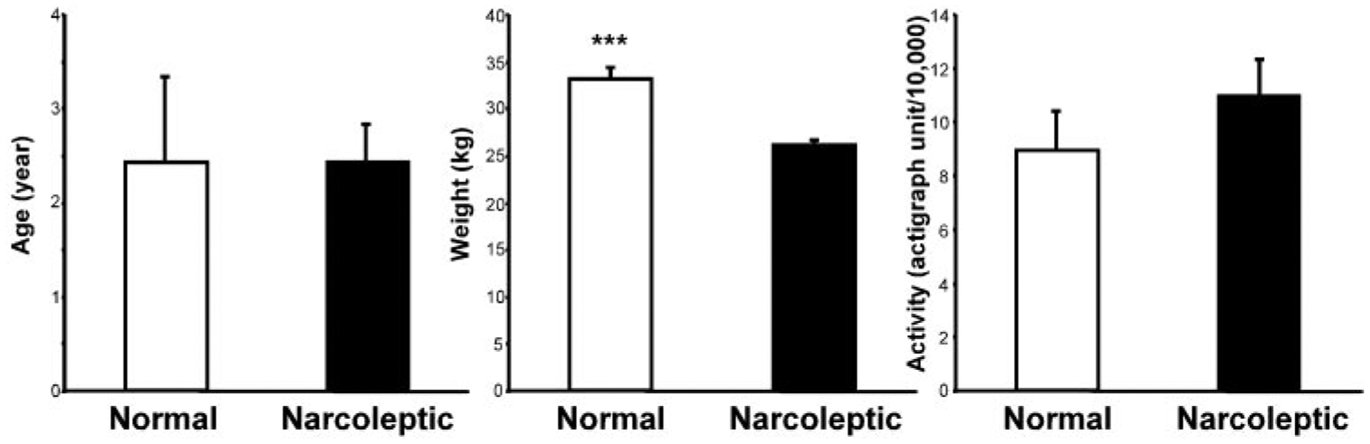
Hypocretins (orexins) are recently discovered hypothalamic neuropeptides that have been implicated in the etiology of narcolepsy. The normal behavioral functions of these peptides are unclear, although a role in feeding has been suggested. We measured hypocretin-1 (Hcrt-1) in the cerebrospinal fluid of dogs during a variety of behaviors. We found that 48 h without food (24 h beyond normal 24-h fasting period) produced no significant change in Hcrt-1 levels nor did feeding after the deprivation. In contrast, 24 h of sleep deprivation produced on average a 70% increase in Hcrt-1 level compared with baseline levels. The amount of increase was correlated with the level of motor activity during the sleep-deprivation procedure. A 2-h period of exercise in the same dogs produced a 57% increase in Hcrt-1 levels relative to quiet waking levels, with the magnitude of the increase being highly correlated with the level of motor activity. The strong correlation between motor activity and Hcrt-1 release may explain some of the previously reported behavioral, physiological, and pathological phenomena ascribed to the Hcrt system.


Figures

Fig. 1.
Fig. 1.
Cerebrospinal fluid (CSF) hypocretin (Hcrt)-1 level after 48 h without food (n = 6) and immediately (n = 5), 30 min (n = 5), or 1 h (n = 6) after eating following food deprivation in normal dogs. No significant changes were seen (ANOVA). Hcrt-1 level was not increased with food deprivation nor was it decreased with feeding.
Fig. 2.
Fig. 2.
Sleep deprivation induced changes in Hcrt level in CSF. Sleep deprivation was correlated with an increase in Hcrt-1 level. Data represent Hcrt-1 level at the termination of 24-h sleep deprivation (24 h SD) and at 24 h (24 h post) and 48 h (48 h post) after the sleep deprivation. Normal: normal dogs (n = 6); narcoleptic: narcoleptic dogs (n = 7). **P < 0.01 Fisher’s least significant difference (LSD) test compared with baseline.
Fig. 3.
Fig. 3.
Correlation of 24-h total activity level and percentage sleep time during baseline and sleep deprivation. Percentage of sleep is negatively correlated with increased activity in 29 runs with actigraph data.
Fig. 4.
Fig. 4.
Percentage time asleep during baseline and sleep deprivation are negatively correlated with Hcrt-1 level. Overall, 32 data points were collected from 9 animals in which activity was measured and included Hcrt-1 levels in both baseline (n = 16) and those after sleep deprivation (n = 16).
Fig. 5.
Fig. 5.
Twenty-four-hour total activity level during sleep deprivation is positively correlated with Hcrt-1 level (n = 29).
Fig. 6.
Fig. 6.
Changes in Hcrt-1 in CSF after 30–120 min of increased locomotor activity. *P < 0.05, **P < 0.01 Fisher’s LSD test compared with baseline. Exercise produced increased Hcrt-1 levels with narcoleptic dogs showing a more rapid and larger magnitude increase.
Fig. 7.
Fig. 7.
Hcrt-1 level during varying amounts of waking activity in non-sleep-deprived dogs. Data points represent Hcrt-1 levels and activity measures during both baseline and experimental conditions (30-, 60-, and 120-min exercise). A total of 46 data points (26 from normals and 20 from narcoleptics) was collected in which activity was measured. CSF Hcrt-1 level is correlated with the amount of activity before CSF sampling.
Fig. 8.
Fig. 8.
Comparison of age, weight, and activity levels of narcoleptics (n = 14) and normals (n = 7) in the exercise study. ***P < 0.0001, t-test.

Similar articles

Cited by

References

    1. Alam MN, Gong H, Alam T, Jaganath R, McGinty D, and Szymusiak R. Sleep-waking discharge patterns of neurons recorded in the rat perifornical lateral hypothalamic area. J Physiol 538: 619–631, 2002. - PMC - PubMed
    1. Aldrich MS. Diagnostic aspects of narcolepsy. Neurology 50, Suppl 1: S2–S7, 1998. - PubMed
    1. American Physiological Society. Guiding principles for research involving animals and human beings. Am J Physiol Regul Integr Comp Physiol 283: R281–R283, 2002. - PubMed
    1. Bourgin P, Huitron-Resendiz S, Spier AD, Fabre V, Morte B, Criado JR, Sutcliffe JG, Henriksen SJ, and De Lecea L. Hypocretin-1 modulates rapid eye movement sleep through activation of locus coeruleus neurons. J Neurosci 20: 7760–7765, 2000. - PMC - PubMed
    1. Cai XJ, Lister CA, Buckingham RE, Pickavance L, Wilding J, Arch JR, Wilson S, and Williams G. Down-regulation of orexin gene expression by severe obesity in the rats: studies in zucker fatty and zucker diabetic fatty rats and effects of rosiglitazone. Mol Brain Res 77: 131–137, 2000. - PubMed

Publication types

LinkOut - more resources