. 2002 Oct 29;99(22):14524-9.
doi: 10.1073/pnas.172514599. Epub 2002 Oct 10.

Resistance of alpha -synuclein null mice to the parkinsonian neurotoxin MPTP

Affiliations

Resistance of alpha -synuclein null mice to the parkinsonian neurotoxin MPTP

William Dauer et al. Proc Natl Acad Sci U S A. .

Abstract

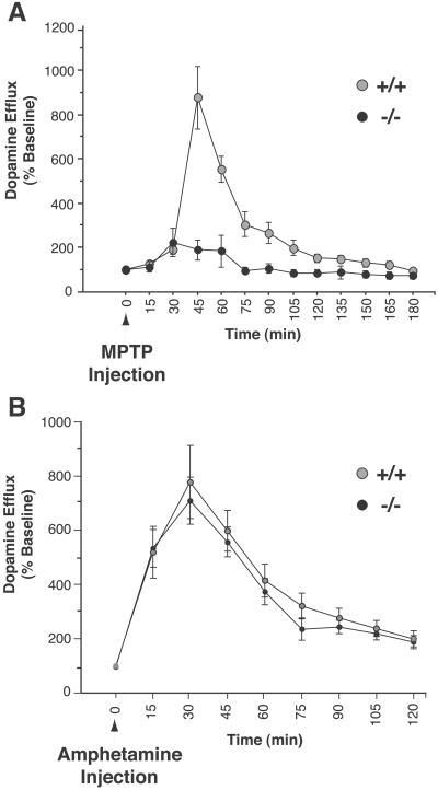
Parkinson's disease (PD) is most commonly a sporadic illness, and is characterized by degeneration of substantia nigra dopamine (DA) neurons and abnormal cytoplasmic aggregates of alpha-synuclein. Rarely, PD may be caused by missense mutations in alpha-synuclein. MPTP, a neurotoxin that inhibits mitochondrial complex I, is a prototype for an environmental cause of PD because it produces a pattern of DA neurodegeneration that closely resembles the neuropathology of PD. Here we show that alpha-synuclein null mice display striking resistance to MPTP-induced degeneration of DA neurons and DA release, and this resistance appears to result from an inability of the toxin to inhibit complex I. Contrary to predictions from in vitro data, this resistance is not due to abnormalities of the DA transporter, which appears to function normally in alpha-synuclein null mice. Our results suggest that some genetic and environmental factors that increase susceptibility to PD may interact with a common molecular pathway, and represent the first demonstration that normal alpha-synuclein function may be important to DA neuron viability.


Figures

Fig 1.
Fig 1.
Targeted disruption of the α-synuclein locus by homologous recombination. (A) Schematic drawing of the wild-type (WT) locus, the targeting construct, and the recombined allele. A null allele was generated by blocking transcription from the α-synuclein allele with a “STOP” cassette (12) inserted upstream of the start ATG. The location of hybridization probes for Southern blot analysis (5′ probe, 3′ probe) are shown. (B) Southern blot analysis of embryonic stem cell clones. DNA was digested with BglII and was hybridized with the 5′ probe. The WT allele produced a 9.5-kb fragment, and the targeted allele produced a 6.5-kb fragment. BglII-digested DNA was also hybridized with the 3′ probe (data not shown). (C) Southern blot analysis of genomic DNA from littermates of a α-synuclein heterozygote mating. Tail DNA was isolated and analyzed as in B. (D) Northern blot analysis of whole brain mRNA. Total RNA was hybridized with α-synuclein cDNA probe containing the entire coding region. The lower panel shows ethidium bromide staining of the 28s ribosomal bands. (E) Western blot analysis of whole brain protein blotted with an α-synuclein antibody (Transduction Laboratories).
Fig 2.
Fig 2.
α-Synuclein−/− dopaminergic neurons are strikingly resistant to MPTP-induced neurodegeneration. α-Synuclein−/− mice and wild-type littermate controls were treated with a chronic (30 mg/kg/d for 5 d) or acute (80 mg/kg for 1 d) MPTP regimen, and were killed 21 d after the final chronic dose or 7 d after the acute dose. n = 4–9 mice per group. (A) Stereological counts of DA TH+ cell bodies after saline (gray bar), chronic MPTP (white bar), or acute MPTP (black bar). (B) TH antibody staining of the SN pars compacta (DA) after the two different MPTP regimens. (C) Quantitation (OD) of the intensity of striatal TH staining; legend as in A. (D) TH antibody staining of the striatum after two different MPTP regimens. (Error bars = SEM.) Both MPTP regimens produced significant reductions of TH+ DA neurons and striatal immunostaining in wild-type mice, but no significant changes in α-synuclein−/− mice (P < 0.05, Newman–Keuls post hoc).
Fig 3.
Fig 3.
The amount and activity of the DAT is normal in α-synuclein mutant mice. (A and B) Analysis of DAT in the striatum of wild type and α-synuclein mutant mice using DAT antibody staining (A) and [3H]mazindol (30 nM) autoradiography (B) revealed no genotype-related differences. Immunofluorescence was measured by obtaining all images at the same empirically defined exposure that contained no saturated pixels, and all images were taken at the same rostrocaudal striatal plane. (C) [3H]DA uptake in ventral midbrain cultures.
Fig 4.
Fig 4.
Dissociation between MPTP- and rotenone-induced death of α-synuclein null DA neurons. Ventral midbrain cultures were treated for two days with 10 or 50 μM MPP+ (A) and 20 or 100 nM rotenone (B). α-Synuclein mutant DA neurons displayed significant resistance to death at both concentrations of MPP+, but did not show resistance to rotenone; there were significantly fewer α-synuclein−/− TH+ neurons at the 20 nM rotenone dose (P < 0.05, Newman–Keuls post hoc). For each condition, n = 5–10 plates per genotype. Values are represented as a percentage of untreated controls. (Error bars = SEM.)
Fig 5.
Fig 5.
MPTP-induced DA efflux is deficient in α-synuclein null mice. (A) In vivo microdialysis of striatal dopamine efflux after a single i.p injection of 30 mg/kg MPTP. Gray circle, wild type; black circle, α-synuclein−/−. (B) α-Synuclein mutant mice display normal amphetamine-induced DA release. Legend as in A. Microdialysis values are represented as a percentage of the average of four baseline measurements taken in the hour preceding MPTP injection; each animal was used as its own baseline control. There were no significant differences in baseline values. n = 3–8 mice per genotype.

Comment in

Similar articles

Cited by

References

    1. Scott W. K., Nance, M. A., Watts, R. L., Hubble, J. P., Koller, W. C., Lyons, K., Pahwa, R., Stern, M. B., Colcher, A., Hiner, B. C., et al. (2001) J. Am. Med. Assoc. 286, 2239-2244. - PubMed
    1. Kruger R., Vieira-Saecker, A. M., Kuhn, W., Berg, D., Muller, T., Kuhnl, N., Fuchs, G. A., Storch, A., Hungs, M., Woitalla, D., et al. (1999) Ann. Neurol. 45, 611-617. - PubMed
    1. Martin E. R., Scott, W. K., Nance, M. A., Watts, R. L., Hubble, J. P., Koller, W. C., Lyons, K., Pahwa, R., Stern, M. B., Colcher, A., et al. (2001) J. Am. Med. Assoc. 286, 2245-2250. - PMC - PubMed
    1. Polymeropoulos M. H., Lavedan, C., Leroy, E., Ide, S. E., Dehejia, A., Dutra, A., Pike, B., Root, H., Rubenstein, J., Boyer, R., et al. (1997) Science 276, 2045-2047. - PubMed
    1. Kruger R., Kuhn, W., Muller, T., Woitalla, D., Graeber, M., Kosel, S., Przuntek, H., Epplen, J. T., Schols, L. & Riess, O. (1998) Nat. Genet. 18, 106-108. - PubMed

Publication types

MeSH terms