. 2002 Oct 29;99(22):14047-52.
doi: 10.1073/pnas.212229599. Epub 2002 Oct 10.

A nucleolar TAR decoy inhibitor of HIV-1 replication

Affiliations

A nucleolar TAR decoy inhibitor of HIV-1 replication

Alessandro Michienzi et al. Proc Natl Acad Sci U S A. .

Abstract

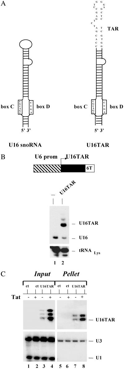
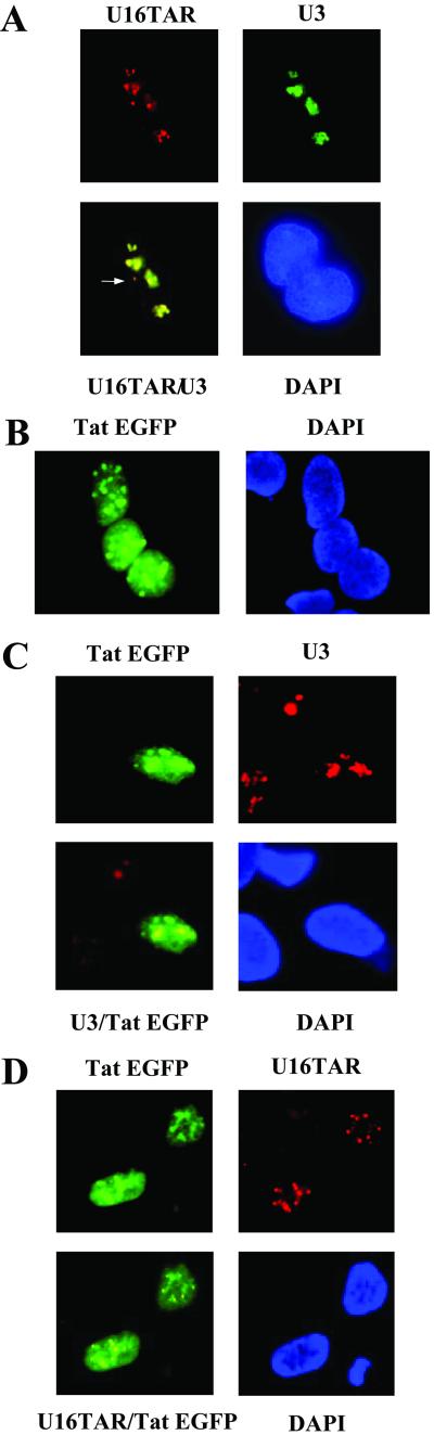
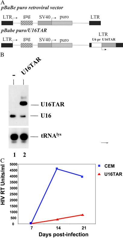
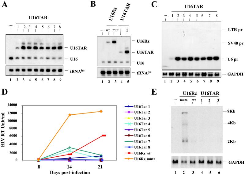
Tat is a critical regulatory factor in HIV-1 gene expression. It mediates the transactivation of transcription from the HIV-1 LTR by binding to the transactivation response (TAR) element in a complex with cyclin T1. Because of its critical and early role in HIV gene expression, Tat and its interaction with the TAR element constitute important therapeutic targets for the treatment of HIV-1 infection. Based on the known nucleolar localization properties of Tat, we constructed a chimeric small nucleolar RNA-TAR decoy that localizes to the nucleoli of human cells and colocalizes in the nucleolus with a Tat-enhanced GFP fusion protein. When the chimeric RNA was stably expressed in human T lymphoblastoid CEM cells it potently inhibited HIV-1 replication. These results demonstrate that the nucleolar trafficking of Tat is critical for HIV-1 replication and suggests a role for the nucleolus in HIV-1 viral replication.


Figures

Fig 1.
Fig 1.
U16TAR RNA design and intracellular expression. (A) Schematic representation of the U16 snoRNA (Left) and U16TAR RNA (Right). The apical loop of the U16 snoRNA was replaced by a minimal, functional TAR sequence. (B) Intracellular U16TAR expression. The U16TAR sequence was inserted within a U6 snoRNA expression cassette (pTZ/U6+1) generating the transcription unit U6+1/U16TAR (Upper). The U6 expression cassette allows intracellular expression mediated by RNA pol III. Six thymidines were added immediately downstream of the U16TAR sequence to function as an RNA pol III termination signal. The 293 cells were transiently transfected with 10 μg of the U6+1/U16TAR plasmid. After 48 h total RNA was isolated, electrophoresed in a 6% polyacrylamide-7 M urea gel, and blotted onto a nylon membrane (Lower). The U16 snoRNA-specific probe was used to simultaneously detect the U16TAR and the endogenous U16 snoRNAs. An additional probe was used to detect endogenous tRNAformula image. Lane 1 contains total RNA isolated from untransfected 293 cells. Lane 2 contains total RNA extracted from 293 cells transfected with U6+1/U16TAR. (C) U16TAR stable association with fibrillarin. The 293 cells were transiently transfected with 10 μg of the U6+1/U16TAR plasmid and/or 4 μg of the pLEGFP-C1/Tat plasmid. After 48 h total cell extract was prepared and immunoselected with antifibrillarin antibody. Total RNAs were isolated from the input cell extract and the pellet and electrophoresed in a 6% polyacrylamide-7 M urea gel followed by blotting onto a nylon membrane. A 32P-labeled probe was used to detect U16TAR. A probe complementary to the endogenous U1 snoRNA was used to detect this snoRNA, which is not associated with fibrillarin. A probe complementary to endogenous U3 snoRNA, which is fibrillarin-associated, was used to detect this RNA. Lane 1 contains total RNA extracted from the untransfected 293 cells. Lane 2 contains total RNA extracted from 293 cells transiently transfected with the pLEGFP-C1/Tat plasmid. Lanes 3 and 4 contain total RNAs extracted from 293 cells transfected with U6+1/U16TAR without or with the pLEGFP-C1/Tat plasmid. Lane 5 contains total RNA extracted from the immunoselected pellet of untransfected 293 cells. Lane 6 contains total RNA extracted from the immunoselected pellet of 293 cells transiently transfected with pLEGFP-C1/Tat plasmid. Lanes 7 and 8 contain total RNAs extracted from immunoselected pellets of 293 cells transfected with U6+1/U16TAR without or with the pLEGFP-C1/Tat plasmid.
Fig 2.
Fig 2.
Intracellular localization of U16TAR and colocalization with Tat-EGFP. The 293 cells were grown on coverslips and transiently transfected with 10 μg of the U6+1/U16TAR plasmid and/or 4 μg of pLEGFP-C1/Tat plasmid. After 48 h the cells were fixed in 4% para-formaldehyde dissolved in 1× PBS and in situ hybridizations were carried out. (A) The 293 cells transiently transfected with the U6+1/U16TAR plasmid. Hybridization was performed by using a U16TAR RNA-specific probe conjugated with the CY3 fluorophore (red fluorescence, Upper Left) and a U3 snoRNA-specific probe (for a nucleolar control), conjugated with Oregon green fluorophore (green fluorescence, Upper Right). The yellow signal depicts overlapping of the two hybridization signals and confirms the U16TAR nucleolar localization (Lower Left). The blue staining nuclei (4′-6′-diamino-2-phenyindole, DAPI) are indicated (Lower Right). In some of the 293 cells a fraction of the U16TAR signal (red) does not overlap with the U3 snoRNA (green) signal (white arrow, Lower Left), suggesting a small amount of localization at a site other than the nucleolus when U16TAR is overexpressed (data not shown). (B) The 293 cells transiently transfected with the pLEGFP-C1/Tat plasmid. The Tat EGFP green fluorescence was detected in the nucleus of 293 cells (Left) (confirmed by DAPI staining, Right). (C) The 293 cells transiently transfected with 4 μg of pLEGFP-C1/Tat plasmid. Hybridization was performed by using a U3 snoRNA-specific probe conjugated with the CY3 fluorophore (red fluorescence, Upper Right). Tat EGFP is detectable via the green fluorescence (Upper Left). The yellow signal depicts overlapping signals of the green fluorescence from the Tat EGFP fusion protein and the red fluorescence of the U3 snoRNA hybridization (Lower Left). (D) The 293 cells transiently transfected with pLEGFP-C1/Tat and U6+1/U16TAR plasmids. Hybridization was performed by using a U16TAR-specific probe conjugated with CY3 (red fluorescence, Upper Right). Tat EGFP is detected by the green fluorescence (Upper Left). The yellow signal in the nucleoli indicates an overlap between the red fluorescence of the U16TAR probe and the green fluorescence of the Tat EGFP fusion protein (Lower Left).
Fig 3.
Fig 3.
Delivery and intracellular activity of the U16TAR RNA in a pool of transfected CEM cells. (A) Schematic representation of the pBabe Puro retroviral vector. The U6+1/U16TAR cassette was inserted within the U3 region of the 3′ LTR (pBabe Puro/U16TAR). SV40, simian virus 40. (B) Pooled transfected CEM cells expressing the U16TAR RNA. CEM cells were transduced with the pBabe/U16TAR construct, and a pooled population of puromycin-resistant cells was selected. Total RNA was isolated from these cells, electrophoresed in a 6% polyacrylamide-7 M urea gel, and blotted onto a nylon membrane. Hybridization with the U16 snoRNA-specific probe allowed simultaneous detection of the U16TAR RNA and the endogenous U16 snoRNAs. Hybridization to endogenous tRNAformula image was used as a loading control. Lane 1 contains total RNA extracted from untransduced CEM cells. Lane 2 contains total RNA extracted from CEM cells transduced with pBabe/U16TAR. (C) U16TAR anti-HIV-1 activity. Parental CEM cells or the pooled transductants expressing U16TAR were challenged with HIV-1 NL4–3 at an moi of 0.01. At 7, 14, and 21 days postinfection supernatants were collected from the cell culture and analyzed by HIV reverse transcriptase (RT) assay.
Fig 4.
Fig 4.
Selection of stably transduced clones expressing U16TAR RNA and HIV-1 challenge assays. Individual clones stably expressing the various constructs were isolated by limiting dilution of the pool of transduced CEM cells. Northern blot analyses were performed after electrophoresis of total RNAs in a 6% polyacrylamide-7 M urea gel (A and B) or in a 1.2% agarose-formaldehyde gel (C). (A) Hybridizations were carried out by using a specific U16 snoRNA probe to detect the U16TAR RNAs and the endogenous U16 snoRNA. Probing for endogenous tRNAformula image serves as a loading control. Lane 1 contains RNA extracted from untransduced CEM cells. Lanes 2–9 contain total RNAs extracted from clones of transduced CEM clones expressing U16TAR. (B) Hybridizations were carried out under the same condition used for A. Lane 1 contains RNA extracted from untransduced CEM cells. Lanes 2 and 3 contain RNAs extracted from individual CEM clones expressing the U16Rz WT and mutant, respectively (30). Lanes 4 and 5 contain RNAs extracted from individual CEM clones expressing U16TAR (clones 1 and 2). (C) Hybridizations were carried out by using a U16TAR-specific probe and a probe for glyceraldehyde phosphate dehydrogenase (GAPDH) mRNA (loading control). Lane 1 contains RNA extracted from untransduced CEM cells. Lanes 2–9 contain total RNAs extracted from individual CEM clones expressing U16TAR. SV40, simian virus 40. (D) Individual CEM clones expressing the U16TAR RNA are resistant to HIV-1 infection. Cells expressing U16TAR were challenged with HIV-1 NL4–3 at an moi of 0.01. At 8, 14, and 21 days postinfection supernatants were collected from the cultures and analyzed by HIV-1 reverse transcriptase assays. CEM clones expressing a nucleolar-localized hammerhead ribozyme (WT or a nonfunctional mutant) (U16Rz WT and mutant, ref. 30) were used as controls. The data presented represent an average of four independent challenge experiments, and the standard errors for each point are around 5% maximum. (E) Total RNAs extracted from challenged cells 21 days postinfection (see D) were electrophoresed in a 1% agarose-formaldehyde gel, blotted onto a nylon membrane, and hybridized with an HIV-1 probe or a probe for GAPDH mRNA. Lane 1 contains RNA from uninfected CEM cells. Lanes 2 and 3 contain total RNAs extracted from HIV-1-infected CEM cells expressing the U16Rz mutant or U16Rz WT RNAs, respectively. Lanes 4–6 contain RNAs extracted from HIV-1-infected CEM cells expressing U16TAR RNA.

Similar articles

Cited by

References

    1. Jeang K. T., Xiao, H. & Rich, E. A. (1999) J. Biol. Chem. 274, 28837-28840. - PubMed
    1. Garber M. E. & Jones, K. A. (1999) Curr. Opin. Immunol. 11, 460-465. - PubMed
    1. Kao S. Y., Calman, A. F., Rich, E. A. & Peterlin, B. M. (1987) Nature 330, 489-493. - PubMed
    1. Laspia M. F., Wendel, P. & Mathews, M. B. (1993) J. Mol. Biol. 232, 732-746. - PubMed
    1. Marciniak R. A. & Sharp, P. A. (1991) EMBO J. 10, 4189-4196. - PMC - PubMed

Publication types

MeSH terms