. 2002 Oct 29;99(22):14338-43.
doi: 10.1073/pnas.212290499. Epub 2002 Oct 10.

Protection of the intestinal mucosa by intraepithelial gamma delta T cells

Affiliations

Protection of the intestinal mucosa by intraepithelial gamma delta T cells

Yaping Chen et al. Proc Natl Acad Sci U S A. .

Abstract

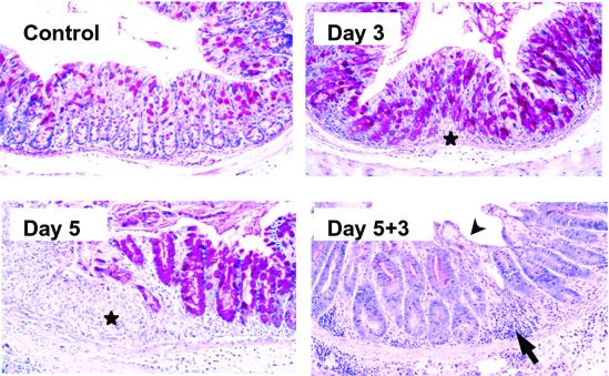
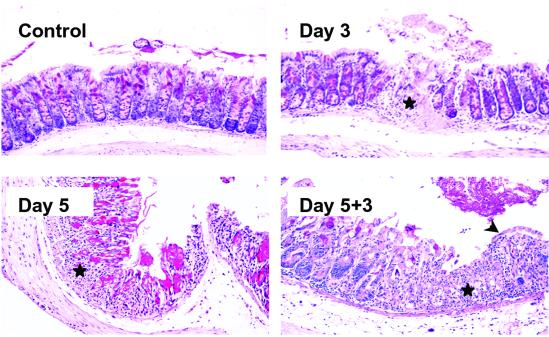
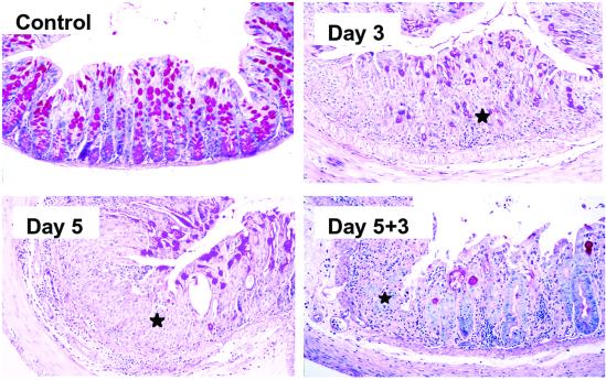
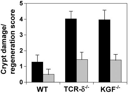
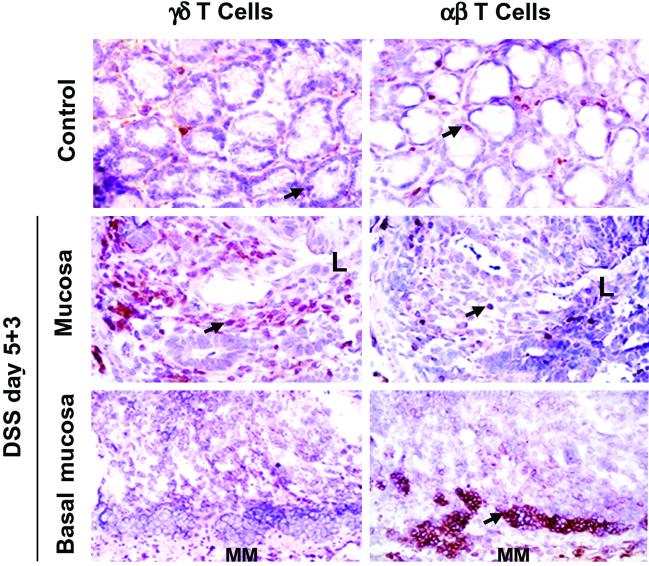
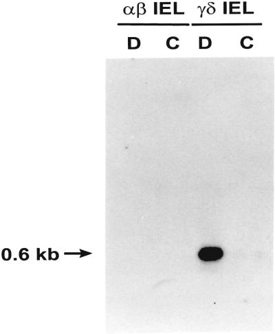
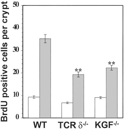
gammadelta intraepithelial T lymphocytes (IEL) represent a major T cell population within the intestine of unclear functional relevance. The role of intestinal gammadelta IEL was evaluated in the dextran sodium sulfate (DSS) induced mouse colitis model system. Large numbers of gammadelta T cells, but not alphabeta T cells, were localized at sites of DSS-induced epithelial cell damage. gammadelta IEL in DSS treated mice expressed keratinocyte growth factor (KGF), a potent intestinal epithelial cell mitogen. gammadelta cell-deficient mice (TCRdelta(-/-)) and KGF-deficient mice (KGF(-/-)), but not alphabeta cell-deficient mice (TCRalpha(-/-)), were more prone than wild-type mice to DSS-induced mucosal injury and demonstrated delayed tissue repair after termination of DSS treatment. Termination of DSS treatment resulted in vigorous epithelial cell proliferation in wild-type mice but not in TCRdelta(-/-) mice or KGF(-/-) mice. These results suggest that gammadelta IEL help preserve the integrity of damaged epithelial surfaces by providing the localized delivery of an epithelial cell growth factor.


Figures

Fig 1.
Fig 1.
Histopathology of the colon in C57BL/6 mice subjected to DSS treatment. Representative areas of normal mucosa, ulcerated mucosa after 3 days and 5 days of DSS treatment, and mucosal regeneration 3 days after termination of DSS treatment. Ulcerated areas are shown by stars, mononuclear cell infiltration is shown by an arrow, and areas of surface re-epithelialization are shown by an arrowhead. Similar results were obtained from at least four independent experiments. (Original magnification = ×200.)
Fig 2.
Fig 2.
Heightened susceptibility of TCRδ−/− mice to DSS-induced colitis. Representative areas of normal mucosa, ulcerated mucosa after 3 days and 5 days of DSS treatment, and mucosal regeneration 3 days after termination of DSS treatment. Ulcerated areas are indicated by stars, and areas of surface re-epithelialization are indicated by an arrowhead. TCRδ−/− mice had consistently more severe colonic damage compared with wild type. Evidence of regeneration, and in particular surface re-epithelialization, was scant 3 days after termination of DSS treatment. Similar results were obtained in three independent experiments. (Original magnification = ×200.)
Fig 3.
Fig 3.
KGF−/− mice suffer from a severe form of DSS colitis. Representative areas of normal mucosa, ulcerated mucosa after 3 days of DSS treatment, ulcerated mucosa after 5 days of DSS treatment, and mucosal regeneration 3 days after termination of DSS treatment. Ulcerated areas are indicated by stars. Almost complete ulceration of the colonic mucosa occurred after 3 days of DSS treatment. Repair of colonic lesions was significantly impaired compared with that in wild-type mice, and was similar to that observed in TCRδ−/− mice. Similar results were obtained in three independent experiments. (Original magnification = ×200.)
Fig 4.
Fig 4.
Histological grading of tissue damage and regeneration. Bars represent the ratio of the colonic damage score divided by the tissue regeneration score 3 days (black bars) and 14 days (gray bars) after termination of DSS treatment. TCRδ−/− mice and KGF−/− have significantly more colonic damage and less tissue regeneration compared with wild-type mice. Data are expressed as the mean and SD of a minimum of 12 sections representing the distal and medial colon of at least 3 mice per group. Data are representative of three experiments.
Fig 5.
Fig 5.
Localization of γδ T cells and αβ T cells in the colon of DSS-treated wild-type mice. Arrowheads point to representative positive cells. In wild-type mice recovering from DSS-colitis, γδ T cells were found in large numbers within areas of mucosal damage, whereas αβ T cells were detected predominantly in aggregates near the basal mucosa. Within damaged crypts, the number of γδ T cells averaged 24.06 ± 14.40, and the number of αβ T cells was 1.72 ± 1.67 (t test, P < 0.001, n = 18). Increased numbers of γδ T cells were detected as early as 3 days after initiation of DSS treatment (data not shown). L, the orientation of the section shown relative to the lumen; MM, muscularis mucosae. (Original magnification = ×400.)
Fig 6.
Fig 6.
KGF expression in the intestine of DSS-treated mice. KGF mRNA expression by purified intestinal γδ IEL isolated from DSS-treated C57BL/6 mice. D, IEL recovered from the intestine 3 days after termination of DSS treatment; C, IEL isolated from control untreated mice. Similar results were obtained in two independent experiments.
Fig 7.
Fig 7.
Decreased intestinal epithelial cell proliferation in TCRδ−/− mice and KGF−/− mice recovering from DSS-induced colitis. A minimum of 4 representative longitudinal sections of the large intestine for each animal group were enumerated for BrdUrd-positive cells. The data are presented for untreated mice (white bars) and for mice 3 days after termination of DSS treatment (gray bars). Data are expressed as the mean BrdUrd-positive cells per crypt and SD; **, P < 0.01.

Similar articles

Cited by

References

    1. Trejdosiewicz L. K., Calabrese, A., Smart, C. J., Oakes, D. J., Howdle, P. D., Crabtree, J. E., Losowsky, M. S., Lancaster, F. & Boylston, A. W. (1991) Immunology 84, 440-444. - PMC - PubMed
    1. Fukashima K., Masuda, T., Ohtani, H., Sasaki, I., Funayama, Y., Matsuno, S. & Nagura, H. (1991) Gastroenterology 101, 670-678. - PubMed
    1. Giacommelli R., Parzanese, I., Frieri, G., Passacantando, A., Pizzuto, F., Pimpo, T., Cipriani, P., Viscido, A., Caprilli, R. & Tonietti, G. (1994) Clin. Exp. Immunol. 98, 83-88. - PMC - PubMed
    1. Komano H., Fujiura, Y., Kawaguchi, M., Matsumoto, S., Hashimoto, Y., Obana, S., Mombaerts, P., Tonegawa, S., Yamamoto, H., Itohara, S., et al. (1995) Proc. Natl. Acad. Sci. USA 92, 6147-6151. - PMC - PubMed
    1. Soderstrom K., Bucht, A., Halapi, E., Gronberg, A., Magnusson, I. & Kiessling, R. (1996) J. Immunol. 156, 2331-2339. - PubMed

Publication types

MeSH terms

Substances

LinkOut - more resources