. 2002 Oct;130(2):675-87.
doi: 10.1104/pp.002550.

The calcium-binding activity of a vacuole-associated, dehydrin-like protein is regulated by phosphorylation

Affiliations

The calcium-binding activity of a vacuole-associated, dehydrin-like protein is regulated by phosphorylation

Bruce J Heyen et al. Plant Physiol. 2002 Oct.

Abstract

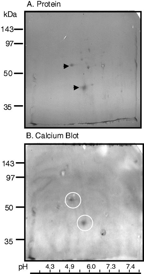
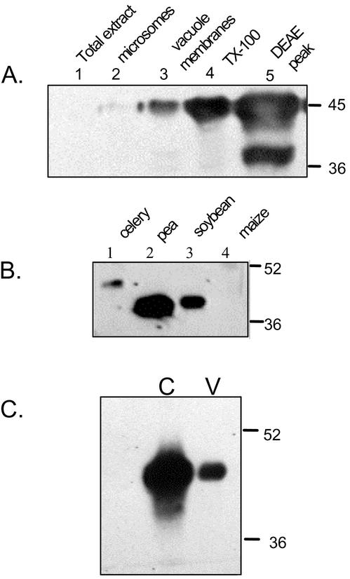
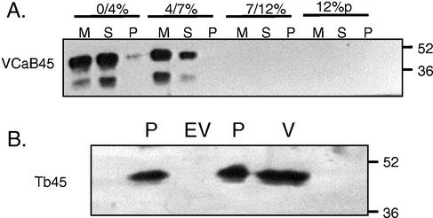
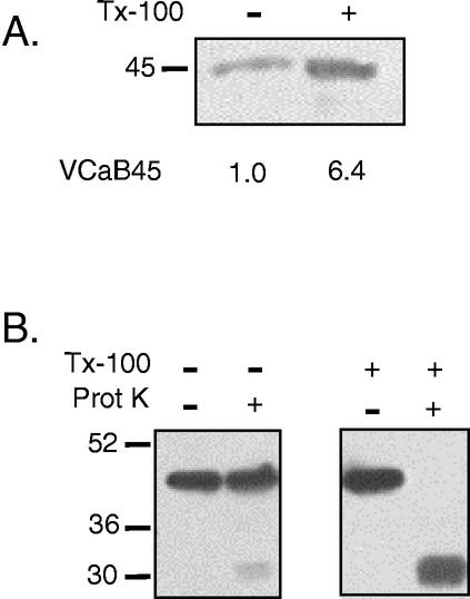
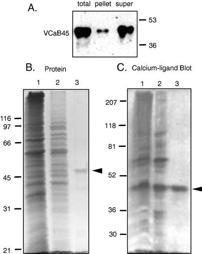
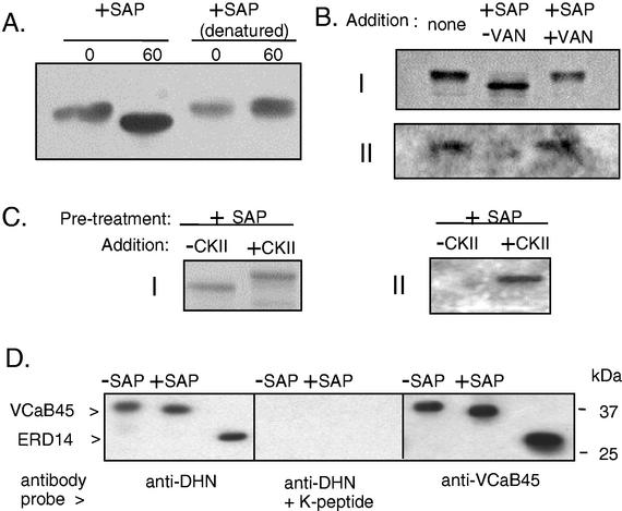
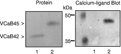
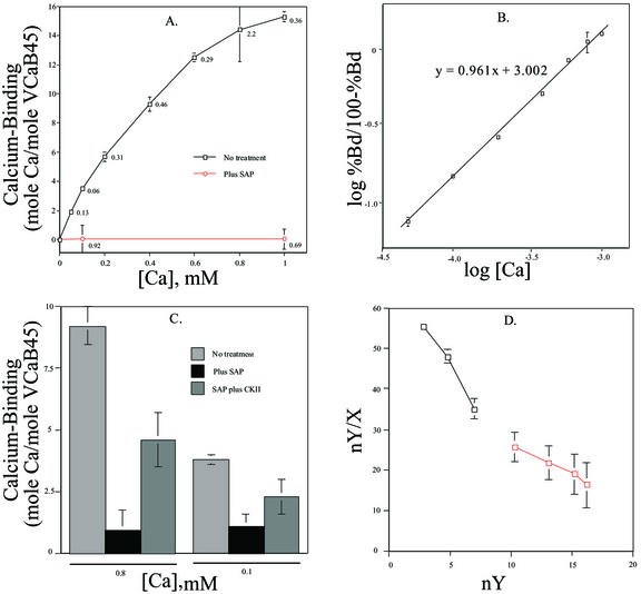
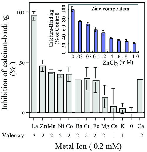
A vacuole membrane-associated calcium-binding protein with an apparent mass of 45 kD was purified from celery (Apium graveolens). This protein, VCaB45, is enriched in highly vacuolate tissues and is located within the lumen of vacuoles. Antigenically related proteins are present in many dicotyledonous plants. VCaB45 contains significant amino acid identity with the dehydrin family signature motif, is antigenically related to dehydrins, and has a variety of biochemical properties similar to dehydrins. VCaB45 migrates anomalously in sodium dodecyl sulfate-polyacrylamide gel electrophoresis having an apparent molecular mass of 45 kD. The true mass as determined by matrix-assisted laser-desorption ionization time of flight was 16.45 kD. VCaB45 has two characteristic dissociation constants for calcium of 0.22 +/- 0.142 mM and 0.64 +/- 0.08 mM, and has an estimated 24.7 +/- 11.7 calcium-binding sites per protein. The calcium-binding properties of VCaB45 are modulated by phosphorylation; the phosphorylated protein binds up to 100-fold more calcium than the dephosphorylated protein. VCaB45 is an "in vitro" substrate of casein kinase II (a ubiquitous eukaryotic kinase), the phosphorylation resulting in a partial activation of calcium-binding activity. The vacuole localization, calcium binding, and phosphorylation of VCaB45 suggest potential functions.


Figures

Figure 1
Figure 1
Two-dimensional analysis of 0.2% (w/w) Triton X-100-extracted vacuole membranes. After the Triton X-100 treatment, the soluble phase was separated first by isoelectric focusing and then by SDS-PAGE (O' Farrell, 1975). The pH gradient was established in the first dimension with 0.5% (w/v) ampholytes (3.5–5 pH range), 0.75% (w/v) ampholytes (4–6 pH range), and 0.75% ampholytes (5–7 pH range). After electrophoresis and transfer to nitrocellulose, the blot was first probed with 45calcium (B, as described) and later stained with Ponceau S to detect protein (A). The arrows in A indicate the protein spots that correspond to the calcium-binding activity detected in B. The white circles in B indicate calcium-binding activity. The pH values were obtained by slicing a parallel first dimension gel, incubating the slices in deionized water, and then measuring the pH with an pH meter. The average value of VCaB45 was deduced to be 5.2 ± 0.2 (average of five determinations). Molecular mass standards are indicated on the left.
Figure 2
Figure 2
A, Enrichment of VCaB45 during purification. Equal amounts of each fraction (2 μg protein) were fractionated by SDS-PAGE. A western blot was probed with a 1:2,000 (v/v) dilution of antibody raised against purified VCaB45. Lane 1, Whole celery (Apium graveolens) homogenate (VCaB45 is not detectable at this exposure); lane 2, microsomal membranes; lane 3, vacuolar membrane fraction; lane 4, Triton X-100 extract of vacuole membrane fraction (soluble vacuolar proteins); lane 5, peak fraction from DEAE-Sepharose column. B, Various plant tissues contain proteins of similar molecular mass that are immunologically related to VCaB45. Celery vacuolar membranes (0.05 μg, lane 1) and tissue homogenates of pea (Pisum sativum; lane 2), soybean (Glycine max; lane 3), and maize (Zea mays; lane 4) seedlings (9 μg each), were probed with anti-VCaB45. C, VCaB45 accumulates to high levels in cortical tissues of celery petioles. Cortical (C) and vascular (V) tissues were isolated separately and homogenized directly into 2× SDS-PAGE buffer. Gels were loaded with equivalent amounts of protein (10 μg) and blots were probed with anti-VCaB45. Molecular mass standards (in kD) are indicated on the right.
Figure 3
Figure 3
A, VCaB45 is enriched in low-density fractions and is released by 0.2% (w/w) Triton X-100. Membranes obtained from dextran step gradients (0%/4% [w/v] dextran interface, 4%/7% [w/v] dextran interface, etc., see Randall, 1992) were permeabilized with 0.2% (w/w) Triton X-100 (for 30 min at 4 C) and then centrifuged at 214,200g for 40 min. Equivalent portions of untreated membranes (M), the Triton X-100-solubilized supernatants (S), and membrane pellets (P) were separated by SDS-PAGE. The western blot was probed with anti-VCaB45. B, VCaB45-related protein (designated Tb45) is depleted in evacuolated tobacco protoplasts and is enriched in isolated vacuoles. Protoplasts (P) were produced from tobacco BY2 cells and vacuoles were selectively removed by ultracentrifugation resulting in evacuolated protoplasts (EV). In a separate preparation, vacuoles (V) were isolated and purified from protoplasts (P). Identical quantities of protein were resolved by SDS-PAGE. Tb45 indicates a tobacco protein immunologically related to celery VCaB45. Blots were probed with anti-VCaB45. Molecular mass standards (in kD) are indicated on the right.
Figure 4
Figure 4
Localization of VCaB45 in vacuole membranes. A, Supernatants of vacuole membranes treated with or without 0.2% (w/w) Triton X-100, were obtained after centrifugation at 100,000g for 30 min. Supernatants were separated by SDS-PAGE, blotted, and probed with anti-VCaB45. Note that to visualize VCaB45 in untreated membrane supernatants it was necessary to load twice the proportion of that loaded for Triton-treated supernatants. The numerical data for VCaB45 represent densitometric analysis (arbitrary units) of the western blot, factoring the different gel loads. Acid phosphatase activity of the supernatants of membranes treated either with or without Triton X-100 were 17 and 2 A405 min−1 μl extract−1, respectively. B, Vacuole membranes were treated with or without 0.2% (w/w) Triton X-100 and simultaneously with or without 2 mg mL−1 proteinase K. After a 30-min incubation at 4°C, a portion of the entire sample was separated by SDS-PAGE, blotted, and probed with anti-VCaB45. Molecular mass standards (mass in kD) are shown at the left.
Figure 5
Figure 5
Comparison of calcium-binding activity and total protein obtained after heat treatment. A, Celery VCaB45 remains soluble after heat treatment. The Triton X-100 extract (Total) was heat treated (20 min at 80°C–90°C) and then chilled to 4°C (10 min). A supernatant and a pellet were obtained after centrifugation of the extract at 100,000g for 30 min. Equal portions of all fractions were separated by SDS-PAGE and blots probed with anti-VCaB45. Greater than 90% of total protein precipitated after the heat treatment (not shown). B, Lane 1 contained 22.5 μg of protein; the other lanes contained a corresponding portion equivalent to the membrane volume containing 22.5 μg of protein. B, One-millimeter-thick gel stained with Coomassie Brilliant Blue. C, The same samples (but twice the amounts of protein were loaded compared with the gel in B) separated on a 2-mm-thick SDS-PAGE gel and 45calcium ligand-blotted. Lanes 1, 2, and 3 are equivalent fractions in both B and C. Lane 1, 0%/4% (w/v) dextran membranes; lane 2, Triton-extracted supernatant; lane 3, heat-treated supernatant. Though run on different gels, the major Coomassie-staining band in B, lane 3 corresponds to the calcium-binding band (arrow indicates VCaB45) in C, lane 3 (by protein staining of the polyvinylidene difluoride [PVDF] blot after the calcium blot; data not shown). Molecular mass standards (B, stainable; C, prestained; Bio-Rad Laboratories, Hercules, CA) are indicated on the left. Arrowhead indicates VCaB45.
Figure 6
Figure 6
Expression of VCaB45 protein in celery during low temperature (LT), ABA, and drought (DR) treatments. Older celery plants were treated at 6°C for 1 week and younger seedlings at 5°C for 2 d, 100 μm ABA (in 0.01% [v/v] Tween 20, 0.26% [v/v] methanol, sprayed on leaves 2 successive d) for 2 d, and drought stress (not watered for 11 d). Age indicated is the time after planting (germination is approximately 2 weeks after planting). The minus signs indicate the respective controls for each of the treatments. Total proteins were extracted as described in “Materials and Methods.” A, Blots were probed with anti-VCaB45. Left, Older plants; performed in a different experiment and the blots were developed separately from those shown in the younger plants. Both of these experiments were performed at least twice with similar results. B, Coomassie-stained gel indicating total protein from the plants treated in A. The major band present is Rubisco large subunit, mass of approximately 53 kD.
Figure 7
Figure 7
Treatment of purified celery VCaB45 with SAP results in a shift in apparent molecular mass and a decrease in calcium-binding activity. A, VCaB45, purified by heat treatment followed by anion-exchange chromatography, was treated for 60 min with SAP. Controls (0 time) were obtained by adding SAP and immediately adding SDS-PAGE sample buffer and boiling. After SDS-PAGE, blots were probed with anti-VCaB45. B, VCaB45 was treated for 60 min as in A, either without SAP, with SAP, or with SAP plus 200 μm sodium (ortho) vanadate. Reactions were terminated by the addition of hot SDS-PAGE sample buffer and heating at 90°C for 5 min. After SDS-PAGE, gels were stained with Coomassie Brilliant Blue (I) or used for calcium ligand blots (II). C, VCaB45 was dephosphorylated with SAP as in A and B, then was repurified by anion-exchange chromatography, and incubated for 3 h at 30°C in the presence or absence of casein kinase II (CKII). Samples were analyzed by Coomassie staining of gels (I) or by calcium ligand blots (II). D, Gel shifts monitored by anti-VCaB45 and antidehydrin (DHN). The first two lanes of each panel contained VCaB45, whereas the third contained the purified Arabidopsis dehydrin ERD14. VCaB45 was treated with SAP as in B. Competition of the anti-DHN was with 2.5 mg mL−1 K peptide (TGEKKGIMDKIKEKLPGQH). ERD14 (Arabidopsis ecotype Columbia) was expressed in Escherichia coli and purified by heat treatment followed by anion-exchange chromatography.
Figure 8
Figure 8
VCaB45 binds calcium in the ligand blot but the vacuole annexin, VCaB42, does not. VCaB42 was obtained as previously described (Seals and Randall, 1994) and loaded in the first lane of each gel. VCaB45 was eluted from a nitrocellulose spot cut from a two-dimensional gel. The nitrocellulose was boiled in SDS-PAGE buffer and loaded directly into the well with the sample buffer (the second lane of each gel). Left, Coomassie-stained SDS-PAGE. Right, Calcium ligand blot. Position of molecular mass standards (in kD) are indicated.
Figure 9
Figure 9
Calcium binding to purified VCaB45 estimated by equilibrium dialysis. A, Calcium binding was estimated by equilibrium dialysis. Calcium binding to purified VCaB45 (white squares) is the average of three independent experiments from a single preparation of purified VCaB45. The SAP-treated calcium-binding data (white circles) were obtained from two experiments using the same preparation. Error bars represent sds; where not shown, the sds were smaller than the symbol indicating the data point. B, Hill plot (log % VCaB45 bound/100% − % VCaB45 bound, plotted against log [Ca])k derived from the data in A. Line drawn through the 50% bound region had a slope of 0.961, indicating little cooperativity in calcium binding. C, VCaB45 was untreated (gray bar), treated with SAP to dephosphorylate the protein (black bars), or was dephosphorylated followed by rephosphorylation with casein kinase II (hatched bar). Calcium binding was performed at concentrations estimated to measure high-affinity sites (100 μm) or low-affinity sites (800 μm). D, Scatchard plot (nY/X versus nY, where nY is mol calcium bound per mol VCaB45 and X is free calcium) derived from the data shown in A. Kds derived from the shown Scatchard plot were 0.41 and 0.75 mm calcium, while the maximum number of binding sites was 27 (assuming mass of VCaB45 is 16.5 kD). Data from three different preparations of purified VCaB45 (nine experiments) indicated average Kds of 0.22 ± 0.142, 0.64 ± 0.08, and the maximal number of binding sites was 24.7 ± 11.7.
Figure 10
Figure 10
Inhibition of calcium binding by cations. Calcium binding was estimated by equilibrium dialysis. Calcium binding was performed as described, in the presence of 0.2 mm calcium. Competition was examined by adding an additional 0.2 mm of the indicated metal ion. The valence of the metal added is indicated. The bar value for an additional 0.2 mm calcium added was calculated based upon a 2-fold dilution of isotope and the binding data from Figure 9. Inset, Competition of calcium-binding by zinc. sds (where greater than 5%) are indicated. Data are average of three experiments.

Similar articles

Cited by

References

    1. Allen GJ, Sanders D. Calcineurin, a type 2B protein phosphatase, modulates the calcium-permeable slow vacuolar ion channel of stomatal guard cells. Plant Cell. 1995;7:1473–1483. - PMC - PubMed
    1. Boller T, Kende H. Hydrolytic enzymes in the central vacuole of plant cells. Plant Physiol. 1979;63:1123–1132. - PMC - PubMed
    1. Camacho P, Lechleiter JD. Calreticulin inhibits repetitive intracellular calcium waves. Cell. 1995;82:765–771. - PubMed
    1. Campbell KP, MacLennan DH, Jorgensen AO. Staining of calcium-binding proteins, calsequestrin, calmodulin, troponin C, and S-100, with the cationic carbocyanine dye, “stains-all.”. J Biol Chem. 1983a;258:11267–11273. - PubMed
    1. Campbell KP, MacLennan DH, Jorgensen AO. Purification and characterization of calsequestrin from canine cardiac sarcoplasmic reticulum and identification of the 53,000 dalton glycoprotein. J Biol Chem. 1983b;258:1197–1204. - PubMed

Publication types

MeSH terms

LinkOut - more resources