. 2002 Oct;130(2):740-56.
doi: 10.1104/pp.008110.

Molecular characterization of a heteromeric ATP-citrate lyase that generates cytosolic acetyl-coenzyme A in Arabidopsis

Affiliations

Molecular characterization of a heteromeric ATP-citrate lyase that generates cytosolic acetyl-coenzyme A in Arabidopsis

Beth L Fatland et al. Plant Physiol. 2002 Oct.

Abstract

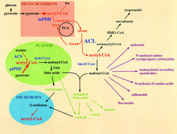
Acetyl-coenzyme A (CoA) is used in the cytosol of plant cells for the synthesis of a diverse set of phytochemicals including waxes, isoprenoids, stilbenes, and flavonoids. The source of cytosolic acetyl-CoA is unclear. We identified two Arabidopsis cDNAs that encode proteins similar to the amino and carboxy portions of human ATP-citrate lyase (ACL). Coexpression of these cDNAs in yeast (Saccharomyces cerevisiae) confers ACL activity, indicating that both the Arabidopsis genes are required for ACL activity. Arabidopsis ACL is a heteromeric enzyme composed of two distinct subunits, ACLA (45 kD) and ACLB (65 kD). The holoprotein has a molecular mass of 500 kD, which corresponds to a heterooctomer with an A(4)B(4) configuration. ACL activity and the ACLA and ACLB polypeptides are located in the cytosol, consistent with the lack of targeting peptides in the ACLA and ACLB sequences. In the Arabidopsis genome, three genes encode for the ACLA subunit (ACLA-1, At1g10670; ACLA-2, At1g60810; and ACLA-3, At1g09430), and two genes encode the ACLB subunit (ACLB-1, At3g06650 and ACLB-2, At5g49460). The ACLA and ACLB mRNAs accumulate in coordinated spatial and temporal patterns during plant development. This complex accumulation pattern is consistent with the predicted physiological needs for cytosolic acetyl-CoA, and is closely coordinated with the accumulation pattern of cytosolic acetyl-CoA carboxylase, an enzyme using cytosolic acetyl-CoA as a substrate. Taken together, these results indicate that ACL, encoded by the ACLA and ACLB genes of Arabidopsis, generates cytosolic acetyl-CoA. The heteromeric organization of this enzyme is common to green plants (including Chlorophyceae, Marchantimorpha, Bryopsida, Pinaceae, monocotyledons, and eudicots), species of fungi, Glaucophytes, Chlamydomonas, and prokaryotes. In contrast, all known animal ACL enzymes have a homomeric structure, indicating that a evolutionary fusion of the ACLA and ACLB genes probably occurred early in the evolutionary history of this kingdom.


Figures

Figure 1
Figure 1
Subcellular compartmentation of acetyl-CoA metabolism in plants. Because acetyl-CoA is impermeable to membranes, it is envisioned that it is generated independently in each compartment where it is required (cytosol, mitochondria, plastids, and peroxisomes). Cytosolic ACL is depicted together with a postulated citrate cycle that would provide citrate from the mitochondria (red). In the cytosol, acetyl-CoA can be carboxylated by acetyl-CoA carboxylase to form malonyl-CoA; alternately, two molecules of acetyl-CoA can undergo condensation to form the isoprenoid-precursor acetoacetyl-CoA. Both of these intermediates can give rise to a wide variety of metabolites. Plastidic pyruvate dehydrogenase complex and acetyl-CoA synthetase contribute to plastidic acetyl-CoA, which can be carboxylated by acetyl-CoA carboxylase and hence converted to fatty acids. In the peroxisomes, acetyl-CoA is generated during the β oxidation of fatty acids.
Figure 2
Figure 2
Immunological characterization of the Arabidopsis ACL subunits. Western-blot analysis of protein extracts from Arabidopsis siliques, probed with anti-ACLA-1 or anti-ACLB-2 sera. These antisera react with 45- and 65-kD polypeptides, respectively.
Figure 3
Figure 3
Plant ACL is composed of two subunits. Arabidopsis cDNAs ACLA-1 and ACLB-2 were cloned into the yeast expression vectors, pYX042 and pYX012, immediately downstream of the TPI promoter. The resulting transgenes were integrated individually or in combination at the leu2 and ura3 loci of S. cerevisiae strain αD273. A, Western blot of yeast proteins reacted with anti-ACLA-1 serum. B, Western blot of yeast proteins reacted with anti-ACLB-2 serum. C, ACL activity in extracts from parental strain αD273 and its derivatives that carry the indicated transgenes. The data represents the mean and sd of three separate extractions. Only the S. cerevisiae strain that carries and expresses both transgenes has ACL activity.
Figure 4
Figure 4
Arabidopsis ACL is a heteromeric complex. Protein extract from Arabidopsis siliques was subjected to gel-filtration chromatography on a Superdex 200 column. Individual fractions were collected and assayed for ACL activity, which elutes at a volume corresponding to a Mr of 500 kD (A), and SDS-PAGE/western blots:ACLA subunit (B, top) and ACLB subunit (B, bottom). ACL activity elution profile corresponds closely to the elution profile of the ACLA and ACLB subunits. C, Arabidopsis seedling extract (100 μg of protein), fractionated by nondenaturing gel electrophoresis and subjected to western-blot analysis. Blots were probed for ACLA (left) or ACLB (right). Consistent with ACLA and ACLB being organized in a protein complex, the ACLA and ACLB subunits migrate to the same position on the native gel.
Figure 5
Figure 5
ACL is a cytosolic enzyme. Chloroplasts, mitochondria, and peroxisomes were purified from pea seedling extract by a combination of differential centrifugation and Percoll-density gradient centrifugation. The specific activities of ACL, NADP-dependent glyceraldehyde 3-P dehydrogenase (NADP-GAPDH), cytochrome c oxidase, hydroxypyruvate reductase (HPR), and phosphoenolpyruvate (PEP) carboxylase were determined (A). Aliquots from each fraction, containing 50 μg of protein, were subjected to western-blot analysis for immunological detection of ACLA, ACLB, BCCP1 subunit of the chloroplastic acetyl-CoA carboxylase, the A subunit of methylcrotonyl-CoA carboxylase (MCC-A), and catalase (B).
Figure 6
Figure 6
Temporal changes in accumulation of ACLA and ACLB mRNAs in Arabidopsis. RNA (20 μg lane−1) was hybridized with ACLA-1 and ACLB-2 32P-labeled antisense RNA probes. RNA was isolated from expanding leaves (L), flower buds (B), flowers (F), and developing siliques at the indicated DAF. The data presented in this figure were gathered from a single experiment; near identical data were obtained in two replicates.
Figure 7
Figure 7
Spatial distribution of ACLA and ACLB mRNAs in Arabidopsis. Histological tissue sections were hybridized with antisense or sense (control) ACLA-1, ACLB-2, or ACC1, 35S-labeled RNA probes. Slides are stained with toluidine blue to visualize the tissue. Black spots are silver grains reflecting the location of ACLA, ACLB, or ACC1 mRNAs. Sense controls, which were conducted for each type of section, had negligible background (not shown). Hybridizations were repeated three times with similar results. ACLA, ACLB, and ACC1 mRNAs coaccumulate in particular cell types during development. Accumulation is high in the epidermis and trichomes of expanding leaves (A, E, and I), the tapetal cells of anthers of stage 10 flowers (B, F, and J), epidermal cells of growing organs (petals and ovaries) of flowers stage 11 (C, G, and K), and inner integuments of ovules the day preceding testal (seed coat) deposition (D, H, and L). Hybridization to ACLA mRNA only is shown in M through AA; results with ACLB and ACC1 are similar. Ovules of siliques at 3 DAF (M), 5 DAF (N), 7 DAF (O), 9 DAF (P), and 12 DAF (Q). Seedlings 1 d after imbibition (R), 2 d after imbibition (S and T), 3 d after imbibition (U), and 4 d after imbibition (V). Ovary of flower stage 12 (W) and stage 13 (X); nectaries and abscission zones of petal and sepals of flower stage 12 (Y). Pedicel of flower stage 13 (Z). Upper one-third of silique 2 DAF (AA). a, Anther; ap, apical meristem; ce, curled embryo; cot, cotyledon; f, filament; ge, globular embryo; he, heart embryo; ii, inner integument of ovule; l, leaf; me, mature embryo; n, nectary; o, ovule; oi, outer integument of ovule; ov, ovary; p, petal; pa, petal abscission zone; r, receptacle; ro, root; rtp, root tip; sa, sepal abscission zone; sp, sepal; stg, stigma; t, tapetum; te, torpedo embryo; tri, trichome; vb, vascular bundle; w, silique wall. Bars = 50 μm in A through O, Q, T through W, and Y through AA; bars = 150 μm in P, R, and X; and bars = 25 μm in S.
Figure 7
Figure 7
Spatial distribution of ACLA and ACLB mRNAs in Arabidopsis. Histological tissue sections were hybridized with antisense or sense (control) ACLA-1, ACLB-2, or ACC1, 35S-labeled RNA probes. Slides are stained with toluidine blue to visualize the tissue. Black spots are silver grains reflecting the location of ACLA, ACLB, or ACC1 mRNAs. Sense controls, which were conducted for each type of section, had negligible background (not shown). Hybridizations were repeated three times with similar results. ACLA, ACLB, and ACC1 mRNAs coaccumulate in particular cell types during development. Accumulation is high in the epidermis and trichomes of expanding leaves (A, E, and I), the tapetal cells of anthers of stage 10 flowers (B, F, and J), epidermal cells of growing organs (petals and ovaries) of flowers stage 11 (C, G, and K), and inner integuments of ovules the day preceding testal (seed coat) deposition (D, H, and L). Hybridization to ACLA mRNA only is shown in M through AA; results with ACLB and ACC1 are similar. Ovules of siliques at 3 DAF (M), 5 DAF (N), 7 DAF (O), 9 DAF (P), and 12 DAF (Q). Seedlings 1 d after imbibition (R), 2 d after imbibition (S and T), 3 d after imbibition (U), and 4 d after imbibition (V). Ovary of flower stage 12 (W) and stage 13 (X); nectaries and abscission zones of petal and sepals of flower stage 12 (Y). Pedicel of flower stage 13 (Z). Upper one-third of silique 2 DAF (AA). a, Anther; ap, apical meristem; ce, curled embryo; cot, cotyledon; f, filament; ge, globular embryo; he, heart embryo; ii, inner integument of ovule; l, leaf; me, mature embryo; n, nectary; o, ovule; oi, outer integument of ovule; ov, ovary; p, petal; pa, petal abscission zone; r, receptacle; ro, root; rtp, root tip; sa, sepal abscission zone; sp, sepal; stg, stigma; t, tapetum; te, torpedo embryo; tri, trichome; vb, vascular bundle; w, silique wall. Bars = 50 μm in A through O, Q, T through W, and Y through AA; bars = 150 μm in P, R, and X; and bars = 25 μm in S.
Figure 8
Figure 8
Comparisons of the primary structure of ACL. Comparisons of the amino acid sequences of the Arabidopsis ACLA and ACLB proteins with ACL, SCS, and CS from other organisms used ClustalW (Thompson et al., 1994). The degree of similarity in sequences is color-coded as follows: similar residues, green; identical residues, salmon. Conserved motifs include the ACL-SCS family signature 3 (PROSITE accession no. PS01217; residues 270–294 of Arabidopsis ACLA); the ACL-SCS family active site with His phosphorylated by ATP (black arrow; PROSITE accession no. PS00399; residues 259–275 of Arabidopsis ACLB); and the Gly-rich ACL-SCS family signature 1 (PROSITE accession no. PS01216; residues 174–203 of Arabidopsis ACLB). These motifs partially encompass the putative ATP-binding site (red bar), and a potential CoA-binding site (blue bar). Other conserved residues across ACL and SCS are Lys 3, Lys 58, Glu 116, and Asp 213 of Arabidopsis ACLA, conserved in the ATP-grasp domains (Fraser et al., 1999; Sanchez et al., 2000), and Gln 24, Pro 46, Ala 86, Arg 175, Asp 212, and Glu 232 of ACLB shown to be critical in the active site in SCS (Wolodko et al., 1994; Fraser et al., 1999; purple arrows). Ala 97 in Arabidopsis ACLB is the plant nonconservative substitution for otherwise conserved Glu. Residues forming the oxaloacetate-binding site in CS (Karpusas et al., 1990) and conserved in ACL are indicated by yellow arrows.
Figure 9
Figure 9
Phylogenetic relationships between ACLA and ACLB and homologous domains in SCS and CS. A through C, Phylogenetic trees were constructed from aligned sequences using maximum likelihood and parsimony with 100 bootstrap resampling methods of the PHYLIP 3.573 software package (Felsenstein, 1989). A, ACLA compared with SCS-β. B, ACLB compared with SCS-α. C, ACLB compared with CS. D, Scheme representing the possible evolutionary history of ACL.

Similar articles

Cited by

References

    1. Altschul SF, Gish W, Miller W, Myers EW, Lipman DJ. Basic local alignment search tool. J Mol Biol. 1990;215:403–410. - PubMed
    1. Anderson MD, Che P, Song J, Nikolau BJ, Wurtele ES. 3-Methylcrotonyl-CoA carboxylase is a component of the mitochondrial leucine catabolic pathway in plants. Plant Physiol. 1998;118:1127–1138. - PMC - PubMed
    1. Anderson M, Roberts JA. Arabidopsis: Annual Plant Reviews. Vol. 1. Boca Raton, FL: CRC Press; 1998.
    1. Arnold GW, Hill JJ. Chemical factors affecting the selection of food plants by ruminants. In: Harborne J, editor. Phytochemical Ecology. New York: Academic Press; 1972. pp. 71–101.
    1. Back K, He S, Kim KU, Shin DH. Cloning and bacterial expression of sesquiterpene cyclase, a key branch point enzyme for the synthesis of sesquiterpenoid phytoalexin capsidiol in UV-challenged leaves of Capsicum annuum. Plant Cell Physiol. 1998;39:899–904. - PubMed

Publication types

MeSH terms

Associated data