. 2002 Oct;130(2):784-95.
doi: 10.1104/pp.007781.

Inventory and functional characterization of the HAK potassium transporters of rice

Affiliations

Inventory and functional characterization of the HAK potassium transporters of rice

María A Bañuelos et al. Plant Physiol. 2002 Oct.

Abstract

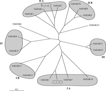
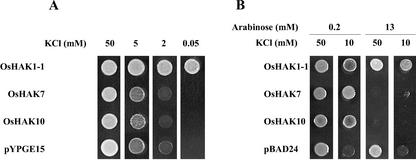
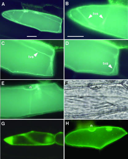
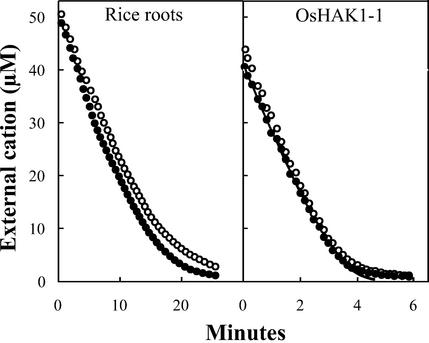
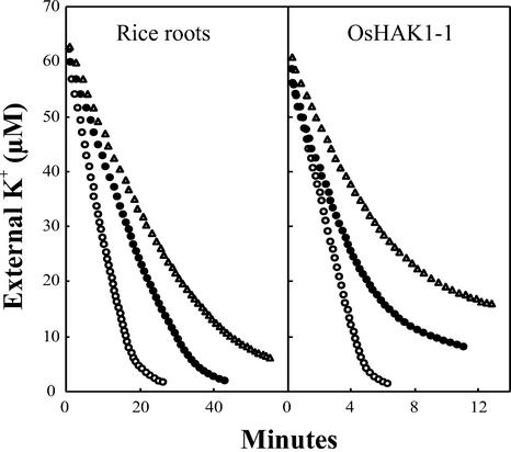
Plants take up large amounts of K(+) from the soil solution and distribute it to the cells of all organs, where it fulfills important physiological functions. Transport of K(+) from the soil solution to its final destination is mediated by channels and transporters. To better understand K(+) movements in plants, we intended to characterize the function of the large KT-HAK-KUP family of transporters in rice (Oryza sativa cv Nipponbare). By searching in databases and cDNA cloning, we have identified 17 genes (OsHAK1-17) encoding transporters of this family and obtained evidence of the existence of other two genes. Phylogenetic analysis of the encoded transporters reveals a great diversity among them, and three distant transporters, OsHAK1, OsHAK7, and OsHAK10, were expressed in yeast (Saccharomyces cerevisiae) and bacterial mutants to determine their functions. The three transporters mediate K(+) influxes or effluxes, depending on the conditions of the experiment. A comparative kinetic analysis of HAK-mediated K(+) influx in yeast and in roots of K(+)-starved rice seedlings demonstrated the involvement of HAK transporters in root K(+) uptake. We discuss that all HAK transporters may mediate K(+) transport, but probably not only in the plasma membrane. Transient expression of the OsHAK10-green fluorescent protein fusion protein in living onion epidermal cells targeted this protein to the tonoplast.


Figures

Figure 1
Figure 1
Schematic representation of 14 rice HAK genes. The black bars represent the open reading frames and the positions and lengths of introns are indicated by cuts and gray bars. Interruption of the black bars with two parallel lines denotes that the sequence is incomplete.
Figure 2
Figure 2
Phylogenetic tree of rice cv Nipponbare HAK transporters and barley transporters HvHAK1 and HvHAK2. Alignments of the sequences were performed with the ClustalX program. The scale bar corresponds to a distance of 10 changes per 100 amino acid positions. Accession numbers: HvHAK1, T04379; and HvHAK2, AAF36491; other accession numbers are given in Table I.
Figure 3
Figure 3
Alignments of the N-terminal sequences of barley HvHAK1, P. australis PceHAK1A, and OsHAK1 transporters. Accession numbers: HvHAK1, T04379; and PceHAK1A, AB055631.
Figure 4
Figure 4
Growth of OsHAK1-1-, OsHAK7-, and OsHAK10-transformed yeast (A) and bacterial (B) mutants at different K+ concentrations. The yeast trk1 trk2 mutant was transformed with the empty plasmid pYPGE15 or with the plasmid containing the tested cDNAs under the control the PGK1 promoter. The E. coli strain TKW4205, deficient in the K+ transport systems Kdp, TrkA, and Kup, was transformed with the empty plasmid pBAD24 or with the plasmid containing the tested cDNAs under the control of an Ara-responsive promoter (Guzman et al., 1995). Testing growth media contained the K+ concentrations (A) or the K+ and Ara concentrations (B) recorded in each case.
Figure 5
Figure 5
K+ loss in the E. coli strain TKW4205 transformed with plasmid pBAD24 containing the OsHAK1-1, OsHAK7, or OsHAK10 cDNAs. Time courses of the K+ contents of cells induced in 13 mm Ara and suspended in K+-free minimal medium, pH 5.5, supplemented with 4.9% (w/v) sorbitol, and 10 mm propionic acid. Black circles, Cells transformed with pBAD24; white circles, cells transformed with OsHAK1-1; black triangles, cells transformed with OsHAK7; white triangles, cells transformed with OsHAK10.
Figure 6
Figure 6
Tonoplast localization of HAK10:GFP. Transient expressions in onion epidermal cells of a OsHAK10:GFP translational fusion protein was visualized by epifluorescence microscopy. A, GFP fluorescence concentrated to the tonoplast. The large vacuole of onion epidermal cells occupies most of the cell volume. B through D, Captions of different focal planes of the cell imaged in A, showing transvacuolar strands of cytoplasm (tvs). Note that the transvacuolar strands seen in C and D are lined by two tonoplast membranes. E, The tonoplast follows the cell contour except in the perinuclear region (n) where the vacuole detaches from the cell surface. F, Clear field caption of the cell shown in E depicting the perinuclear region (n). As controls, transient expression assays in onion epidermal cells of GFP and AtNHX1:GFP were carried out in parallel. G, In the control cell expressing GFP, the fluoresce concentrated in the cytoplasm and was manifestly absent in the vacuole. H, In the control cell expressing AtNHX1:GFP, the fluorescence concentrated to the tonoplast showing the perinuclear region (n). Bar = 25 μm.
Figure 7
Figure 7
Uptake of K+ or Rb+ by roots of K+-starved rice seedlings and by the yeast trk1 trk2 mutant expressing OsHAK1-1. Depletion in the external medium of K+ (white circles) or Rb+ (black circles) was followed with a selective electrode, whose response was calibrated at intervals in the same experiment by atomic emission spectrophotometry. The line drawn in the right panel corresponds to an exact Michaelis-Menten relationship between rate and Rb+ concentration. The Kms calculated for the parts of the curves that follow the Michaelis-Menten equation are: in rice roots, 11 μm K+ or Rb+, and in yeast, 6 μm K+ or Rb+. The corresponding Vmaxs are: 3.2 nmol mg−1 min−1 in roots and 10 nmol mg−1 min−1 in yeast (dry weight in both cases).
Figure 8
Figure 8
Inhibition by NH4+ of the depletion of the external K+ by roots of K+-starved rice seedlings and by the yeast trk1 trk2 mutant expressing OsHAK1-1. Conditions as in Figure 7. NH4+ concentrations: control, white circles; 250 μm, black circles; and 500 μm, triangles.

Similar articles

Cited by

References

    1. Apse MP, Aharon GS, Snedden WA, Blumwald E. Salt tolerance conferred by overexpression of a vacuolar Na+/H+ antiport in Arabidopsis. Science. 1999;285:1256–1258. - PubMed
    1. Bakker EP. Low-affinity K+uptake systems. In: Bakker EP, editor. Alkali Cation Transport Systems in Prokaryotes. Boca Raton, FL: CRC Press; 1993. pp. 253–276.
    1. Bañuelos MA, Klein RD, Alexander-Bowman SJ, Rodríguez-Navarro A. A potassium transporter of the yeast Schwanniomyces occidentalis homologous to the Kup system of Escherichia colihas a high concentrative capacity. EMBO J. 1995;14:3021–3027. - PMC - PubMed
    1. Bañuelos MA, Madrid R, Rodríguez-Navarro A. Individual functions of the HAK and TRK potassium transporters of Schwanniomyces occidentalis. Mol Microbiol. 2000;37:671–679. - PubMed
    1. Barbier-Brygoo H, Vinauger M, Colcombet J, Ephritikhine G, Frachisse J-M, Maurel C. Anion channels in higher plants: functional characterization, molecular structure and physiological role. Biochim Biophys Acta. 2000;1465:199–218. - PubMed

Publication types

MeSH terms