. 2002 Oct;130(2):796-807.
doi: 10.1104/pp.006262.

Differential expression of two distinct phenylalanine ammonia-lyase genes in condensed tannin-accumulating and lignifying cells of quaking aspen

Affiliations

Differential expression of two distinct phenylalanine ammonia-lyase genes in condensed tannin-accumulating and lignifying cells of quaking aspen

Yu-Ying Kao et al. Plant Physiol. 2002 Oct.

Abstract

Lignins, along with condensed tannins (CTs) and salicylate-derived phenolic glycosides, constitute potentially large phenylpropanoid carbon sinks in tissues of quaking aspen (Populus tremuloides Michx.). Metabolic commitment to each of these sinks varies during development and adaptation, and depends on L-phenylalanine ammonia-lyase (PAL), an enzyme catalyzing the deamination of L-phenylalanine to initiate phenylpropanoid metabolism. In Populus spp., PAL is encoded by multiple genes whose expression has been associated with lignification in primary and secondary tissues. We now report cloning two differentially expressed PAL cDNAs that exhibit distinct spatial associations with CT and lignin biosynthesis in developing shoot and root tissues of aspen. PtPAL1 was expressed in certain CT-accumulating, non-lignifying cells of stems, leaves, and roots, and the pattern of PtPAL1 expression varied coordinately with that of CT accumulation along the primary to secondary growth transition in stems. PtPAL2 was expressed in heavily lignified structural cells of shoots, but was also expressed in non-lignifying cells of root tips. Evidence of a role for Pt4CL2, encoding 4-coumarate:coenzyme A ligase, in determining CT sink strength was gained from cellular co-expression analysis with PAL1 and CTs, and from experiments in which leaf wounding increased PAL1 and 4CL2 expression as well as the relative allocation of carbon to CT with respect to phenolic glycoside, the dominant phenolic sink in aspen leaves. Leaf wounding also increased PAL2 and lignin pathway gene expression, but to a smaller extent. The absence of PAL2 in most CT-accumulating cells provides in situ support for the idea that PAL isoforms function in specific metabolic milieus.


Figures

Figure 1
Figure 1
Northern-blot analysis of PAL, 4CL, and COMT transcript levels in various tissues of aspen. Ten micrograms of total RNA from each tissue was resolved on a 1% (w/v) denaturing agarose gel that was photographed before being blotted and cross-linked onto a nylon membrane. Duplicate blots were hybridized with 32P-labeled full-length PtPAL1 and PtPAL2 cDNAs, stripped between hybridizations, and consecutively probed with 32P-labeled full-length Pt4CL1, Pt4CL2, and PtCOMT cDNAs at high stringency.
Figure 2
Figure 2
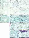
In situ localization of PtPAL1 and PtPAL2 mRNAs and histochemical detection of CTs and lignin in aspen stem tissues. Transverse stem sections (10-μm thickness) were hybridized with digoxygenin (DIG)-labeled antisense PAL1 (A–C) or PAL2 (D–F) RNA probes and photographed in bright field. Transverse stem sections (75-μm thickness) were stained with dimethylaminocinnamaldehyde (DMACA; G–I), vanillin-HCl (J), or were nitroso-derivatized (K) for the detection of CTs, or were stained with phloroglucinol for the detection of lignin (L and M). Shown are first internode (A, D, and G), third internode (B and E), fifth internode (H, J, and L), and 10th internode (C, F, I, K, and M). Scale bar = 200 μm (A, D, and G) or 100 μm (all other panels). co, Cortex; cz, cambial zone; e, epidermis; h, hypodermis; id, idioblast, pf, phloem fibers; ph, phloem; pi, pith; xy, xylem.
Figure 3
Figure 3
In situ localization of PtPAL1, PtPAL2, and Pt4CL2 mRNAs and histochemical detection of CTs in developing aspen leaves. Sections (10-μm thickness) were hybridized with DIG-labeled antisense PAL1 (A, C, and E), PAL2 (B), or 4CL2 (D and F) RNA probes, or stained with toluidine blue (TB) for the detection of total phenolics (H and J). Sections (75-μm thickness) were stained with DMACA (G) or vanillin-HCl (I) for the detection of CTs. Shown are third leaf midvein and lamina (A, B, C, D, G and H) and 10th leaf lamina (E, F, I and J). Scale bar = 200 μm (A and B) or 50 μm (C–J). e, Epidermis; h, hypodermis; pa, palisade; sp, spongy mesophyll; xy, xylem.
Figure 4
Figure 4
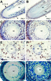
In situ localization of PtPAL1 and PtPAL2 mRNAs and histochemical detection of CTs in aspen root tips. Longitudinal tangential sections (A and B) and transverse sections (C–F) 10 μm in thickness were hybridized with DIG-labeled antisense PAL1 (A, C, and E) or PAL2 (B, D, and F) RNA probes. Transverse sections (75-μm thickness) were stained with DMACA for the detection of CTs (G and H). Arrows in A indicate locations of transverse sections represented in C, D, and G. Transverse sections E, F, and H correspond to a distance approximately 2 mm from root tips. Scale bar = 200 μm (A and B) or 100 μm (C–H). co, Cortex; e, epidermis; ph, phloem; rc, root cap; vc, vascular cylinder; xy, xylem.
Figure 5
Figure 5
Local and systemic wounding effects on PAL transcript and CT levels in aspen. A, PAL, 4CL, and COMT transcript levels in leaves and stems of aspen that were wounded, sprayed with 1 mm salicylic acid in 0.01% (v/v) Triton X-100 (SA) or with 0.01% (v/v) Triton X-100 as a control for SA treatment (Triton) on leaves 11 and 12, or were dark acclimated for 72 h. Wounding and spray treatments were repeated after 2 h and tissues harvested 24 h after the initial treatment for RNA analysis as described in Figure 2. B, Effects of a more severe 24- to 48-h wounding treatment on PAL1 and PAL2 transcript levels in injured leaves (11th and 12th) and uninjured nearby (eighth–10th) or distant (third–sixth) stem internodes.

Similar articles

Cited by

References

    1. Allina SM, Aviva P-H, Theilmann DA, Ellis BE, Douglas CJ. 4-Coumarate:coenzyme A ligase in hybrid poplar. Plant Physiol. 1998;116:743–754. - PMC - PubMed
    1. Allwood EG, Davies DR, Gerrish C, Ellis BE, Bolwell GP. Phosphorylation of phenylalanine ammonia-lyase: evidence for a novel protein kinase and identification of the phosphorylated residue. FEBS Lett. 1999;457:47–52. - PubMed
    1. Appert C, Logemann E, Hahlbrock K, Schmid J, Amrhein N. Structural and catalytic properties of the four phenylalanine ammonia-lyase isoenzymes from parsley (Petroselinum crispum Nym.) Eur J Biochem. 1994;225:491–499. - PubMed
    1. Bolwell GP. A role for phosphorylation in the down-regulation of phenylalanine ammonia-lyase in suspension-cultured cells of French bean. Phytochemistry. 1992;31:4081–4086.
    1. Bolwell GP, Cramer CL, Lamb CJ, Schuch W, Dixon RA. l- Phenylalanine ammonia-lyase from Phaseolus vulgaris: modulation of the levels of active enzyme by trans-cinnamic acid. Planta. 1986;169:97–107. - PubMed

MeSH terms

Associated data