. 2002 Oct;130(2):808-22.
doi: 10.1104/pp.003491.

SHORT INTEGUMENTS1/SUSPENSOR1/CARPEL FACTORY, a Dicer homolog, is a maternal effect gene required for embryo development in Arabidopsis

Affiliations

SHORT INTEGUMENTS1/SUSPENSOR1/CARPEL FACTORY, a Dicer homolog, is a maternal effect gene required for embryo development in Arabidopsis

Teresa A Golden et al. Plant Physiol. 2002 Oct.

Abstract

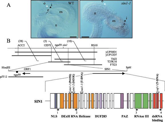
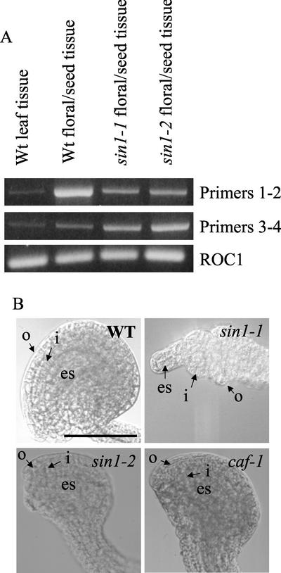
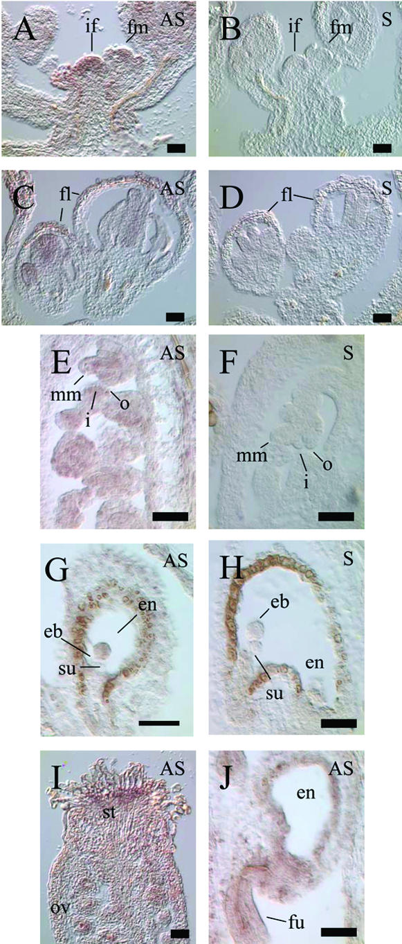
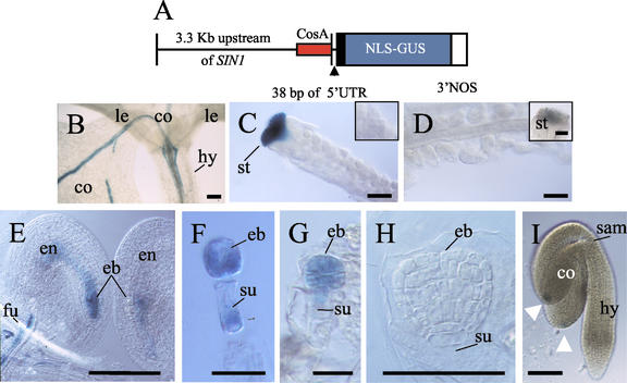
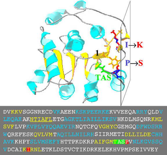
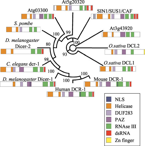
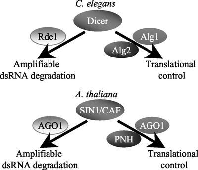
The importance of maternal cells in controlling early embryogenesis is well understood in animal development, yet in plants the precise role of maternal cells in embryogenesis is unclear. We demonstrated previously that maternal activity of the SIN1 (SHORT INTEGUMENTS1) gene of Arabidopsis is essential for embryo pattern formation and viability, and that its postembryonic activity is required for several processes in reproductive development, including flowering time control and ovule morphogenesis. Here, we report the cloning of SIN1, and demonstrate its identity to the CAF (CARPEL FACTORY) gene important for normal flower morphogenesis and to the SUS1 (SUSPENSOR1) gene essential for embryogenesis. SIN1/SUS1/CAF has sequence similarity to the Drosophila melanogaster gene Dicer, which encodes a multidomain ribonuclease specific for double-stranded RNA, first identified by its role in RNA silencing. The Dicer protein is essential for temporal control of development in animals, through the processing of small RNA hairpins that in turn inhibit the translation of target mRNAs. Structural modeling of the wild-type and sin1 mutant proteins indicates that the RNA helicase domain of SIN1/SUS1/CAF is important for function. The mRNA was detected in floral meristems, ovules, and early embryos, consistent with the mutant phenotypes. A 3.3-kb region 5' of the SIN1/SUS1/CAF gene shows asymmetric parent-of-origin activity in the embryo: It confers transcriptional activation of a reporter gene in early embryos only when transmitted through the maternal gamete. These results suggest that maternal SIN1/SUS1/CAF functions early in Arabidopsis development, presumably through posttranscriptional regulation of specific mRNA molecules.


Figures

Figure 1
Figure 1
SIN1 gene structure and its mutant phenotype in the ovule. A, In wild-type ovules, the outer integument cell layers entirely cover the inner integument that encloses the embryo sac that contains the egg. Most mutant ovules show uncoordinated growth of both the inner and outer integuments and the nucellus, such that the embryo sac, with the egg, is extruded. o, Outer integuments; i, inner integuments; es, embryo sac; n, nucellus. B, Map of the chromosomal region overlapping SIN1. RS10, nga59, 12D7L, and ACC2 are DNA sequence markers. Numbers within parentheses are numbers of crossovers between La-er and Columbia (Col) chromosomes. yUP20D1 and yUP12D7 are yeast (Saccharomyces cerevisiae) artificial chromosome clones; T4J2, T25K16, and F7I23 are bacterial artificial chromosomes (BACs); and pJT12 is a plasmid subclone of Arabidopsis chromosome I. The positions of the only two identified open reading frames (ORFs) in this region (ORF1 and SIN1) are represented by large arrows. The lower diagram shows intron-exon boundaries of SIN1 (exons are boxed), with different protein-coding domains color coded. The two primer sets (1 and 2, 3 and 4) used for reverse transcriptase (RT)-PCR (Fig. 2A) are represented by arrows corresponding to the primers location in the SIN1 gene.
Figure 2
Figure 2
Analysis of sin1 and caf mutants. A, RT-PCR analysis of SIN1/SUS1/CAF mRNA expression. The top row represents signal from RT-PCR with a primer set that amplifies +457 to +1,763 (1 and 2), and the middle row is signal with a set that amplifies +4,545 to +6,138 (3 and 4). The bottom row is signal from the constitutively expressed mRNA of the ROC1 gene. SIN1/SUS1/CAF mRNA is expressed at a higher level in floral tissue than in leaves, and it is not significantly lower in the sin1 point mutations. The relative levels of the two RT-PCR-amplified bands from mutant and wild-type flowers were not reproducibly different. B, Ovule morphology affected by the sin1 and caf mutant alleles in La-er. o, Outer integuments; i, inner integuments; es, embryo sac. Bar = 100 microns.
Figure 3
Figure 3
Localization of SIN1/SUS1/CAF transcript by in situ hybridization. A and B, Inflorescence and early floral meristems, showing strong expression in the inflorescence meristem and in the center of the floral meristem. C and D, Developing flowers, with expression in the center of the flower. E and F, RNA accumulated in all cell types of the early ovule. G and H, Expression in early embryo is located in the embryo proper, and is not detected in the suspensor or the endosperm. I, Stigmatic tissue shows strong expression, and message is detected in immature ovules. J, Strong expression is coincident with vascular elements of the funiculus in seeds with heart stage embryos. Sections hybridized with full-length cDNA antisense probe (A, C, E, G, I, and J) are indicated by AS, whereas sections hybridized with the sense probe (B, D, F, H, and J) are indicated by S. if, Inflorescence meristem; fm, floral meristem; fl, developing flowers; mm, megaspore mother cell; o, outer integuments; i, inner integuments; eb, embryo; en, endosperm; su, suspensor; st, stigmatic tissue; ov, ovules; fu, funiculus. All bars = 100 microns.
Figure 4
Figure 4
Expression pattern of the SIN1/SUS1/CAF upstream genomic region, assayed by promoter GUS fusion. A, The SIN1/SUS1/CAF putative promoter construct pSP2, containing 3.3 kb of sequence upstream of the SIN1/SUS1/CAF gene (with 38 bp of the 5′-UTR), fused to the GUS reporter gene. The minimal promoter region from the cosmid CosA (which rescues the caf-1 mutant phenotype) is identified in red (Jacobsen et al., 1999). B, Two-leaf stage seedling (10 d post-germination) showing GUS activity in developing veins of the cotyledons and some expression in the hypocotyl. C, GUS expression is only detectable in the stigmatic tissue, and undetected in the initiating integuments at floral stage 11 (inset). D, Mature ovules at floral stage 13 show no detectable activity, and a reduction in GUS activity is seen in the stigma (inset). E and F, Post-fertilization ovules showing staining in the zygote and the endosperm. GUS activity is also present in the funiculus, which was observed throughout seed development. F, GUS expression in a dissected two-cell embryo, with activity in both the embryo and suspensor. G, GUS expression in octant embryo, but now absent from the suspensor. H, No detectable GUS activity in late globular or later embryonic stages (data not shown). I, Linear to cotyledon stage embryo showing GUS expression at the tip of the cotyledon (white arrowheads). co, Cotyledon; le, leaf; hy, hypocotyl; st, stigma; eb, embryo; en, endosperm; fu, funiculus; su, suspensor; sam, SAM. All bars = 100 microns, except F and G, which = 50 microns.
Figure 5
Figure 5
Structural model of the SIN1/SUS1/CAF helicase domain. Yeast translation initiation factor yIF4A was used as a template for alignment of the SIN1/SUS1/CAF helicase domain residues (inset shows the amino acid sequence, with mutated residues boxed). α-helices and β-strands are light-blue and yellow ribbons, respectively. The peptide backbone of the TAS motif is in green. The side chains of residues in the mutated positions are shown; wild-type residues are dark blue and mutated residues are red. The side chains of those residues in the first β-strand (underlined in the inset) that clash with the mutated residues are in yellow. The plane, formed by an α-helix and two β-strands, predicted to be involved in RNA binding and unwinding, is indicated.
Figure 6
Figure 6
Phylogenetic analysis of known Dicer-like proteins. A neighbor-joining circle tree, constructed by using full-length protein sequences of SIN1/SUS1/CAF and all known Dicer homologs. The predicted domain structures for Dicer-like proteins were identified by Pfam and Prosite searches. Sequence accessions are: human Dicer-1 (gi14748177), Schizosaccharomyces pombe CAB37423 (gi2130449), Arabidopsis F5024.210 and At5g20320 (gi15241323), Arabidopsis T15B3.8 and At3g43920 (gi7594553), SIN1/SUS1/CAF and At1g01040 (gi11559646), Arabidopsis T17B22.1 and Atg03300 (gi6714410), C. elegans dcr-1 (gi630692), D. melanogaster Dicer-1 (gi17738129), D. melanogaster Dicer-2 (gi16215719), Oryza sativa DCL1 (gi18087887), Mus musculus mDCR-1 (gi20385913), and O. sativa DCL2 (gi:20804934). Analysis using only the conserved helicase or RNase III domains produced identical tree structures.
Figure 7
Figure 7
Models of SIN1/SUS1/CAF function in vivo. Based on parallel genetic evidence in C. elegans (Olsen and Ambros, 1999; Grishok et al., 2001; Ruvkun, 2001), we propose that in Arabidopsis, SIN1/SUS1/CAF interacts with AGO1 and/or PNH to regulate developmentally important genes through control of translation initiation. In addition, we propose that SIN1/SUS1/CAF mediates degradation of dsRNA in conjunction with AGO1 (Fagard et al., 2000).

References

    1. Altschul SF, Madden TL, Schaffer AA, Zhang J, Zhang Z, Miller W, Lipman DJ. Gapped BLAST and PSI-BLAST: a new generation of protein database search programs. Nucleic Acids Res. 1997;25:3389–3402. - PMC - PubMed
    1. Baroux C, Spillane C, Grossniklaus U. Genomic imprinting during seed development. In: Dunlap JC, Wu C-t, editors. Advances in Genetics: Homology Effects. San Diego: Academic Press; 2002. pp. 165–214. - PubMed
    1. Bateman A, Birney E, Durbin R, Eddy SR, Howe KL, Sonnhammer EL. The Pfam protein families database. Nucleic Acids Res. 1999;27:263–266. - PMC - PubMed
    1. Bernstein E, Caudy A, Hammond S, Hannon G. Role for a bidentate ribonuclease in the initiation step of RNA interference. Nature. 2001;409:363–366. - PubMed
    1. Birney E, Thompson JD, Gibson TJ. PairWise and SearchWise: finding the optimal alignment in a simultaneous comparison of a protein profile against all DNA translation frames. Nucleic Acids Res. 1996;24:2730–2739. - PMC - PubMed

Publication types

MeSH terms

Associated data