. 2002 Oct;130(2):977-88.
doi: 10.1104/pp.004267.

Endoplasmic microtubules configure the subapical cytoplasm and are required for fast growth of Medicago truncatula root hairs

Affiliations

Endoplasmic microtubules configure the subapical cytoplasm and are required for fast growth of Medicago truncatula root hairs

Björn J Sieberer et al. Plant Physiol. 2002 Oct.

Abstract

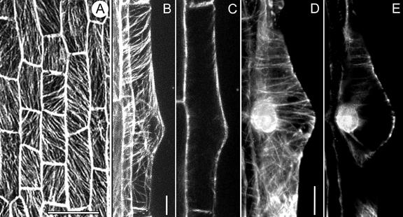
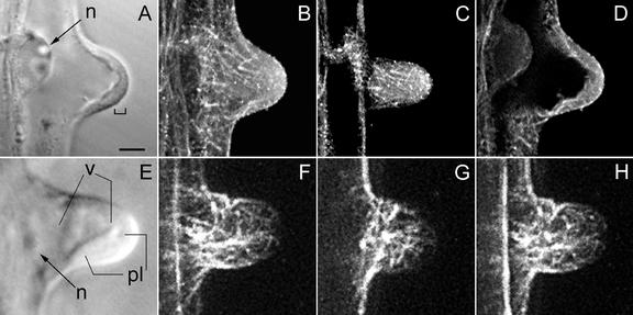
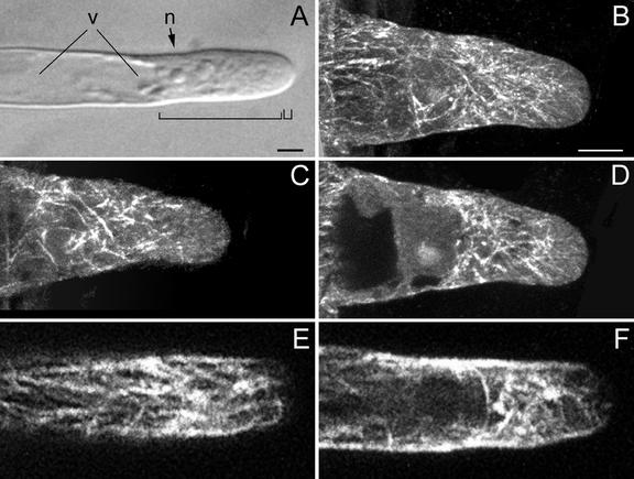
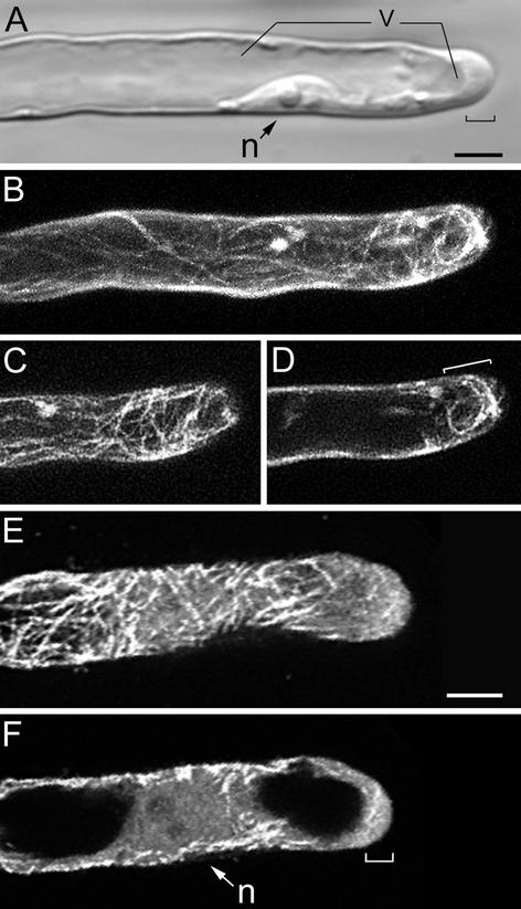
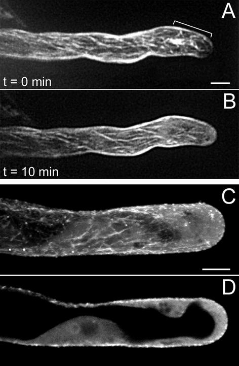
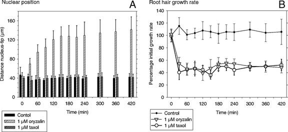
To investigate the configuration and function of microtubules (MTs) in tip-growing Medicago truncatula root hairs, we used immunocytochemistry or in vivo decoration by a GFP linked to a MT-binding domain. The two approaches gave similar results and allowed the study of MTs during hair development. Cortical MTs (CMTs) are present in all developmental stages. During the transition from bulge to a tip-growing root hair, endoplasmic MTs (EMTs) appear at the tip of the young hair and remain there until growth arrest. EMTs are a specific feature of tip-growing hairs, forming a three-dimensional array throughout the subapical cytoplasmic dense region. During growth arrest, EMTs, together with the subapical cytoplasmic dense region, progressively disappear, whereas CMTs extend further toward the tip. In full-grown root hairs, CMTs, the only remaining population of MTs, converge at the tip and their density decreases over time. Upon treatment of growing hairs with 1 microM oryzalin, EMTs disappear, but CMTs remain present. The subapical cytoplasmic dense region becomes very short, the distance nucleus tip increases, growth slows down, and the nucleus still follows the advancing tip, though at a much larger distance. Taxol has no effect on the cytoarchitecture of growing hairs; the subapical cytoplasmic dense region remains intact, the nucleus keeps its distance from the tip, but growth rate drops to the same extent as in hairs treated with 1 microM oryzalin. The role of EMTs in growing root hairs is discussed.


Figures

Figure 1
Figure 1
CMTs in trichoblasts before bulge formation (A) and during bulge formation (B–E), visualized with a CLSM in scanning steps of 1 μm. A through C, GFP-MBD; D and E, Immunocytochemistry. A, CMTs are obliquely or longitudinally oriented to the long axis of the root. Bar = 50 μm. B, Full-stack projection of CMTs. CMTs loop through the tip of the bulge and are transversely or slightly helically oriented to the long axis of the root in the epidermal part. Bar = 10 μm. C, Projection of four median sections. There are no detectable EMTs in this developmental stage. D, Full-stack projection of CMTs; for explanation see B. Bar = 10 μm. E, Note position of the nucleus. For explanation see C.
Figure 2
Figure 2
MTs during transition from a bulge into a growing root hair, visualized with a CLSM in scanning steps of 1 μm (B–D and F–H). B through D, Immunocytochemistry; F through H, GFP-MBD. In this developmental stage, polar growth is initiated. A, Corresponding bright-field image to B through D; the bracket indicates a thin layer of cytoplasm at the tip. B, Full-stack projection of MTs. C, Projection of three peripheral sections showing CMTs. A few CMTs still reach the very tip and are net-axially oriented below the tip region. D, Projection of four median sections; EMTs start to appear at this stage of hair development. E, Bright-field image of a hair at a somewhat later stage than shown in A. The cytoplasmic layer in the tip region has increased in length. F, Full-stack projection of MTs. G, Projection of three peripheral sections. Single CMTs still reach the very tip. H, Projection of four median sections showing EMTs. There is a concomitant increase in the length of the subapical cytoplasmic dense region and the density of EMTs. A through D show an earlier stage than E through H. Magnification is the same in all images. Bar = 20 μm. n, Nucleus; v, vacuole; pl, cytoplasmic layer.
Figure 3
Figure 3
MTs in growing root hairs visualized with a CLSM in scanning steps of 1 μm (B–F). B through D, Immunocytochemistry; E and F, GFP-MBD. A, Bright-field image of a living hair; the small bracket indicates the vesicle-rich region and the large bracket indicates the subapical cytoplasmic dense region. n, Nucleus; v, central vacuole. Bar = 10 μm. B, Full-stack projection of MTs. C, Projection of three sections of the cell periphery showing net-axially aligned CMTs. CMTs do not reach the very tip. D, Projection of four median sections showing EMTs. EMTs are abundant close to the nucleus and in the lower part of the subapical cytoplasmic dense region. The density of EMTs in the upper part of the subapical cytoplasmic dense region is low. Only a few EMTs reach the very tip. E, Projection of three sections of the cell periphery showing CMTs. For explanation see C. F, Projection of four median sections; for explanation see D. Magnification in B through F is the same. Bar in B = 10 μm.
Figure 4
Figure 4
MTs in growth-arresting hairs visualized with a CLSM in scanning steps of 1 μm (B–F). B through D, GFP-MBD; E and F, immunocytochemistry. At this developmental stage, the subapical cytoplasmic dense region has almost disappeared and the distance tip-nucleus has increased. A, Bright-field image of a living hair; the bracket indicates the remaining subapical cytoplasmic dense region and the smooth (vesicle rich) region at the very tip. Bar = 10 μm (A–D). B, Full-stack projection of MTs. C, Projection of three peripheral sections showing CMTs. CMTs are net-axially oriented. D, Projection of four median sections. Region of EMTs has decreased in length (bracket). E, Projection of three peripheral sections showing CMTs. For explanation see C. Bar = 10 μm. F, Projection of four median sections; for explanation see D. n, Nucleus; v, vacuole.
Figure 5
Figure 5
MTs in full-grown root hairs visualized with a CLSM in scanning steps of 1 μm. B and C, Immunocytochemistry; D through F, GFP-MBD. A, Bright-field image of a living hair. Note position of the nucleus (n). Bar = 10 μm. B, Full-stack projection of CMTs. CMTs, the only remaining population of MTs in this developmental stage, are longitudinally oriented; they converge at the very tip. Bar = 10 μm (B–F). C, Projection of two median sections. Full-grown hairs have no EMTs. D, Full-stack projection of CMTs. The hair has stopped growth recently and the density of CMTs, which are net-axially oriented, is similar to previous developmental stages. E, Projection of two median sections; for explanation see C. F, Full-stack projection of CMTs in a hair of a later stage than shown in D. The density of CMTs is lower than in hairs that have just terminated growth.
Figure 6
Figure 6
MTs in growing root hairs treated with 1 μm oryzalin. A and B, GFP-MBD; C and D, immunocytochemistry. A, Hair before treatment; a full-stack projection shows abundant MTs in the subapical region (bracket). B, Ten minutes after treatment. The subapical region contains less MTs and the MTs (cortical) in the shank of the hair are unchanged. C, Hair 15 min after treatment; a full-stack projection shows CMTs. D, A projection of four median sections of the hair in C shows that EMTs and the subapical cytoplasmic dense region have completely disappeared, but the vesicle-rich region still is present at the very tip (see also Fig. 7, B and C). Bar in A = 10 μm; bar in C = 10 μm.
Figure 7
Figure 7
The effect of treatment with oryzalin or taxol on the cytoarchitecture of growing root hairs. A, Before treatment. B, Thirty minutes after 1 μm oryzalin. C, Sixty minutes after 1 μm oryzalin. D, Before treatment. E, Thirty minutes after 1 μm taxol. F, Sixty minutes after 1 μm taxol. Bright-field images from living root hairs. Large bracket indicates the subapical cytoplasmic dense region, and the small bracket indicates the smooth (vesicle-rich) region. n, Nucleus. Magnification is the same in all images. Bar = 20 μm.
Figure 8
Figure 8
Nuclear position (A) and root hair growth rate (B) in growing M. truncatula root hairs after control treatment and treatment with 1 μm oryzalin or 1 μm taxol. In controls and taxol-treated hairs, the nucleus kept a distance of 30 to 40 μm to the tip over time, but it was significantly increased in oryzalin (A). Growth rate in the hairs remained high in controls, but dropped by approximately 60% in oryzalin and taxol (B). Results for each treatment are presented as the means of 10 hairs with their sd. Results are representative for three independent replicates for each treatment.

Similar articles

Cited by

References

    1. Anthony RG, Hussey PJ. Dinitroaniline herbicide resistance and the microtubule cytoskeleton. Trends Plant Sci. 1999;4:112–116. - PubMed
    1. Baluska F, Salaj J, Mathur J, Braun M, Jasper F, Samaj J, Chua NH, Barlow PW, Volkmann D. Root hair formation: F-actin-dependent tip growth is initiated by local assembly of profilin-supported F-actin meshworks accumulated within expansin-enriched bulges. Dev Biol. 2000;227:618–632. - PubMed
    1. Baskin TI, Miller DD, Vos JW, Wilson JE, Hepler PK. Cryofixing single cells and multicellular specimens enhances structure and immunocytochemistry for light microscopy. J Microsc. 1996;182:149–161. - PubMed
    1. Bibikova TN, Blancaflor EB, Gilroy S. Microtubules regulate tip growth and orientation in root hairs of Arabidopsis thaliana. Plant J. 1999;17:657–665. - PubMed
    1. Boisson-Dernier A, Chabaud M, Garcia F, Bécard G, Rosenberg C, Barker DG. Agrobacterium rhizogenes-transformed roots of Medicago truncatula for the study of nitrogen-fixing and endomycorrhizal symbiotic associations. Mol Plant-Microbe Interact. 2001;14:695–700. - PubMed

Publication types

MeSH terms

LinkOut - more resources