

High prevalence of osteoporotic vertebral fractures in patients with Crohn's disease
- PMID: 12377802
- PMCID: PMC1773437
- DOI: 10.1136/gut.51.5.654
High prevalence of osteoporotic vertebral fractures in patients with Crohn's disease
Abstract
Background and aims: Osteopenia and osteoporosis are frequent in Crohn's disease. However, there are few data on related vertebral fractures. Therefore, we evaluated prospectively the prevalence of osteoporotic vertebral fractures in these patients.
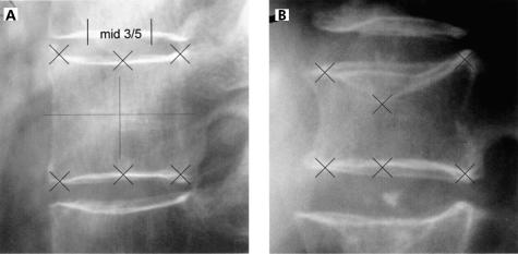
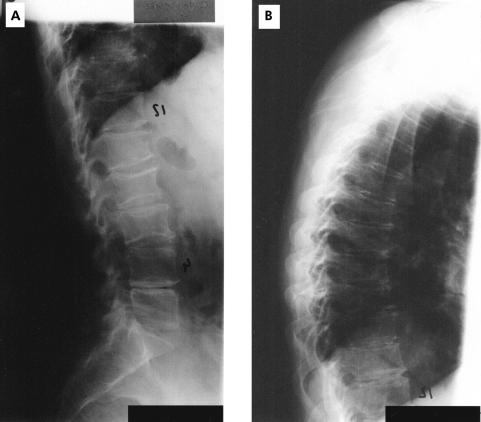
Methods: A total of 293 patients were screened with dual energy x ray absorptiometry of the lumbar spine (L1-L4) and proximal right femur. In 156 patients with lumbar osteopenia or osteoporosis (T score <-1), x ray examinations of the thoracic and lumbar spine were performed. Assessment of fractures included visual reading of x rays and quantitative morphometry of the vertebral bodies (T4-L4), analogous to the criteria of the European Vertebral Osteoporosis Study.
Results: In 34 (21.8%; 18 female) of 156 Crohn's disease patients with reduced bone mineral density, 63 osteoporotic vertebral fractures (50 fx. (osteoporotic fracture with visible fracture line running into the vertebral body and/or change of outer shape) and 13 fxd. (osteoporotic fracture with change of outer shape but without visible fracture line)) were found, 50 fx. in 25 (16%, 15 female) patients and 13 fxd. in nine (5.8%, three female) patients. In four patients the fractures were clinically evident and associated with severe back pain. Approximately one third of patients with fractures were younger than 30 years. Lumbar bone mineral density was significantly reduced in patients with fractures compared with those without (T score -2.50 (0.88) v -2.07 (0.66); p<0.025) but not at the hip (-2.0 (1.1) v -1.81 (0.87); p=0.38). In subgroups analyses, no significant differences were observed.
Conclusions: In patients with Crohn's disease and reduced bone mineral density, the prevalence of vertebral fractures-that is, manifest osteoporosis-was strikingly high at 22%, even in those aged less than 30 years, a problem deserving further clinical attention.
Figures
References
-
- Semeao EJ, Stallings VA, Peck SN, et al. Vertebral compression fractures in pediatric patients with Crohn’s disease. Gastroenterology 1997;112:1710–13. - PubMed
-
- Gokhale R, Favus MJ, Karrison Th, et al. Bone mineral density in children with inflammatory bowel disease. Gastroenterology 1998;114:902–11. - PubMed
-
- Vogelsang H, Ferenci P, Woloszeznk W, et al. Bone disease in vitamin D deficient patients with Crohn’s disease. Dig Dis Sci 1989;34:1094–9. - PubMed
MeSH terms
LinkOut - more resources
Full Text Sources
Medical