

Expression of p53 and C-myc genes and its clinical relevance in the hepatocellular carcinomatous and pericarcinomatous tissues
- PMID: 12378623
- PMCID: PMC4656568
- DOI: 10.3748/wjg.v8.i5.822
Expression of p53 and C-myc genes and its clinical relevance in the hepatocellular carcinomatous and pericarcinomatous tissues
Abstract
Aim: To investigate the possible roles of p53 and C-myc genes in the primary hepatocellular carcinogenesis and the relationship between the liver hyperplastic nodule (LHN) and hepatocellular carcinoma(HCC).
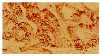
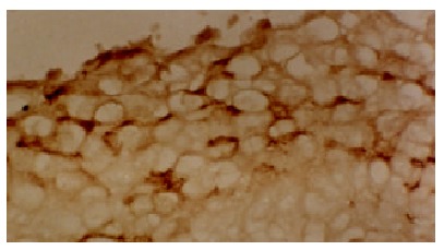
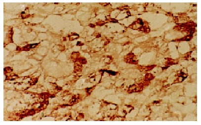
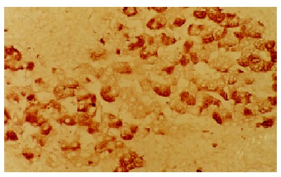
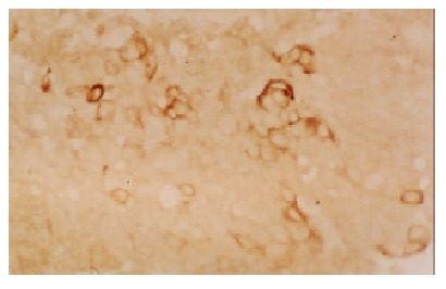
Methods: The expression of p53 and C-myc genes was detected immunohistochemically in 73 and 60 cases of HCC and pericarcinomatous tissues, respectively.
Results: The positive expression of p53 in HCC was significantly higher than that in pericarcinomatous tissues (P<0.05). In pericarcinomatous tissues, the p53 expression was observed only in LHN, but not in liver cirrhosis (LC) and normal liver tissues. The positive expression rate of C-myc in HCC or LHN was significantly higher than that in LC or normal liver tissues (P<0.05 and P<0.01), however, no significant difference was found between HCC and LHN (P>0.05). The positive expression rate of p53 and C-myc in HCC was correlated with the histological differentiation, that in the poorly differentiated was significantly higher than that in well differentiated samples (P<0.05).
Conclusion: The overexpression of p53 and C-myc genes might play a role in the carcinogenesis of HCC; And LHN seems a preneoplastic lesion related to hepatocarcinogenesis; No evidence supports that LC contribute directly to the hepatocarcinogenesis.
Figures
Similar articles
-
The Differential Immunohistochemical Expression of p53, c-Jun, c-Myc, and p21 Between HCV-related Hepatocellular Carcinoma With and Without Cirrhosis.Appl Immunohistochem Mol Morphol. 2016 Feb;24(2):75-87. doi: 10.1097/PAI.0000000000000155. Appl Immunohistochem Mol Morphol. 2016. PMID: 25710583
-
Expression of c-erbB-2 and glutathione S-transferase-pi in hepatocellular carcinoma and its adjacent tissue.World J Gastroenterol. 2005 Jul 28;11(28):4404-8. doi: 10.3748/wjg.v11.i28.4404. World J Gastroenterol. 2005. PMID: 16038042 Free PMC article.
-
Generation of combined hepatocellular-cholangiocarcinoma through transdifferentiation and dedifferentiation in p53-knockout mice.Cancer Sci. 2021 Aug;112(8):3111-3124. doi: 10.1111/cas.14996. Epub 2021 Jun 27. Cancer Sci. 2021. PMID: 34051011 Free PMC article.
-
Small hepatocellular carcinoma: its relationship to multistep hepatocarcinogenesis.Pathol Int. 1995 Mar;45(3):175-84. doi: 10.1111/j.1440-1827.1995.tb03440.x. Pathol Int. 1995. PMID: 7787987 Review.
-
It takes a team: a gain-of-function story of p53-R249S.J Mol Cell Biol. 2019 Apr 1;11(4):277-283. doi: 10.1093/jmcb/mjy086. J Mol Cell Biol. 2019. PMID: 30608603 Free PMC article. Review.
Cited by
-
LncRNA CSMD1-1 promotes the progression of Hepatocellular Carcinoma by activating MYC signaling.Theranostics. 2020 Jun 12;10(17):7527-7544. doi: 10.7150/thno.45989. eCollection 2020. Theranostics. 2020. PMID: 32685003 Free PMC article.
-
Study on the mechanism of epidermal growth factor-induced proliferation of hepatoma cells.World J Gastroenterol. 2003 Feb;9(2):271-5. doi: 10.3748/wjg.v9.i2.271. World J Gastroenterol. 2003. PMID: 12532446 Free PMC article.
-
The molecular features of lung cancer stem cells in dedifferentiation process-driven epigenetic alterations.J Biol Chem. 2024 Dec;300(12):107994. doi: 10.1016/j.jbc.2024.107994. Epub 2024 Nov 14. J Biol Chem. 2024. PMID: 39547513 Free PMC article. Review.
-
Effect of c-myc, Ki-67, MMP-2 and VEGF expression on prognosis of hepatocellular carcinoma patients undergoing tumor resection.World J Gastroenterol. 2004 May 15;10(10):1533-6. doi: 10.3748/wjg.v10.i10.1533. World J Gastroenterol. 2004. PMID: 15133868 Free PMC article.
-
The prevalence of the mutation in codon 249 of the P53 gene in patients with hepatocellular carcinoma (HCC) in Turkey.J Gastrointest Cancer. 2010 Sep;41(3):185-9. doi: 10.1007/s12029-010-9140-5. J Gastrointest Cancer. 2010. PMID: 20306157
References
Publication types
MeSH terms
Substances
LinkOut - more resources
Full Text Sources
Medical
Research Materials
Miscellaneous
