

Assessing periarticular bone mineral density in patients with early psoriatic arthritis or rheumatoid arthritis
- PMID: 12379525
- PMCID: PMC1753932
- DOI: 10.1136/ard.61.11.1007
Assessing periarticular bone mineral density in patients with early psoriatic arthritis or rheumatoid arthritis
Abstract
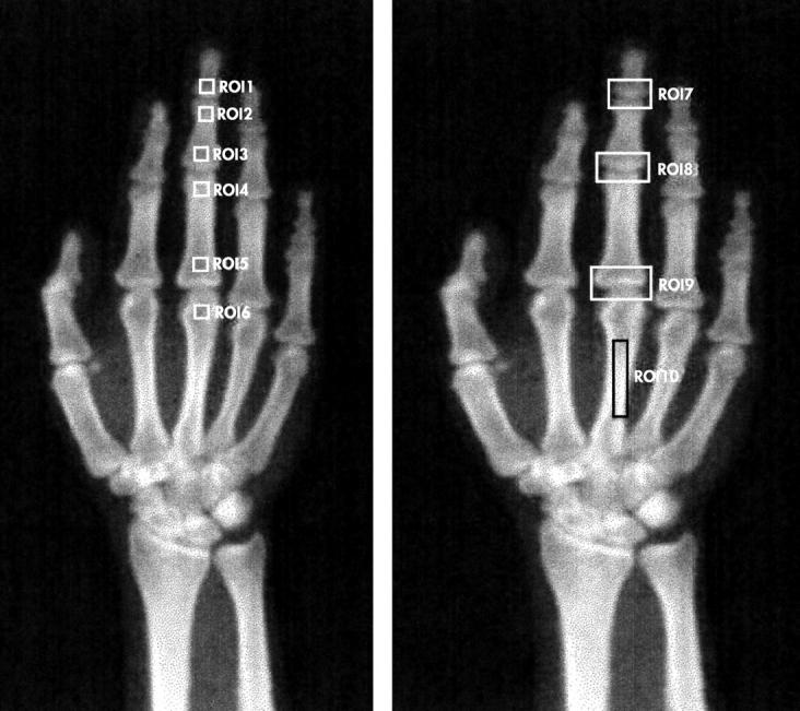
Background: Periarticular osteoporosis is an early finding in the hands of patients with rheumatoid arthritis (RA), due to release of bone resorbing cytokines from the inflamed synovium. There has been disagreement as to whether periarticular bone loss occurs in psoriatic arthritis (PsA). Bone mineral density (BMD) can now be measured accurately using dual energy x ray absorptiometry (DEXA). Recently, DEXA has been used to measure periarticular BMD at predefined regions of interest (ROIs) around the joints.
Objectives: Firstly, to compare periarticular BMD around the finger joints of patients with early RA or PsA. Secondly, to determine whether periarticular bone loss is related to joint inflammation and radiological erosions in RA and PsA.
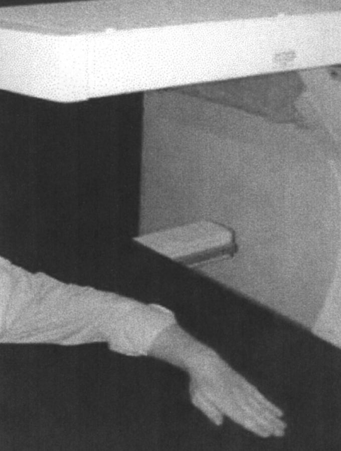
Methods: Seventeen patients with RA and 15 with PsA were recruited, all with disease duration of less than five years. All finger joints were examined by one person for swelling, or tenderness, or both. Hand radiographs were scored for the presence of erosions. Periarticular BMD was measured at 10 predetermined ROIs using a Hologic QDA-4500A fan-beam densitometer.
Results: Patients with PsA were less likely to be positive for rheumatoid factor (RF) (13% v 94%) and more likely to be men (60% v 23%) than patients with RA. There were no other clinical differences between patients with RA or PsA. Patients with RA had significantly lower BMD at each of the ROIs than those with PsA (p<0.05). However, these differences disappeared after adjusting for age and sex. Among patients with RA, those with a higher total number of swollen and/or tender hand joints had significantly lower periarticular BMD at the metocarpophalangeal (MCP) and proximal interphalangeal (PIP) joints. No such association was found for patients with PsA.
Conclusions: In early disease, periarticular bone loss occurred both in patients with RA and those with PsA. Among patients with RA, periarticular osteoporosis was related to measures of joint inflammation. There was no association between joint inflammation and periarticular bone loss in patients with PsA, which lends support to the hypothesis that the primary site of inflammation in PsA is extrasynovial.
Figures
References
Publication types
MeSH terms
LinkOut - more resources
Full Text Sources
Other Literature Sources
Medical
Research Materials
Miscellaneous
