. 2002 Nov;70(11):5997-6004.
doi: 10.1128/IAI.70.11.5997-6004.2002.

Nitric oxide limits the expansion of antigen-specific T cells in mice infected with the microfilariae of Brugia pahangi

Affiliations

Nitric oxide limits the expansion of antigen-specific T cells in mice infected with the microfilariae of Brugia pahangi

Richard A O'Connor et al. Infect Immun. 2002 Nov.

Abstract

Infection of BALB/c mice with the microfilariae (Mf) of the filarial nematode Brugia pahangi results in an antigen-specific proliferative defect that is induced by high levels of NO. Using carboxyfluorescein diacetate succinimydl ester and cell surface labeling, it was possible to identify a population of antigen-specific T cells from Mf-infected BALB/c mice that expressed particularly high levels of CD4 (CD4(hi)). These cells proliferated in culture only when inducible NO synthase was inhibited and accounted for almost all of the antigen-specific proliferative response under those conditions. CD4(hi) cells also expressed high levels of CD44, consistent with their status as activated T cells. A similar population of CD4(hi) cells was observed in cultures from Mf-infected gamma interferon receptor knockout (IFN-gammaR(-/-)) mice. Terminal deoxynucleotidyltransferase-mediated dUTP-biotin nick end labeling staining revealed that the CD4(+) T cells from Mf-infected wild-type mice were preferentially susceptible to apoptosis compared to CD4(+) T cells from IFN-gammaR(-/-) mice. These studies suggest that the expansion of antigen-specific T cells in Mf-infected mice is limited by NO.


Figures
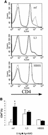
FIG. 1.
FIG. 1.
iNOS inhibition allows the Ag-driven expansion of CD4hi T cells in vitro. (A) The histograms show the CD4 staining profile of splenocytes from Mf-infected (top), L3-infected (middle), and uninfected control (bottom) BALB/c mice after 96 h of Ag-stimulated culture in the presence (dashed line) or absence (solid line) of 500 mM AMG. Note the tertiary peak of CD4hi staining in cells from Mf-infected mice. The graphs show individual animals from each group, but the results were consistent within the groups (Fig. 1B) and in replicate experiments. (B) iNOS inhibition significantly increased the percentage of total lymphocytes expressing high levels of CD4 only in Ag-stimulated splenocytes from Mf-infected animals. The results show the mean plus standard deviation of five mice per group. Open bars, cells cultured in the presence of 10 μg of B. pahangi Ag per ml; solid bars, cells cultured in Ag plus 500 mM AMG; ∗, significantly different from unsupplemented cultures; HBSS, Hanks balanced salt solution.
FIG. 2.
FIG. 2.
The CD4hi population contains Ag-reactive cells. (A) CFSE-labeled splenocytes from infected and uninfected BALB/c mice were restimulated in vitro with Ag in the presence of 500 mM AMG. After 96 h of culture, dividing cells (CFSE low) were clearly visible among the total CD4+ population (solid line). However, the majority of divisions had occurred within the CD4hi population (boldface line) and not the CD4-normal population (dashed line). Note the overlay between CD4hi cells (boldface line) and the total CD4+ population (solid line). The FACS plot shows cells from an individual animal, but the results were consistent within groups (Fig. 2B) and between experiments. (B) Percentages of CD4hi T cells that have divided in response to Ag in cultures from Mf- or L3-infected mice or control mice supplemented with 500 mM AMG. All data represent the mean plus standard deviation of five mice per group. ∗, significantly different from uninfected control mice; HBSS, Hanks balanced salt solution.
FIG. 3.
FIG. 3.
CD4hi T cells upregulate expression of CD44. Ag-stimulated splenocytes from Mf-infected BALB/c mice were cultured in the presence (AG+AMG) or absence (AG) of 500 mM AMG for 96 h prior to being stained with CD4 and CD44. The FACS plots shown were gated on CD4+ lymphocytes, and the quadrants were set to show CD4hi CD44hi cells (upper right quadrant). The data represent cells from individual animals, but the results were consistent within groups and in replicate experiments.
FIG. 4.
FIG. 4.
IFN-γR-mediated signaling is necessary to suppress T-cell expansion. Splenocytes from Mf-infected (Mf) and uninfected (hbss [Hanks balanced salt solution]) wild-type (WT; 129Sv) and IFN-γR−/− (KO) mice were restimulated with Ag in vitro for 96 h prior to being stained for CD4 expression and FACS analysis. The CD4+ population was significantly expanded in cultures derived from Mf-infected IFN-γR−/− compared to Mf-infected wild-type mice. All data represent the mean plus standard deviation of five mice per group. ∗, significantly different from equivalent wild-type counterparts.
FIG. 5.
FIG. 5.
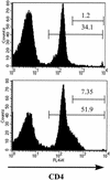
Increased expansion of the CD4hi population among cells from Mf-infected IFN-γR−/− mice. Splenocytes from Mf-infected wild-type mice (top) and IFN-γR−/− mice (bottom) were cultured for 96 h in the presence of Ag and then stained for CD4 expression. The gates were set to show the percentage of splenocytes that were CD4+ and the percentage of cells that were CD4hi and reveal the greater expansion of both CD4+ and CD4hi T cells in the absence of IFN-γR-mediated signaling. The plots show cells from individual animals, but the results were consistent within groups and between experiments.
FIG. 6.
FIG. 6.
Expansion of CD4 T cells is associated with lower levels of T-cell apoptosis. Cells from Mf-infected (Mf) and uninfected (HBSS [Hanks balanced salt solution]) IFN-γR−/− mice were restimulated with Ag in vitro for 48 h prior to TUNEL staining and FACS analysis. The levels of apoptosis in the CD4+ population from Mf-infected wild-type and IFN-γR−/− mice or from uninfected control mice were measured by TUNEL staining. CD4+ T cells from Mf-infected IFN-γR−/− mice displayed significantly lower levels of apoptosis than those from their wild-type counterparts (P < 0.05). All data represent the mean plus standard deviation of five animals per group. Solid bars, wild-type mice; open bars, IFN-γR−/− mice. *, significant difference in levels of apoptosis between cells from Mf-infected IFN-γR−/− mice and wildtype mice.
FIG. 7.
FIG. 7.
CD4hi cells display elevated levels of Fas expression. Cells from Mf-infected (Mf) and L3-infected (L3) animals were restimulated with Ag in the presence of 500 μM AMG for 96 h prior to surface staining and FACS analysis. CD4-normal and CD4hi cells were gated separately, revealing significantly increased MFI of anti-Fas staining on CD4hi T cells compared to CD4-normal cells in both experimental groups. All data represent the mean plus standard deviation of five animals per group. Open bars, CD4 cells; solid bars, CD4hi cells.

Similar articles

Cited by

References

    1. Allen, J. E., R. A. Lawrence, and R. M. Maizels. 1996. APC from mice harbouring the filarial parasite, Brugia malayi, prevent cellular proliferation but not cytokine production. Int. Immunol. 8:143-151. - PubMed
    1. Allen, J. E., and P. Loke. 2001. Divergent roles for macrophages in lymphatic filariasis. Parasite Immunol. 23:345-352. - PubMed
    1. Blass, S. L., E. Pure, and C. A. Hunter. 2001. A role for CD44 in the production of IFN-γ and immunopathology during infection with Toxoplasma gondii. J. Immunol. 166:5726-5732. - PubMed
    1. Chen, D., R. J. McKallip, A. Zeytun, Y. Do, C. Lombard, J. L. Robertson, T. W. Mak, P. S. Nagarkatti, and M. Nagarkatti. 2001. CD44-deficient mice exhibit enhanced hepatitis after concanavilin A injection: evidence for involvement of CD44 in activation-induced cell death. J. Immunol. 166:5889-5897. - PubMed
    1. Chu, C.-Q., S. Wittmer., and D. K. Dalton. 2000. Failure to suppress the expansion of the activated CD4 T cell population in interferon gamma-deficient mice leads to exacerbation of experimental autoimmune enchephalomyelitis. J. Exp. Med. 192:123-128. - PMC - PubMed

Publication types