. 2002 Nov;70(11):6206-14.
doi: 10.1128/IAI.70.11.6206-6214.2002.

Acquisition of regulators of complement activation by Streptococcus pyogenes serotype M1

Affiliations

Acquisition of regulators of complement activation by Streptococcus pyogenes serotype M1

Vinod Pandiripally et al. Infect Immun. 2002 Nov.

Abstract

Opsonization of bacteria by complement proteins is an important component of the immune response. The pathogenic bacterium Streptococcus pyogenes has evolved multiple mechanisms for the evasion of complement-mediated opsonization. One mechanism involves the binding of human regulators of complement activation such as factor H (FH) and FH-like protein 1 (FHL-1). Acquisition of these regulatory proteins can limit deposition of the opsonin C3b on bacteria, thus decreasing the pathogen's susceptibility to phagocytosis. Binding of complement regulatory proteins by S. pyogenes has previously been attributed to the streptococcal M and M-like proteins. Here, we report that the S. pyogenes cell surface protein Fba can mediate binding of FH and FHL-1. We constructed mutant derivatives of S. pyogenes that lack Fba, M1 protein, or both proteins and assayed the strains for FH binding, susceptibility to phagocytosis, and C3 deposition. Fba expression was found to be sufficient for binding of purified FH as well as for binding of FH and FHL-1 from human plasma. Plasma adsorption experiments also revealed that M1(+) Fba(+) streptococci preferentially bind FHL-1, whereas M1(-) Fba(+) streptococci have similar affinities for FH and FHL-1. Fba was found to contribute to the survival of streptococci incubated with human blood and to inhibit C3 deposition on bacterial cells. Streptococci harvested from log-phase cultures readily bound FH, but binding was greatly reduced for bacteria obtained from stationary-phase cultures. Bacteria cultured in the presence of the protease inhibitor E64 maintained FH binding activity in stationary phase, suggesting that Fba is removed from the cell surface via proteolysis. Western analyses confirmed that E64 stabilizes cell surface expression of Fba. These data indicate that Fba is an antiopsonic, antiphagocytic protein that may be regulated by cell surface proteolysis.


Figures
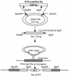
FIG. 1.
FIG. 1.
Construction of fba mutants. The fba gene was amplified via PCR with genomic DNA isolated from GAS strain 90-226 as the template. The oligonucleotide primers were designed to create BamHI restriction sites at the ends of the amplified fragment. The amplicon was digested with BamHI, gel purified, and ligated to BamHI-digested pSportI to create the plasmid pSport-fba. A 730-bp HindIII-to-BglII fragment containing an internal fragment of fba was then subcloned into the suicide vector pFW5 (42). pFW5 carries the aad9 (spectinomycin resistance) gene and does not replicate in GAS. Plasmid pFW5Δfba was introduced into GAS strains 90-226 and 90-226 emm1::Km by electroporation. Southern blot and PCR analyses were performed to verify integration of pFW5Δfba into the chromosomal fba gene.
FIG. 2.
FIG. 2.
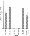
Inactivation of fba inhibits FH binding. Bacterial cells were harvested from exponential-phase cultures and adsorbed to wells of microtiter plates. The immobilized cells were incubated with FH followed by goat anti-human FH serum and an alkaline phosphatase-labeled secondary antibody. FH binding is expressed relative to that of the wild-type strain 90-226. Data are from three independent experiments, wherein each assay was performed in triplicate. Error bars represent standard deviations. Strains: M1+ Fba+, 90-226; M1 Fba+, 90-226 emm1::Km; M1 Fba, DC276; M1+ Fba, DC283.
FIG. 3.
FIG. 3.
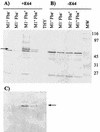
Western analysis of cell surface proteins from strain 90-226 and its isogenic M1 and Fba derivatives. Cell surface proteins were extracted from stationary-phase cultures of GAS, fractionated by SDS-PAGE, and transferred to nitrocellulose membranes. The phenotypes of the strains are listed above the blots. Proteins used in blots A and C were extracted from streptococci cultured to stationary phase in THY containing 25 μM of the cysteine protease inhibitor E64. Proteins for blot B were extracted from THY-grown cultures. (A and B) Blots were incubated with Fba antiserum. The arrow in panel A indicates the position of the 58-kDa band present in extracts from Fba+ strains that was not present in extracts from Fba strains. Cell surface Fba was not detectable in cultures grown in the absence of E64. Lanes: THY, mock extraction performed with sterile culture medium; MW, molecular mass standards, with masses indicated in kilodaltons. (C) FH binding to Fba is shown. The same extracts used for blot A were transferred to a membrane, blocked, and successively incubated with 10 μg of FH/ml, FH antiserum, and a labeled secondary antibody. The arrow indicates the position of the same protein band indicated by the arrow in panel A.
FIG. 4.
FIG. 4.
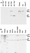
Adsorption of FH and FHL-1 from human plasma. (A) GAS strain 90-226 and its isogenic M1 and Fba derivatives were incubated with human plasma, harvested by centrifugation, and washed extensively. Bound plasma proteins were eluted and subjected to Western analysis with FH antiserum. The phenotypes of the strains are listed above the blot. Adsorption experiments were performed with plasma or serum from four different sources. The topmost arrow to the right of the figure indicates the 150-kDa FH band. The lower arrow indicates a 49-kDa band determined by N-terminal sequencing to be FH or FHL-1. Lanes: Plasma, loaded with human plasma; FH, loaded with purified FH; M+ Fba+∗, mock adsorption performed with bacteria in the absence of plasma; None∗∗, mock adsorption performed with plasma in the absence of bacteria. (B) Plasma adsorptions were performed in the presence of protease inhibitors. Plasma adsorptions were performed with wild-type GAS in the absence of protease inhibitors (None) or in the presence of 25 μM E64, 1 mM PMSF, 25 μM E64 and 1 mM PMSF, 25 μM EDTA, or a cocktail of protease inhibitors (PIC; Sigma-Aldrich) as indicated.
FIG. 5.
FIG. 5.
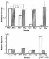
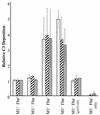
(A) Survival of GAS in human blood and plasma. GAS strains were grown to early- to mid-log phase, and 102 CFU of each strain were added to heparinized human blood (open bars) or human plasma (hatched bars). A portion of each culture was plated to determine the input CFU. Cultures were incubated at 37 C for 3 h. Portions of each culture were again plated to determine the number of surviving CFU. The growth index of each culture was calculated by dividing the number of surviving CFU by the number of input CFU. For each experiment, the growth index for strain 90-226 (M1+ Fba+) was assigned a value of 1. Growth indices for the mutant strains are expressed relative to strain 90-226. The data in the figure were obtained from four independent experiments. Error bars represent standard deviations. (B) Complementation of the fba mutation. Experiments were performed as described for panel A. The M1+ Fba strain is DC283. M1+ Fba (pYT1143) is DC283 carrying the Fba expression plasmid pYT1143 (48). Values are the averages from two experiments.
FIG. 6.
FIG. 6.
C3 deposition on streptococci. Bacterial cells were suspended in VBS containing 10 mM EGTA and 5 mM MgCl2. The suspensions were mixed with an equal volume of human serum or heat-inactivated human serum (HIS) and incubated at room temperature for 30 min. Bacteria were harvested, washed, and suspended in carbonate buffer. The cells were then diluted in carbonate buffer to final OD560s of 0.025 (open bars), 0.05 (hatched bars), and 0.1 (filled bars) and applied to wells of microtiter plates. C3 deposition was detected with a monoclonal anti-human C3d antibody.
FIG. 7.
FIG. 7.
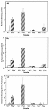
Fba is removed from the cell surface in stationary phase. (A) FH binding activity is lost in stationary phase. Wells of microtiter plates were coated with GAS grown to mid-exponential phase (hatched bars) or stationary phase (open bars). FH binding was assayed and is presented as described in the legend to Fig. 2. (B) E64 stabilizes FH binding activity. Microtiter plates were coated with GAS grown to stationary phase in THY medium (open bars) or THY medium containing 25 μM of E64 (hatched bars). (C) E64 stabilizes Fba expression. Wells were coated with GAS grown to stationary phase in THY medium (open bars) or THY medium containing 25 μM of E64 (hatched bars). Fba expression was assayed with Fba antiserum. All data are from two independent experiments in which assays were performed in triplicate (panels A and B) or duplicate (panel C).

Similar articles

Cited by

References

    1. Akesson, P., K. H. Schmidt, J. Cooney, and L. Bjorck. 1994. M1 protein and protein H: IgGFc- and albumin-binding streptococcal surface proteins encoded by adjacent genes. Biochem. J. 300:877-886. - PMC - PubMed
    1. Berge, A., and L. Bjorck. 1995. Streptococcal cysteine proteinase releases biologically active fragments of streptococcal surface proteins. J. Biol. Chem. 270:9862-9867. - PubMed
    1. Berge, A., B. M. Kihlberg, A. G. Sjoholm, and L. Bjorck. 1997. Streptococcal protein H forms soluble complement-activating complexes with IgG, but inhibits complement activation by IgG-coated targets. J. Biol. Chem. 272:20774-20781. - PubMed
    1. Caparon, M. G., and J. R. Scott. 1991. Genetic manipulation of pathogenic streptococci. Methods Enzymol. 204:556-586. - PubMed
    1. Chaussee, M. S., R. L. Cole, and J. P. M. van Putten. 2000. Streptococcal erythrogenic toxin B abrogates fibronectin-dependent internalization of Streptococcus pyogenes by cultured mammalian cells. Infect. Immun. 68:3226-3232. - PMC - PubMed

Publication types

MeSH terms