. 2002 Nov;70(11):6373-82.
doi: 10.1128/IAI.70.11.6373-6382.2002.

Staphylococcus aureus aconitase inactivation unexpectedly inhibits post-exponential-phase growth and enhances stationary-phase survival

Affiliations

Staphylococcus aureus aconitase inactivation unexpectedly inhibits post-exponential-phase growth and enhances stationary-phase survival

Greg A Somerville et al. Infect Immun. 2002 Nov.

Abstract

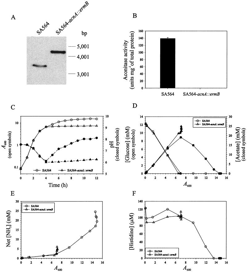
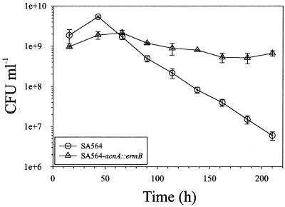
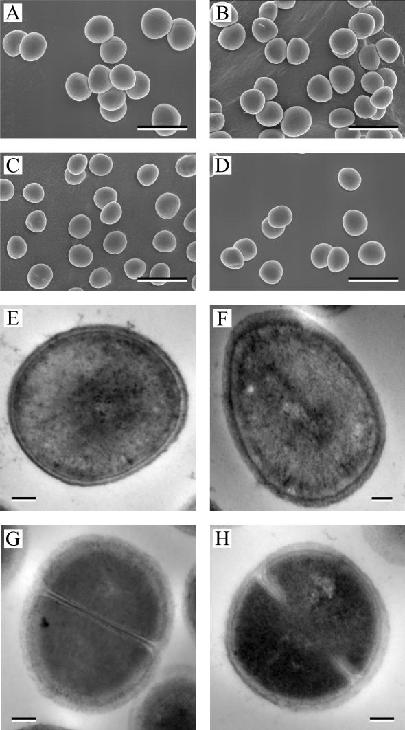
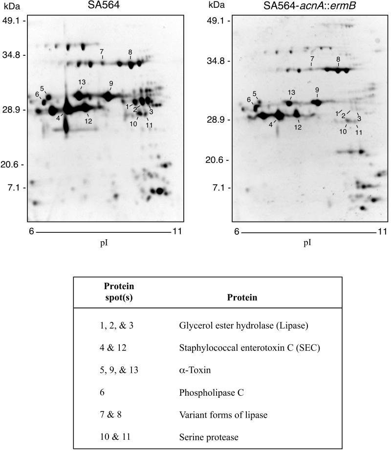
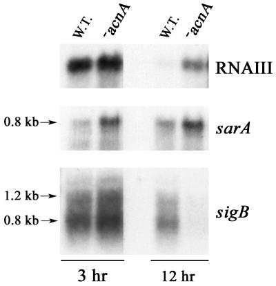
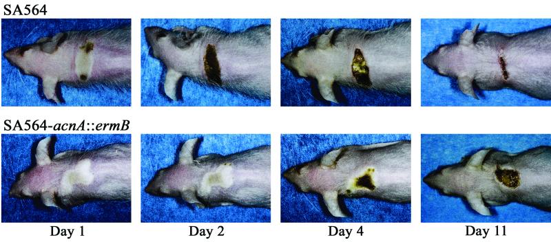
Staphylococcus aureus preferentially catabolizes glucose, generating pyruvate, which is subsequently oxidized to acetate under aerobic growth conditions. Catabolite repression of the tricarboxylic acid (TCA) cycle results in the accumulation of acetate. TCA cycle derepression coincides with exit from the exponential growth phase, the onset of acetate catabolism, and the maximal expression of secreted virulence factors. These data suggest that carbon and energy for post-exponential-phase growth and virulence factor production are derived from the catabolism of acetate mediated by the TCA cycle. To test this hypothesis, the aconitase gene was genetically inactivated in a human isolate of S. aureus, and the effects on physiology, morphology, virulence factor production, virulence for mice, and stationary-phase survival were examined. TCA cycle inactivation prevented the post-exponential growth phase catabolism of acetate, resulting in premature entry into the stationary phase. This phenotype was accompanied by a significant reduction in the production of several virulence factors and alteration in host-pathogen interaction. Unexpectedly, aconitase inactivation enhanced stationary-phase survival relative to the wild-type strain. Aconitase is an iron-sulfur cluster-containing enzyme that is highly susceptible to oxidative inactivation. We speculate that reversible loss of the iron-sulfur cluster in wild-type organisms is a survival strategy used to circumvent oxidative stress induced during host-pathogen interactions. Taken together, these data demonstrate the importance of the TCA cycle in the life cycle of this medically important pathogen.


Figures

FIG. 1.
FIG. 1.
Characteristics of the S. aureus aconitase mutant. (A) Southern blot confirmation of acnA disruption. Genomic DNA was isolated from strains SA564 and SA564-acnA::ermB, digested with EcoRI and EcoRV (the acnA-containing fragment is ∼3,400 bp), and analyzed by Southern blotting. The ermB cassette was 1,280 bp, resulting in the ∼4.6-kb band observed in the mutant strain. (B) Aconitase activities of strains SA564 and SA564-acnA::ermB after 6 h of growth. (C) The wild-type strain SA564 and the aconitase mutant strain SA564-acnA::ermB were grown in TSB. At the indicated times, an aliquot was removed, appropriate dilutions were made, and the absorbance at 600 nm was measured. (D) Glucose depletion and acetate accumulation and depletion in the culture supernatants of strain SA564 and strain SA564-acnA::ermB plotted as a function of growth. (E) Net ammonia accumulation in the culture supernatants of strains SA564 and SA564-acnA::ermB. (F) An example of postexponential free amino acid depletion from the culture medium. The results presented are representative of at least two independent experiments.
FIG. 2.
FIG. 2.
TCA cycle inactivation enhances stationary-phase survival. Single colonies of SA564 and SA564-acnA::ermB were inoculated into TSB, grown at 37°C, and aerated by being shaken at 225 rpm for up to 2 weeks. At 24-h intervals, aliquots were removed and CFU per milliliter were determined in quadruplicate. The data presented are averages and standard deviations.
FIG. 3.
FIG. 3.
Electron micrographs of SA564 and SA564-acnA::ermB. Scanning electron micrographs (A to D) of wild-type strain SA564 (A and C) or the aconitase mutant strain SA564-acnA::ermB (B and D) grown for either 3 (A and B) or 27 (C and D) h are shown. Transmission electron micrographs (E to H) of strain SA564 (E and G) or strain SA564-acnA::ermB (F and H) grown for either 3 (E and F) or 27 (G and H) h are shown. Bars, 1.5 μm (A to D) and 100 nm (E to H).
FIG. 4.
FIG. 4.
2-D electrophoresis of SA564 and SA564-acnA::ermB culture supernatant proteins. Stationary-phase (12-h) culture supernatants were isolated from bacteria grown in protein-free TSB. Isoelectric focusing conducted with a pH 3 to 10 linear separation range showed that the majority of exoproteins had isoelectric points in the pH 6 to 10 range (data not shown). Therefore, subsequent isoelectric focusing was done with a linear separation range from pH 6 to 11. Protein spots analyzed by MALDI-TOF MS are indicated. The results are representative of multiple experiments using supernatant proteins from two independent isolations.
FIG. 5.
FIG. 5.
Northern blot analysis of RNAIII, sarA, and sigB transcription. Total RNA was isolated from bacteria grown to exponential (3-h) and stationary (12-h) phases; 10 μg of total RNA was used per lane. To ensure that equivalent quantities of RNA were loaded, the RNA was visualized by ethidium bromide staining prior to transfer to a charged nylon membrane (data not shown). Radiolabeled probes specific for RNAIII, sarA, and sigB were used. The results presented are representative of multiple Northern blots with independently isolated total-RNA samples. W.T., wild type; acnA, SA564-acnA::ermB.
FIG. 6.
FIG. 6.
Disease progression in mice infected subcutaneously with SA564 or SA564-acnA::ermB. Ten outbred, immunocompetent, hairless mice (Crl::SKH1-hrBR) were inoculated subcutaneously with 3 × 10⁷ CFU of SA564 or SA564-acnA::ermB. Disease progression was followed for 18 days, and representative results are shown.

Similar articles

Cited by

References

    1. Alen, C., and A. L. Sonenshein. 1999. Bacillus subtilis aconitase is an RNA-binding protein. Proc. Natl. Acad. Sci. USA 96:10412-10417. - PMC - PubMed
    1. Babior, B. M. 2000. Phagocytes and oxidative stress. Am. J. Med. 109:33-44. - PubMed
    1. Baughn, A. D., and M. H. Malamy. 2002. A mitochondrial-like aconitase in the bacterium Bacteroides fragilis: implications for the evolution of the mitochondrial Krebs cycle. Proc. Natl. Acad. Sci. USA 99:4662-4667. - PMC - PubMed
    1. Blumenthal, H. J. 1972. Glucose catabolism in staphylococci, p. 111-135. In J. O. Cohen (ed.), The staphylococci. Wiley-Interscience, New York, N.Y.
    1. Bruckner, R. 1997. Gene replacement in Staphylococcus carnosus and Staphylococcus xylosus. FEMS Microbiol. Lett. 151:1-8. - PubMed