. 2002 Nov;70(11):6399-408.
doi: 10.1128/IAI.70.11.6399-6408.2002.

Modulation of cytosolic Ca(2+) concentration in airway epithelial cells by Pseudomonas aeruginosa

Affiliations

Modulation of cytosolic Ca(2+) concentration in airway epithelial cells by Pseudomonas aeruginosa

Tobias Jacob et al. Infect Immun. 2002 Nov.

Abstract

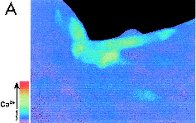
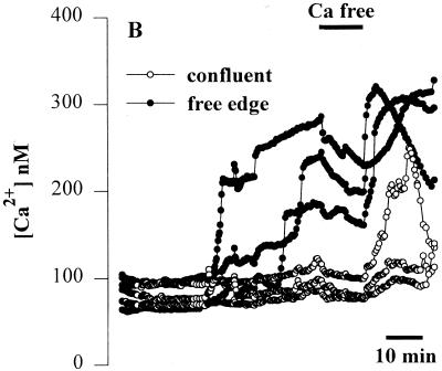
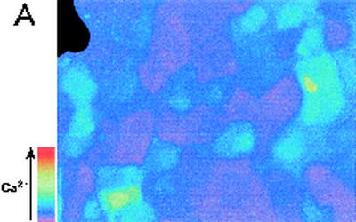
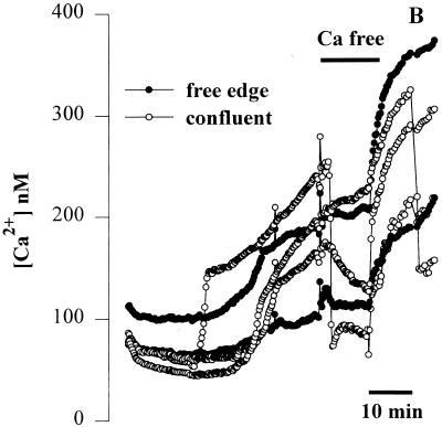
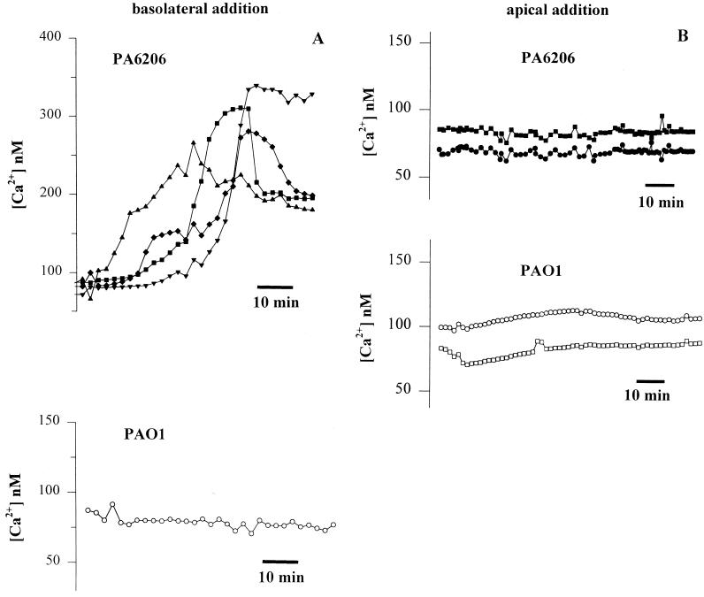
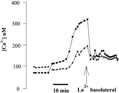
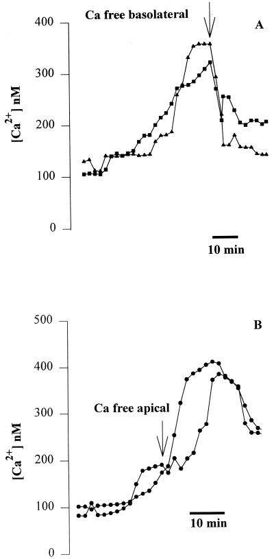
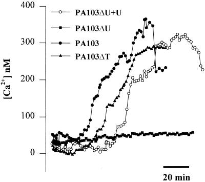
Modulation of cytosolic (intracellular) Ca(2+) concentration (Ca(i)) may be an important host response when airway epithelial cells are exposed to Pseudomonas aeruginosa. We measured Ca(i) in Calu-3 cells exposed from the apical or basolateral surface to cytotoxic and noncytotoxic strains of P. aeruginosa. Apical addition of either noncytotoxic strains or cytotoxic strains failed to affect Ca(i) over a 3-h time period, nor were changes observed after basolateral addition of noncytotoxic strains. In contrast, basolateral addition of cytotoxic strains caused a slow increase in Ca(i) from 100 nM to 200 to 400 nM. This increase began after 20 to 50 min and persisted for an additional 30 to 75 min, at which time the cells became nonviable. P. aeruginosa-induced increases in Ca(i) were blocked by the addition of the Ca channel blocker La(3+) to the basolateral but not to the apical chamber. Likewise, replacing the basolateral but not the apical medium with Ca-free solution prevented P. aeruginosa-mediated changes in Ca(i). With isogenic mutants of PA103, we demonstrated that the type III secretion apparatus, the type III-secreted effector ExoU, and type IV pili were necessary for increased Ca(i). We propose that translocation of ExoU through the basolateral surface of polarized airway epithelial cells via the type III secretion apparatus leads to release of Ca stored in the endoplasmic reticulum and activation of Ca channels in the basolateral membranes of epithelial cells.


Figures

FIG. 1.
FIG. 1.
Effects of apical addition of P. aeruginosa to wounded Calu-3 cells. The cytotoxic strain PA6206 (multiplicity of infection, 50) was added to the apical chamber of confluent Calu-3 cells grown on 0.45-μm pores that had been mechanically wounded 24 h earlier and loaded with fura-2 prior to addition of bacteria. Ratio imaging microscopy was performed during control conditions and then during addition of P. aeruginosa. (A) Pseudocolor image taken after 60 min of exposure to PA6206 showed that Cai increased in cells adjacent to the free edge of the wound (blue), but not in confluent cells located away from the wound edge. (B) Time course of Cai for three cells along the free edge and three cells in the confluent region of the monolayer following addition of apical PA6206. Cai increased first in the cells along the free edge and only later in cells in the confluent region of the monolayer. Normal perfusion solution was replaced with Ca-free solution at the time shown by the black bar at the top of the figure. This treatment slowed the rate of increase of Cai in cells along the free edge but had only small effects on Cai in those cells that had not responded to P. aeruginosa. Similar results were obtained in 30 free edge cells and >85 confluent cells in a total of three experiments.
FIG. 1.
FIG. 1.
Effects of apical addition of P. aeruginosa to wounded Calu-3 cells. The cytotoxic strain PA6206 (multiplicity of infection, 50) was added to the apical chamber of confluent Calu-3 cells grown on 0.45-μm pores that had been mechanically wounded 24 h earlier and loaded with fura-2 prior to addition of bacteria. Ratio imaging microscopy was performed during control conditions and then during addition of P. aeruginosa. (A) Pseudocolor image taken after 60 min of exposure to PA6206 showed that Cai increased in cells adjacent to the free edge of the wound (blue), but not in confluent cells located away from the wound edge. (B) Time course of Cai for three cells along the free edge and three cells in the confluent region of the monolayer following addition of apical PA6206. Cai increased first in the cells along the free edge and only later in cells in the confluent region of the monolayer. Normal perfusion solution was replaced with Ca-free solution at the time shown by the black bar at the top of the figure. This treatment slowed the rate of increase of Cai in cells along the free edge but had only small effects on Cai in those cells that had not responded to P. aeruginosa. Similar results were obtained in 30 free edge cells and >85 confluent cells in a total of three experiments.
FIG. 2.
FIG. 2.
Effects of basolateral addition of P. aeruginosa to wounded Calu-3 cells. The cytotoxic strain PA6206 (multiplicity of infection, 50) was added to the basal chamber of confluent Calu-3 cells grown on 3.0-μm pores that had been mechanically wounded 24 h earlier and loaded with fura-2, and ratio imaging microscopy was performed. (A) Pseudocolor image after 40 min of exposure to PA6206 on the basolateral side of the monolayer. As shown by the blue color, basolateral addition of P. aeruginosa caused increased Cai in cells throughout the monolayer. (B) Time course of Cai following basolateral addition of PA6206 was quantified for two cells at the free edge and three cells in the confluent region of the monolayer. Replacement of the normal medium with Ca-free solution (black bar) resulted in a decrease in Cai in all the cells that had responded to P. aeruginosa. Similar results were obtained in >60 cells from free edges and confluent regions in a total of three experiments.
FIG. 2.
FIG. 2.
Effects of basolateral addition of P. aeruginosa to wounded Calu-3 cells. The cytotoxic strain PA6206 (multiplicity of infection, 50) was added to the basal chamber of confluent Calu-3 cells grown on 3.0-μm pores that had been mechanically wounded 24 h earlier and loaded with fura-2, and ratio imaging microscopy was performed. (A) Pseudocolor image after 40 min of exposure to PA6206 on the basolateral side of the monolayer. As shown by the blue color, basolateral addition of P. aeruginosa caused increased Cai in cells throughout the monolayer. (B) Time course of Cai following basolateral addition of PA6206 was quantified for two cells at the free edge and three cells in the confluent region of the monolayer. Replacement of the normal medium with Ca-free solution (black bar) resulted in a decrease in Cai in all the cells that had responded to P. aeruginosa. Similar results were obtained in >60 cells from free edges and confluent regions in a total of three experiments.
FIG. 3.
FIG. 3.
Effects of apical versus basolateral addition of P. aeruginosa on Cai in confluent Calu-3 cell monolayers. Calu-3 cells were grown to confluence on filters with 3.0-μm pores and loaded with fura-2, and either PA6206 or PAO1 (multiplicity of infection of 50) was added to the basolateral (A) or apical (B) side of the monolayer. (A) The upper panel shows representative results from four typical cells following basolateral addition of PA6206. Similar results were obtained in >35 other cells in the field. Basolateral addition of PA6206 caused a slow, sustained increase in Cai from 80 to 100 nM to 200 to 350 nM. Similar results were obtained on >35 cells in each of a total of six experiments. The lower panel shows results from one representative cell during basolateral exposure to PAO1. None of the 75 cells in the field exhibited any increases in Cai during approximately 60 min of exposure to bacteria. Similar results were obtained on >35 cells in each of a total of six experiments. (B) Typical results from two cells upon apical exposure to PA6206 (multiplicity of infection of 50) (upper panel) and from two cells during apical exposure to PAO1 (multiplicity of infection of 50) (lower panel). Neither PAO1 nor PA6206 had any effect on Cai in any of the >100 cells in the fields over the course of >60 min. Similar results were obtained for >35 cells in three experiments each for apical addition of PAO1 and PA6206.
FIG. 4.
FIG. 4.
PA6206 and thapsigargin have similar effects on Cai. Calu-3 cells were grown as islands on cover glasses, loaded with fura-2, and then exposed to PA6206 (multiplicity of infection, 100). After Cai in the cell at the periphery had increased to its plateau level, thapsigargin (10 μM; THG, arrow) was added. Thapsigargin caused Cai to increase in the cell that had not responded to P. aeruginosa, while Cai increased in the cell in the center that had not yet responded to P. aeruginosa. Similar results were obtained on 25 cells in three similar experiments.
FIG. 5.
FIG. 5.
Basolateral addition of La3+ abolishes basolaterally applied PA6206-induced increases in Cai in Calu-3 cells. Calu-3 cells were grown to confluence on 3.0-μm-pore-size filters, loaded with fura-2, and exposed to PA6206 (multiplicity of infection of 50) from the basolateral surface. After Cai levels had increased, 1 mM La3+ was added to the basolateral surface, resulting in a decrease in Cai back to the baseline. The responses of two typical cells are shown; similar results were obtained with >40 cells in a total of four experiments.
FIG. 6.
FIG. 6.
Effects of exposure of the basolateral and apical surfaces to Ca-free solution after PA6206-induced increases in Cai in Calu-3 cells. Cells were grown to confluence on 3.0-μm-pore filters, loaded with fura-2, and exposed to PA6206 (multiplicity of infection of 50) on the basolateral surface of the monolayer. (A) After Cai had started to increase in two cells, the basolateral medium was replaced with Ca-free solution. This treatment caused Cai to decrease back to the baseline. Similar results were obtained in >50 cells in a total of four experiments. (B) After Cai had started to increase in two cells, the apical medium was replaced with Ca-free solution. This treatment had no effect on the P. aeruginosa-induced Cai response, which continued to increase. Similar results were obtained from >25 cells in a total of four experiments.
FIG. 7.
FIG. 7.
Role of type III-secreted effectors on P. aeruginosa-mediated increases in Cai. Calu-3 cells were grown on cover glasses as islands, then loaded with fura-2, and exposed to PA103, PA103ΔU (lacks ExoU), PA103ΔT (lacks ExoT), or PA103ΔU+U (multiplicity of infection of 50). Responses of typical “free edge” cells from five different Calu-3 islands are shown. The exoU mutant failed to increase Cai, while complementation with exoU expressed from a plasmid (pExoU) restored the ability of PA103ΔU to modulate Cai. Similar results were obtained from >25 cells in three experiments for each bacterial strain.

Similar articles

Cited by

References

    1. Aderem, A., and R. J. Ulevitch. 2000. Toll-like receptors in the induction of the innate immune response. Nature 406:782-787. - PubMed
    1. Allewelt, M., F. T. Coleman, M. Grout, G. P. Priebe, and G. B. Pier. 2000. Acquisition of expression of the Pseudomonas aeruginosa ExoU cytotoxin leads to increased bacterial virulence in a murine model of acute pneumonia and systemic spread. Infect. Immun. 68:3998-4004. - PMC - PubMed
    1. Apodaca, G., M. Bomsel, R. Lindstedt, J. Engel, D. Frank, K. Mostov, and J. Wiener-Kronish. 1995. Characterization of Pseudomonas aeruginosa-induced MDCK cell injury: glycosylation defective host cells are resistant to bacterial killing. Infect. Immun. 63:1541-1551. - PMC - PubMed
    1. Bals, R., X. Wang, R. L. Meegalla, S. Wattler, D. J. Weiner, M. C. Nehls, and J. M. Wilson. 1999. Mouse beta-defensin 3 is an inducible antimicrobial peptide expressed in the epithelia of multiple organs. Infect. Immun. 67:3542-3547. - PMC - PubMed
    1. Comolli, J., A. Hauser, L. Waite, C. Whitchurch, J. Mattick, and J. Engel. 1999. PilU and PilT are required for cytotoxicity and virulence of Pseudomonas aeruginosa. Infect. Immun. 67:3625-3630. - PMC - PubMed

Publication types

Substances

LinkOut - more resources