Modulation of inducible nitric oxide synthase expression by the attaching and effacing bacterial pathogen citrobacter rodentium in infected mice
- PMID: 12379723
- PMCID: PMC130393
- DOI: 10.1128/IAI.70.11.6424-6435.2002
Modulation of inducible nitric oxide synthase expression by the attaching and effacing bacterial pathogen citrobacter rodentium in infected mice
Abstract
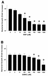
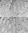
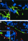
Citrobacter rodentium belongs to the attaching and effacing family of enteric bacterial pathogens that includes both enteropathogenic and enterohemorrhagic Escherichia coli. These bacteria infect their hosts by colonizing the intestinal mucosal surface and intimately attaching to underlying epithelial cells. The abilities of these pathogens to exploit the cytoskeleton and signaling pathways of host cells are well documented, but their interactions with the host's antimicrobial defenses, such as inducible nitric oxide synthase (iNOS), are poorly understood. To address this issue, we infected mice with C. rodentium and found that iNOS mRNA expression in the colon significantly increased during infection. Immunostaining identified epithelial cells as the major source for immunoreactive iNOS. Finding that nitric oxide (NO) donors were bacteriostatic for C. rodentium in vitro, we examined whether iNOS expression contributed to host defense by infecting iNOS-deficient mice. Loss of iNOS expression caused a small but significant delay in bacterial clearance without affecting tissue pathology. Finally, immunofluorescence staining was used to determine if iNOS expression was localized to infected cells by staining for the C. rodentium virulence factor, translocated intimin receptor (Tir), as well as iNOS. Interestingly, while more than 85% of uninfected epithelial cells expressed iNOS, fewer than 15% of infected (Tir-positive) cells expressed detectable iNOS. These results demonstrate that both iNOS and intestinal epithelial cells play an active role in host defense during C. rodentium infection. However, the selective expression of iNOS by uninfected but not infected cells suggests that this pathogen has developed mechanisms to locally limit its exposure to host-derived NO.
Figures


Similar articles
-
Impaired resistance and enhanced pathology during infection with a noninvasive, attaching-effacing enteric bacterial pathogen, Citrobacter rodentium, in mice lacking IL-12 or IFN-gamma.J Immunol. 2002 Feb 15;168(4):1804-12. doi: 10.4049/jimmunol.168.4.1804. J Immunol. 2002. PMID: 11823513
-
Citrobacter rodentium infection causes iNOS-independent intestinal epithelial dysfunction in mice.Can J Physiol Pharmacol. 2006 Dec;84(12):1301-12. doi: 10.1139/y06-086. Can J Physiol Pharmacol. 2006. PMID: 17487239
-
Modulation of intestinal goblet cell function during infection by an attaching and effacing bacterial pathogen.Infect Immun. 2008 Feb;76(2):796-811. doi: 10.1128/IAI.00093-07. Epub 2007 Nov 5. Infect Immun. 2008. PMID: 17984203 Free PMC article.
-
Citrobacter rodentium of mice and man.Cell Microbiol. 2005 Dec;7(12):1697-706. doi: 10.1111/j.1462-5822.2005.00625.x. Cell Microbiol. 2005. PMID: 16309456 Review.
-
Host defences to Citrobacter rodentium.Int J Med Microbiol. 2003 Apr;293(1):87-93. doi: 10.1078/1438-4221-00247. Int J Med Microbiol. 2003. PMID: 12755369 Review.
Cited by
-
Dietary phytate primes epithelial antibacterial immunity in the intestine.Front Immunol. 2022 Oct 19;13:952994. doi: 10.3389/fimmu.2022.952994. eCollection 2022. Front Immunol. 2022. PMID: 36341403 Free PMC article.
-
Toll-like receptor 4 contributes to colitis development but not to host defense during Citrobacter rodentium infection in mice.Infect Immun. 2006 May;74(5):2522-36. doi: 10.1128/IAI.74.5.2522-2536.2006. Infect Immun. 2006. PMID: 16622187 Free PMC article.
-
Active vitamin D (1,25-dihydroxyvitamin D3) increases host susceptibility to Citrobacter rodentium by suppressing mucosal Th17 responses.Am J Physiol Gastrointest Liver Physiol. 2012 Dec 15;303(12):G1299-311. doi: 10.1152/ajpgi.00320.2012. Epub 2012 Sep 27. Am J Physiol Gastrointest Liver Physiol. 2012. PMID: 23019194 Free PMC article.
-
Enteropathogenic Escherichia coli outer membrane proteins induce iNOS by activation of NF-kappaB and MAP kinases.Inflammation. 2004 Dec;28(6):345-53. doi: 10.1007/s10753-004-6645-8. Inflammation. 2004. PMID: 16245077
-
iNOS expression in oral and gastrointestinal tract mucosa.Dig Dis Sci. 2008 Jun;53(6):1437-42. doi: 10.1007/s10620-007-0061-5. Dig Dis Sci. 2008. PMID: 17987386 Review.
References
-
- Asfaha, S., C. J. Bell, J. L. Wallace, and W. K. MacNaughton. 1999. Prolonged colonic epithelial hyporesponsiveness after colitis: role of inducible nitric oxide synthase. Am. J. Physiol. 276:G703-G710. - PubMed
-
- Barthold, S. W., G. L. Coleman, R. O. Jacoby, E. M. Livestone, and A. M. Jonas. 1978. Transmissible murine colonic hyperplasia. Vet. Pathol. 15:223-236. - PubMed
-
- Celli, J., W. Deng, and B. B. Finlay. 2000. Enteropathogenic Escherichia coli (EPEC) attachment to epithelial cells: exploiting the host cell cytoskeleton from the outside. Cell. Microbiol. 2:1-9. - PubMed
Publication types
MeSH terms
Substances
LinkOut - more resources
Full Text Sources
Other Literature Sources

