. 2002 Nov;70(11):6448-55.
doi: 10.1128/IAI.70.11.6448-6455.2002.

Babesia bovis merozoite surface antigen 2 proteins are expressed on the merozoite and sporozoite surface, and specific antibodies inhibit attachment and invasion of erythrocytes

Affiliations

Babesia bovis merozoite surface antigen 2 proteins are expressed on the merozoite and sporozoite surface, and specific antibodies inhibit attachment and invasion of erythrocytes

Juan Mosqueda et al. Infect Immun. 2002 Nov.

Abstract

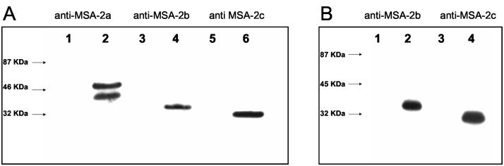
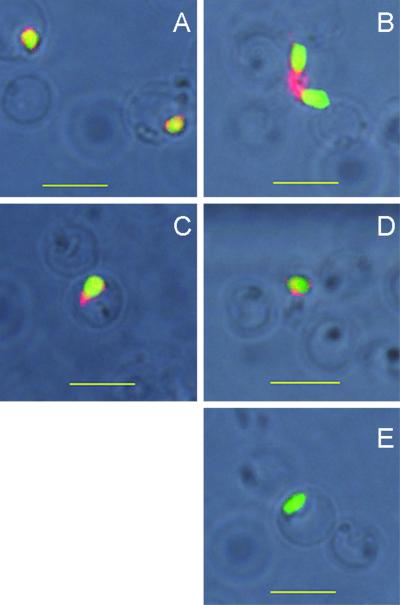
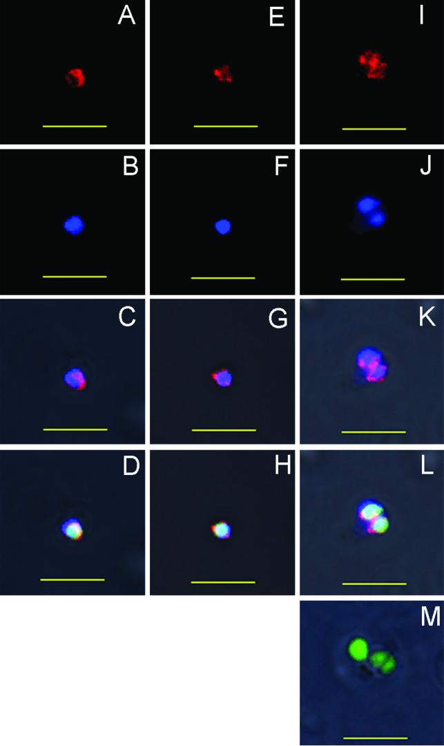
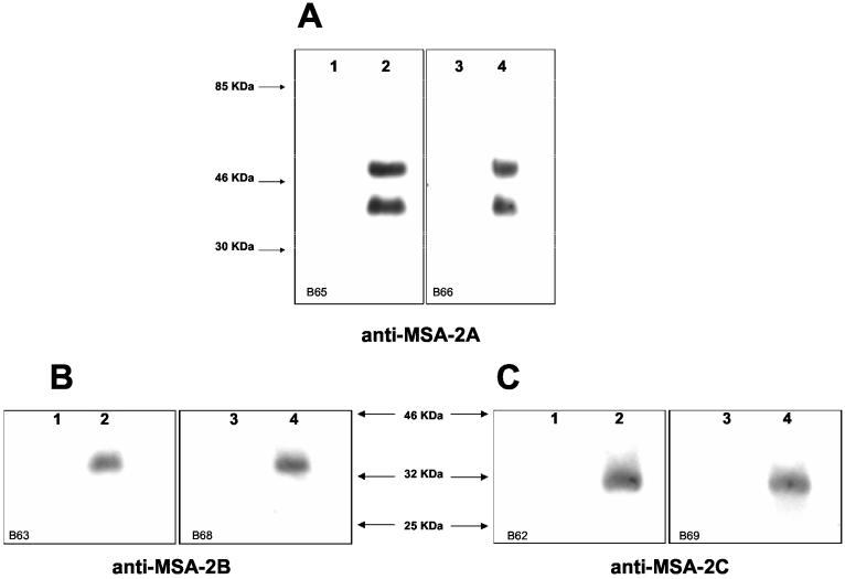
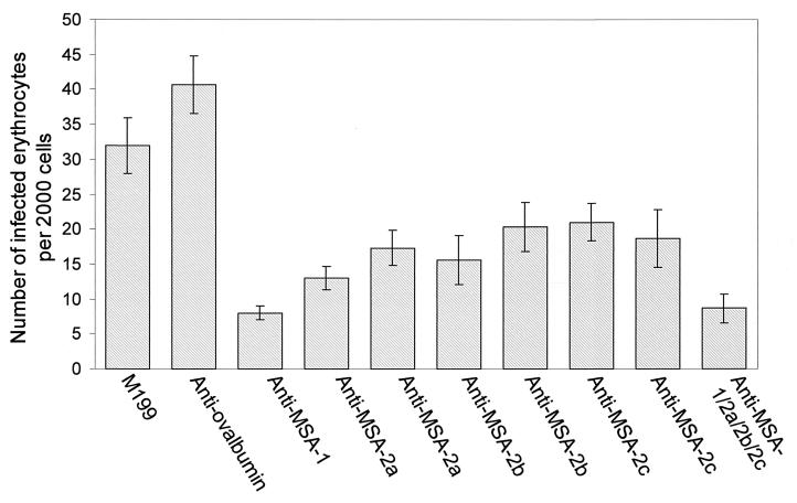
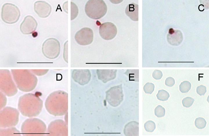
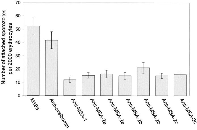
The Babesia bovis merozoite surface antigen 2 (MSA-2) locus encodes four proteins, MSA-2a(1), -2a(2), -2b, and -2c. With the use of specific antibodies, each MSA-2 protein was shown to be expressed on the surface of live extracellular merozoites and coexpression on single merozoites was confirmed. Individual antisera against MSA-2a, MSA-2b, and MSA-2c significantly inhibited merozoite invasion of bovine erythrocytes. As tick-derived sporozoites also directly invade erythrocytes, expression of each MSA-2 protein on the sporozoite surface was examined and verified. Finally, statistically significant inhibition of sporozoite binding to the erythrocytes was demonstrated by using antisera specific for MSA-2a, MSA-2b, and MSA-2c. These results indicate an important role for MSA-2 proteins in the initial binding and invasion of host erythrocytes and support the hypothesis that sporozoites and merozoites use common surface molecules in erythrocyte invasion.


Figures

FIG. 1.
FIG. 1.
Specificity of anti-MSA-2 murine and rabbit sera used in expression analysis. Normal, uninfected erythrocytes (lanes 1, 3, and 5) or B. bovis-infected erythrocytes (lanes 2, 4, and 6) were electrophoresed on sodium dodecyl sulfate-containing polyacrylamide gels and transferred to nitrocellulose membranes. The membranes were incubated with 1:500 dilutions of murine sera specific for MSA-2a, -2b, or -2c (A) or rabbit sera specific for MSA-2b or -2c (B). Molecular size markers are on the left of each panel.
FIG. 2.
FIG. 2.
Expression of MSA-2 proteins on the surface of live B. bovis merozoites. Merozoites were incubated with antisera for MSA-2a (A), -2b (B), or 2c (C) and then with a secondary antibody labeled with tetramethyl rhodamine isothiocyanate, which emits a red fluorescence. Merozoite viability was determined with CFDA, which emits a green fluorescence on live cells. Labeling was observed with the positive-control monoclonal antibody 23/10.36.18 against MSA-1 (D) but not with antisera against A. marginale (E). Bars = 5 μm.
FIG. 3.
FIG. 3.
Coexpression of MSA-2 proteins on the surface of live B. bovis merozoites. Merozoites were incubated with mouse anti-MSA-2a plus rabbit anti-MSA-2b (A to D), mouse anti-MSA-2a plus rabbit anti-MSA-2c (E to H), or mouse anti-MSA-2c plus rabbit anti-MSA-2b (I to L), and then with a rhodamine-labeled anti-murine IgG antibody (red fluorescence) and an Alexa Fluor 350-labeled anti-rabbit IgG antibody (blue fluorescence). Merozoite viability was determined with CFDA, which emits a green fluorescence on live cells. Images are phase contrast and the following channels: individual channel for rhodamine (A, E, and I), individual channel for Alexa Fluor 350 (B, F, and J); combined channels for rhodamine and Alexa Fluor 350 (C, G, and K); and combined channels for rhodamine, Alexa Fluor 350, and fluorescein (D, H, L, and M). Labeling was not observed when merozoites were incubated with a combination of a mouse antiserum against A. marginale recombinant MSP-2 OpAG3 plus a rabbit antiserum against MSP-1 (M). Bars = 5 μm.
FIG. 4.
FIG. 4.
Specificity of anti-MSA-2 bovine sera used for neutralization of merozoites and sporozoites. Normal, uninfected erythrocytes (lanes 1 and 3) or B. bovis-infected erythrocytes (lanes 2 and 4) were electrophoresed on sodium dodecyl sulfate-containing polyacrylamide gels and transferred to nitrocellulose membranes. The membranes were incubated with a 1:500 dilution of sera from calves immunized with recombinant MSA-2a (A), -2b (B), or -2c (C). Molecular size markers are indicated by arrows.
FIG. 5.
FIG. 5.
Antibody-mediated inhibition of merozoite invasion. Shown are the total numbers of infected erythrocytes from 2,000 cells counted at 5 h after merozoite addition. Merozoites were incubated with M199 medium alone; with negative-control bovine antiserum against ovalbumin; with positive-control bovine antiserum against MSA-1; with two different antisera against MSA-2a, -2b, and -2c; or with the combined antisera (MSA-1, -2a, -2b, and -2c). Error bars indicate standard deviations of the results from triplicate cultures.
FIG. 6.
FIG. 6.
Expression of MSA-2 proteins by sporozoites at the time of attachment to erythrocytes. Smears of erythrocyte cultures initiated with sporozoites were incubated with specific mouse antiserum against MSA-2a (A), MSA-2b (B), MSA-2c (C), monoclonal antibody 23/10.36.18 against MSA-1 (D), or negative-control mouse antiserum against A. marginale MSP-2 OpAG3 (E). Erythrocyte cultures treated identically but with the use of extracts from uninfected larvae were incubated with specific mouse antiserum against each MSA-2 protein; anti-MSA-2a is shown in panel F (magnification, ×63,000). The reaction was visualized with AEC, which results in brown staining. Bars = 10 μm.
FIG. 7.
FIG. 7.
Antibody-mediated inhibition of sporozoite attachment to erythrocytes. The total number of parasites attached to 2,000 erythrocytes was counted at 5 h after sporozoite addition. Sporozoites were incubated with M199 medium alone; with negative-control bovine antiserum against ovalbumin; with positive-control bovine antiserum against MSA-1; or with two different antisera against MSA-2a, -2b, or -2c. Error bars indicate standard deviations of the results from triplicate cultures.

Similar articles

Cited by

References

    1. Brown, W. C., and G. H. Palmer. 1999. Designing blood-stage vaccines against Babesia bovis and B. bigemina. Parasitol. Today 15:275-281. - PubMed
    1. Chitnis, C. E., and M. J. Blackman. 2000. Host cell invasion by malaria parasites. Parasitol. Today 16:411-415. - PubMed
    1. Cowman, A. F., D. L. Baldi, M. Duraisingh, J. Healer, K. E. Mills, R. A. O'Donnell, J. Thompson, T. Triglia, M. E. Wickham, and B. S. Crabb. 2002. Functional analysis of Plasmodium falciparum merozoite antigens: implications for erythrocyte invasion and vaccine development. Philos. Trans. R. Soc. Lond. B Biol. Sci. 357:25-33. - PMC - PubMed
    1. Egan, A. F., P. Burghaus, P. Druilhe, A. A. Holder, and E. M. Riley. 1999. Human antibodies to the 19kDa C-terminal fragment of Plasmodium falciparum merozoite surface protein 1 inhibit parasite growth in vitro. Parasite Immunol. 21:133-139. - PubMed
    1. Florin-Christensen, M., C. E. Suarez, S. A. Hines, G. H. Palmer, W. C. Brown, and T. F. McElwain. 2002. The Babesia bovis merozoite surface antigen 2 locus contains four tandemly arranged and expressed genes encoding immunologically distinct proteins. Infect. Immun. 70:3566-3575. - PMC - PubMed

Publication types

Substances