. 2002 Oct 29;99(22):14200-5.
doi: 10.1073/pnas.212519499. Epub 2002 Oct 11.

RB activation defect in tumor cell lines

Affiliations

RB activation defect in tumor cell lines

Cristina Broceño et al. Proc Natl Acad Sci U S A. .

Abstract

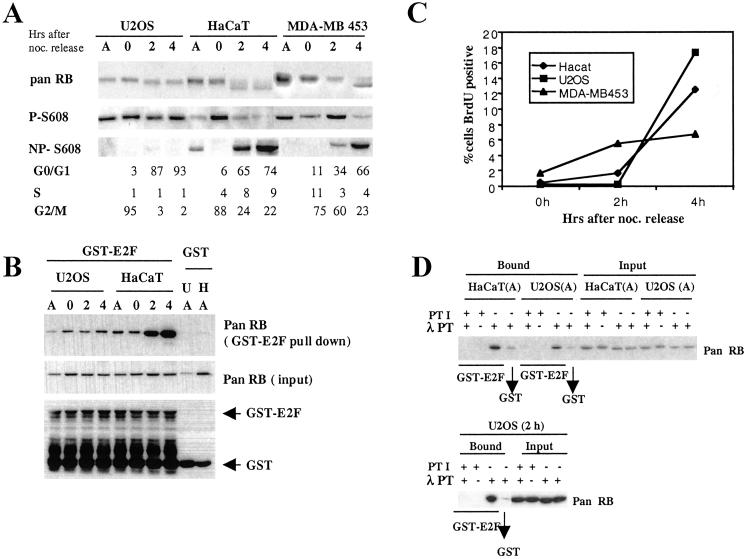
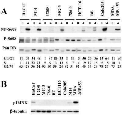
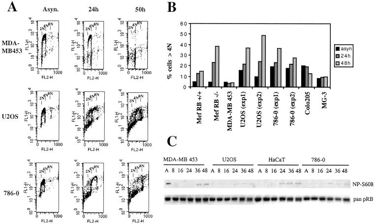
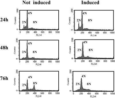
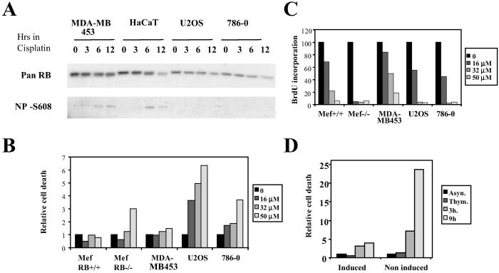
Activation of the retinoblastoma (RB) protein through dephosphorylation arises in cells upon exit from M phase and in response to environmental stresses, including DNA damage. We provide here for the first time evidence that these responses are co-ordinately affected in a subset of tumor derived cell lines. We find that RB dephosphorylation is not apparent in these cells during progression into G(1). Importantly these cells also do not respond with RB activation after DNA damage during S phase. Moreover and as a consequence they display phenotypes classically associated with RB(-) cells, showing accelerated apoptosis after DNA damage and DNA re-replication after spindle-checkpoint activation. A large body of literature provides evidence that controls governing inactivation of RB are lost in tumors. The results presented here indicate that the reverse reaction, namely the activation of RB from an inactive precursor, may also be compromised. Our findings indicate that this type of defect may be coupled with hypersensitivity to DNA damage and an increase in genomic instability in response to spindle-checkpoint activation thus bearing potentially important medical implications.


Figures

Fig 1.
Fig 1.
Lack of RB activation in U2OS osteosarcoma cells. (A) RB phosphorylation analysis. Cell lysates were prepared from asynchronously growing cells (A) or metaphase cells released into G1 for 0, 2, and 4 h and analyzed by using antiretinoblastoma antibodies as follows: pan RB, recognizing all forms of RB; P-Ser-608, recognizing RB phosphorylated on Ser-608; NP-Ser-608, recognizing RB unphosphorylated on Ser-608. A portion of cells was subjected to FACS-based cell-cycle analysis for DNA content. Analysis of three different cell lines (U2OS, HaCaT, and MDA-MB 453) is presented. (B) E2F-binding analysis. Cell lysates were adsorbed to either GST-E2F1 or unfused GST. Bound proteins were analyzed by immunoblot by using pan RB antibody. GST protein equal loading in the experiment is shown by Western blot with an anti-GST antibody. (C) G1 progression. Metaphase cells were released into medium containing BrdUrd. Cells were harvested as indicated, and the percentage of BrdUrd+ cells was determined by FACS. (D) Activation of E2F binding by phosphatase treatment in vitro. Cell lysates from either asynchronous cells or cells released from metaphase for 2 h (2h) were treated with λ-phosphatase (λ PT) in the presence or absence of phosphatase inhibitors (PTI). Lysate was subjected to binding selection (bound) by using GST-E2F1, GST, or loaded directly (input).
Fig 2.
Fig 2.
Changes in RB phosphorylation after metaphase release in selected tumor cell lines. (A) Cell lines randomly selected from the NCI tumor cell panel were treated and analyzed as described for Fig. 1A. Cell lines and their origins were as follows. HCT116, BE, Colo205 (colorectal cancer derived), 786-0 (renal cancer derived), MG-3 and U2OS (osteosarcoma derived) and M14 (melanoma derived). HaCaT and MDA-MB 453 were taken along as RB activation competent controls. (B) Lysate from asynchronously growing tumor cell lines was probed with antibody to human p16INK4a. Each lane contained lysate equivalent to 1 × 10⁵ cells.
Fig 3.
Fig 3.
DNA re-replication upon spindle-checkpoint challenge in RB activation-defective cells. As indicated, cells were cultured in the presence of 50 nM nocodazole for the time period indicated and analyzed for incorporation of BrdUrd and DNA content by FACS. (A) Raw BrdUrd incorporation data in cells as indicated. (B) Numerical representation of FACS-based analysis. Mouse embryo fibroblasts (MEF) RB−/− and MEF RB+/+ represent early passage mouse embryo fibroblasts derived from RB knockout and RB (WT) littermates, respectively. (C) Cell lysates obtained at different time points after nocodazole exposure were analyzed by using antibody for Ser-608 unphosphorylated (NP-Ser-608) or pan RB. Lysate amounts comprising equivalent numbers of cells were loaded in each lane.
Fig 4.
Fig 4.
Expression of constitutively active RB inhibits DNA re-replication after spindle-checkpoint activation. U2OS expressing a constitutively active form of RB under the control of tetracycline were synchronized in S phase by culture in thymidine for 16 h. Cells were subsequently released into medium containing low levels (50 nM) of the spindle inhibitor nocodazole in either the presence (not induced) or absence (induced) of tetracycline. Cells were processed for FACS analysis.
Fig 5.
Fig 5.
RB activation and cell death after cisplatin challenge during S phase. (A) RB activation. RB activation-competent (MDA-MB453 and HaCaT) and -incompetent (U2OS and 786-0) cells were enriched for S phase by culture for 24 h in thymidine-containing medium. Cells were subsequently treated with 50 μM cisplatin for different lengths of time. Samples marked with 0 did not receive cisplatin. Lysates were generated and probed with either a pan RB antibody or antibody specific for RB unmodified on Ser-608. (B) Cell death after cisplatin treatment. S phase-enriched cells were treated with increasing doses of cisplatin for 16 h. Cells were subsequently washed and cultured in growth medium for an additional 3 h and processed for FACS analysis. Death response was assessed by scoring the percentage of cells with a sub-G1 DNA content. (C) Inhibition of S-phase progression after cisplatin treatment. S phase-enriched cells were treated with increasing doses of cisplatin as described in B. They were subsequently washed and cultured in the presence of BrdUrd for 4 h. Cells then were collected, fixed, and analyzed for DNA content and BrdUrd incorporation. (D) U2OS with tetracycline-regulated expression of constitutively active RB were exposed to thymidine for 24 h in the presence or absence of tetracycline; cisplatin at 50 μM was subsequently added to the culture for a further 16 h. Cells then were released into thymidine- and cisplatin-free growth medium for 3 or 9 h and analyzed for death response as in B.

Similar articles

Cited by

References

    1. Zheng L. & Lee, W. H. (2001) Exp. Cell Res. 264, 2-18. - PubMed
    1. Morris E. J. & Dyson, N. J. (2001) Adv. Cancer Res. 82, 1-54. - PubMed
    1. Mittnacht S. (1998) Curr. Opin. Genet. Dev. 8, 21-27. - PubMed
    1. Adams P. D. (2001) Biochim. Biophys. Acta 1471, M123-M133. - PubMed
    1. Harbour J. W. & Dean, D. C. (2000) Genes Dev. 14, 2393-2409. - PubMed

Publication types

Substances

LinkOut - more resources