. 2002 Oct 14;159(1):69-78.
doi: 10.1083/jcb.200206015. Epub 2002 Oct 14.

ARFGAP1 promotes the formation of COPI vesicles, suggesting function as a component of the coat

Affiliations

ARFGAP1 promotes the formation of COPI vesicles, suggesting function as a component of the coat

Jia-Shu Yang et al. J Cell Biol. .

Abstract

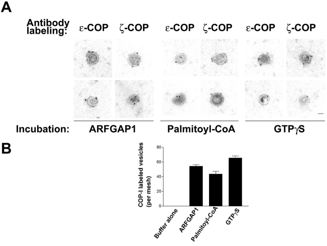
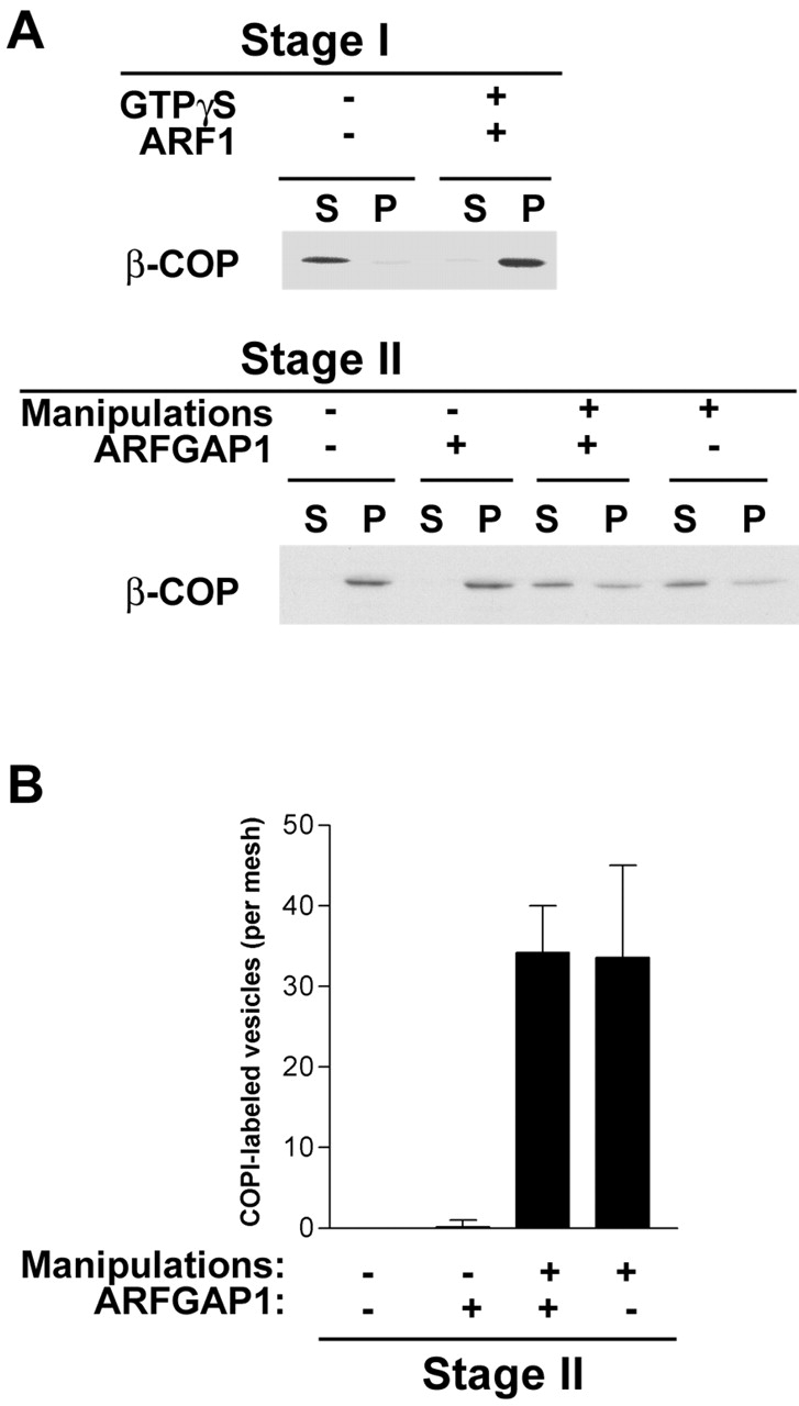
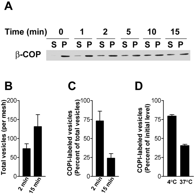
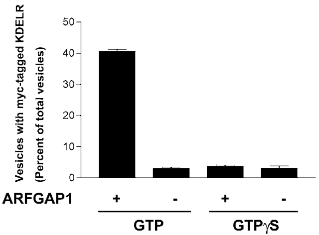
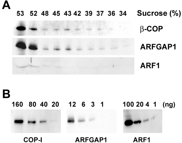
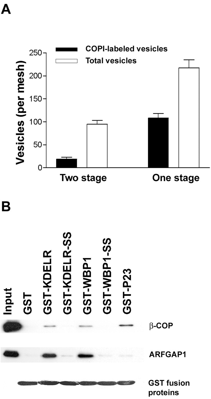
The role of GTPase-activating protein (GAP) that deactivates ADP-ribosylation factor 1 (ARF1) during the formation of coat protein I (COPI) vesicles has been unclear. GAP is originally thought to antagonize vesicle formation by triggering uncoating, but later studies suggest that GAP promotes cargo sorting, a process that occurs during vesicle formation. Recent models have attempted to reconcile these seemingly contradictory roles by suggesting that cargo proteins suppress GAP activity during vesicle formation, but whether GAP truly antagonizes coat recruitment in this process has not been assessed directly. We have reconstituted the formation of COPI vesicles by incubating Golgi membrane with purified soluble components, and find that ARFGAP1 in the presence of GTP promotes vesicle formation and cargo sorting. Moreover, the presence of GTPgammaS not only blocks vesicle uncoating but also vesicle formation by preventing the proper recruitment of GAP to nascent vesicles. Elucidating how GAP functions in vesicle formation, we find that the level of GAP on the reconstituted vesicles is at least as abundant as COPI and that GAP binds directly to the dilysine motif of cargo proteins. Collectively, these findings suggest that ARFGAP1 promotes vesicle formation by functioning as a component of the COPI coat.


Figures

Figure 1.
Figure 1.
The distribution of ARF1, COPI, and GAP in the two-stage incubation system. Golgi membrane was incubated with ARF1 and coatomer in the presence of GTP for the first stage followed by centrifugation to assess their relative distribution on the Golgi membrane, as reflected in the pellet (P), and the remaining fraction, as reflected in the supernatant (S). For the second stage, the pelleted Golgi membrane from the first stage was recovered and then incubated with either GAP or buffer followed by centrifugation to assess again their relative distribution.
Figure 2.
Figure 2.
GAP induces the formation of COPI vesicles. (A) COPI vesicles reconstituted with GAP appear similar to those formed by previous methods of reconstituting COPI vesicles. Supernatant either from the second-stage incubation for conditions involving GAP or palmitoyl-CoA, or from incubating Golgi membrane with cytosol in the presence of GTPγS was applied onto EM grids followed by immunogold labeling using antibodies directed against ɛ- or ζ-COP. Magnification, 120,000. Bar, 50 nm. (B) The level of COPI vesicles formed by reconstitution with GAP is similar to levels formed by previously established methods. A fraction (10% by volume) of the supernatant from the different incubations was applied onto EM grids for immunogold labeling using antibodies direct against ɛ- or ζ-COP, followed by quantitation for vesicles with gold particles.
Figure 4.
Figure 4.
GTPγS blocks the formation of COPI vesicles. (A) GTPγS inhibits the release of COPI from Golgi membranes induced by GAP. The two-stage incubation was performed with the first stage performed in the presence of GTPγS. The second-stage incubation was performed as indicated, followed by centrifugation to derive Golgi membrane (P) and supernatant (S). The distribution of COPI was assessed by immunoblotting for β-COP. (B) GTPγS inhibits the formation of COPI vesicles. A fraction (10% by volume) of the supernatant derived from the different conditions of the second-stage incubation was applied onto EM grids for immunogold labeling using antibodies direct against ɛ- or ζ-COP, followed by quantitation for vesicles with gold particles.
Figure 3.
Figure 3.
Increasing the time of incubation enhances vesicle production and also vesicle uncoating. (A) COPI is released from Golgi membrane with increasing time of the second-stage incubation. The two-stage incubation was performed with the second stage performed for various times, as indicated, followed by centrifugation to segregate Golgi membrane in the pellet (P) and unpelleted material in the supernatant (S). The distribution of COPI was then assessed by immunoblotting for β-COP. (B) More vesicles are formed with increasing time of the second-stage incubation. The two-stage incubation was performed with the second stage performed for times as indicated. A fraction (10% by volume) of the supernatant derived from the second-stage incubation was applied onto EM grids followed by quantitation for vesicles. (C) Fewer COPI vesicles are observed with increasing time of incubation at the second stage. The two-stage incubation was performed with the second stage performed for times as indicated. A fraction (10% by volume) of the supernatant derived from the second-stage incubation was applied onto EM grids for immunogold labeling using antibodies direct against ɛ- or ζ-COP, followed by quantitation for vesicles with gold particles. (D) The isolated supernatant from the second-stage incubation contains fewer COPI vesicles upon further incubation. The starting condition was the supernatant from the second-stage incubation performed for 2 min, as described in C. This fraction was subjected to an additional 30-min incubation at different temperatures as indicated. Samples were then applied onto EM grids for immunogold labeling using antibodies direct against ɛ- or ζ-COP, followed by quantitation for vesicles with gold particles. The values derived were then expressed as fractions of the starting level.
Figure 5.
Figure 5.
GAP in the presence of GTP is required for the efficient sorting of the KDEL receptor into reconstituted vesicles, and GTPγS inhibits this process. The two-stage incubation was performed with the first stage using Golgi membrane derived from CHO cells that had been stably transfected with a myc-tagged KDEL receptor. The first stage was performed with either GTP or GTPγS, followed by the second stage either with or without GAP. For the GTPγS conditions, the additional manipulations of pipette shearing and salt treatment were used so that similar levels of COPI vesicles could be generated for comparison. A fraction (10% by volume) of the supernatant derived from the different conditions was applied onto EM grids for immunogold labeling with antibody directed against the myc epitope, followed by quantitation for vesicles with gold particles.
Figure 6.
Figure 6.
Ultrastructural characterization of vesicles for GAP and ARF1. (A) Vesicles reconstituted in the presence of GTP contain detectable levels of GAP but not ARF1. The two-stage incubation was performed with the second stage using either GAP or palmitoyl-CoA, as indicated. A fraction (10% by volume) of the supernatant derived from the second-stage incubation was then applied onto grids for immunogold EM using antibodies as indicated. Magnification, 120,000. Bar, 50 nm. (B) The level of GAP on COPI vesicles is reduced when reconstitution is performed in the presence of GTPγS. The two-stage incubation was performed with the first stage using either GTP or GTPγ, and the second stage using GAP. A fraction (10% by volume) of the supernatant derived from the second stage was then applied onto grids for immunogold EM with antibody against GAP, followed by quantitation for vesicles with gold particles.
Figure 7.
Figure 7.
Coated vesicles have stoichiometric levels of COP and GAP, and significantly reduced levels of ARF1. For A, the two-stage incubation was scaled up fivefold with the supernatant derived from the second stage and then loaded at the bottom of a continuous sucrose gradient for equilibrium centrifugation followed by fractionation and immunoblotting with antibodies indicated. Quantitation of β-COP, ARF1, and GAP was performed based on immunoblotting using known amounts of purified proteins, as indicated in B.
Figure 8.
Figure 8.
Functional evidence that GAP acts as a component of the COPI coat. (A) Simultaneous recruitment of GAP and COPI is more efficient for vesicle formation than separating the two into distinct incubation steps. A one-stage incubation was performed by incubating Golgi membrane with ARF1, coatomer, and GAP in the presence of GTP for 10 min. The level of vesicles, total or COPI labeled, from this incubation was compared with that from the standard two-stage incubation using the same amount of Golgi membrane and purified components, where the second stage was performed for 10 min. (B) GAP interacts directly with the cytoplasmic domain of COPI cargo proteins in a dilysine-dependent manner. GST fusion proteins that contain different cytoplasmic domains of cargo proteins were gathered onto glutathione-bound beads followed by incubation with either purified coatomer or GAP. Beads were washed and then analyzed by immunoblotting to assess COPI and GAP or by Coomassie blue staining to assess the level of GST fusion proteins on beads.

Similar articles

Cited by

References

    1. Antonny, B., D. Madden, S. Hamamoto, L. Orci, and R. Schekman. 2001. Dynamics of the COPII coat with GTP and stable analogues. Nat. Cell Biol. 3:531–537. - PubMed
    1. Aoe, T., E. Cukierman, A. Lee, D. Cassel, P.J. Peters, and V.W. Hsu. 1997. The KDEL receptor, ERD2, regulates intracellular traffic by recruiting a GTPase-activating protein for ARF1. EMBO J. 16:7305–7316. - PMC - PubMed
    1. Aoe, T., I. Huber, C. Vasudevan, S.C. Watkins, G. Romero, D. Cassel, and V.W. Hsu. 1999. The KDEL receptor regulates the in vivo activity of a GTPase-activating protein for ARF1 by interacting with its non-catalytic domain. J. Biol. Chem. 274:20545–20549. - PubMed
    1. Barlowe, C., L. Orci, T. Yeung, M. Hosobuchi, S. Hamamoto, N. Salama, M.F. Rexach, M. Ravazzola, M. Amherdt, and R. Schekman. 1994. COPII: a membrane coat formed by Sec proteins that drive vesicle budding from the endoplasmic reticulum. Cell. 77:895–907. - PubMed
    1. Boehm, M., R.C. Aguilar, and J.S. Bonifacino. 2001. Functional and physical interactions of the adaptor protein complex AP-4 with ADP-ribosylation factors (ARFs). EMBO J. 20:6265–6276. - PMC - PubMed

Publication types

MeSH terms