. 2002 Oct 14;159(1):147-56.
doi: 10.1083/jcb.200204046. Epub 2002 Oct 14.

Insulin receptor substrate-2 maintains predominance of anabolic function over catabolic function of osteoblasts

Affiliations

Insulin receptor substrate-2 maintains predominance of anabolic function over catabolic function of osteoblasts

Toru Akune et al. J Cell Biol. .

Abstract

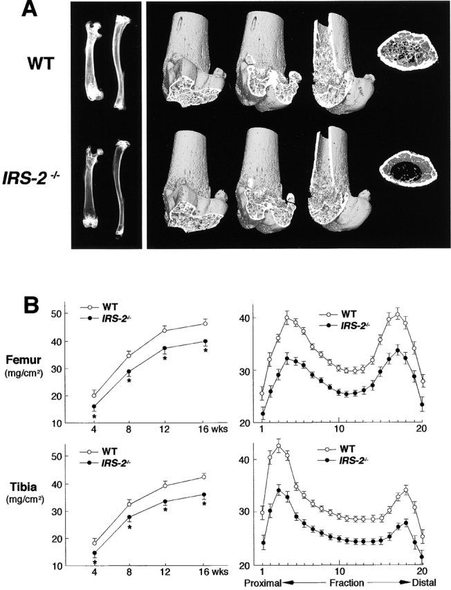
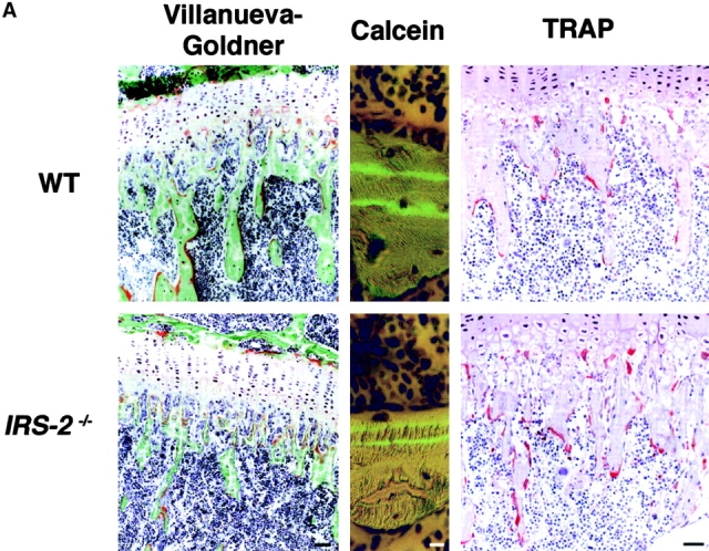
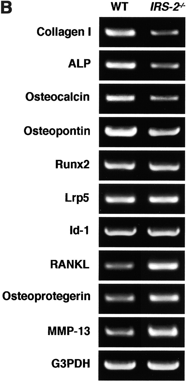
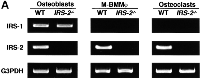
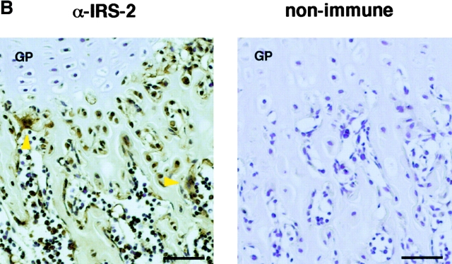
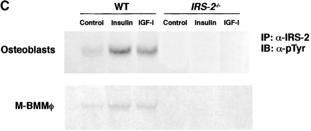
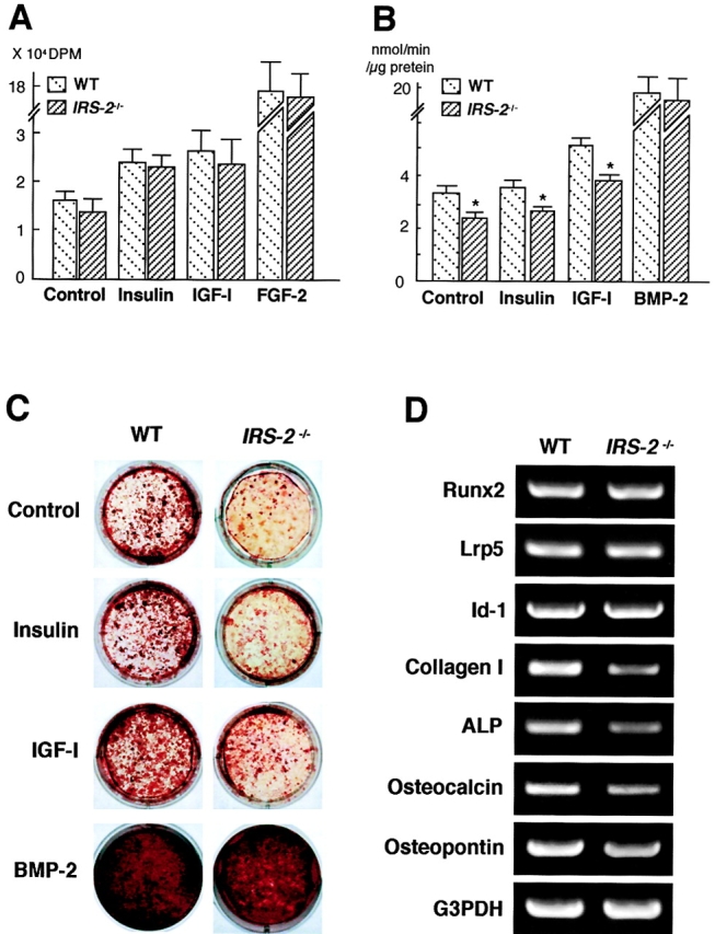
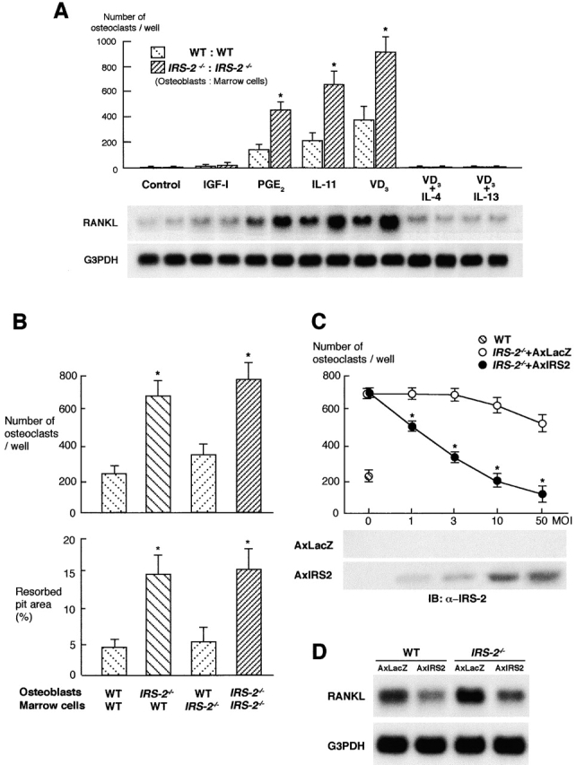
Insulin receptor substrates (IRS-1 and IRS-2) are essential for intracellular signaling by insulin and insulin-like growth factor-I (IGF-I), anabolic regulators of bone metabolism. Although mice lacking the IRS-2 gene (IRS-2-/- mice) developed normally, they exhibited osteopenia with decreased bone formation and increased bone resorption. Cultured IRS-2-/- osteoblasts showed reduced differentiation and matrix synthesis compared with wild-type osteoblasts. However, they showed increased receptor activator of nuclear factor kappaB ligand (RANKL) expression and osteoclastogenesis in the coculture with bone marrow cells, which were restored by reintroduction of IRS-2 using an adenovirus vector. Although IRS-2 was expressed and phosphorylated by insulin and IGF-I in both osteoblasts and osteoclastic cells, cultures in the absence of osteoblasts revealed that intrinsic IRS-2 signaling in osteoclastic cells was not important for their differentiation, function, or survival. It is concluded that IRS-2 deficiency in osteoblasts causes osteopenia through impaired anabolic function and enhanced supporting ability of osteoclastogenesis. We propose that IRS-2 is needed to maintain the predominance of bone formation over bone resorption, whereas IRS-1 maintains bone turnover, as we previously reported; the integration of these two signalings causes a potent bone anabolic action by insulin and IGF-I.


Figures

Figure 1.
Figure 1.
IRS-2 / mice showed osteopenia both in trabecular and cortex bones. (A) Plain X-ray images of femora and tibiae (left) and three- dimensional CT images of distal femora (right) of representative WT and IRS-2 / littermates (8 wk old). (B) BMD of the femora and tibiae of WT and IRS-2 / littermates. (B, left) BMD of the whole femora and tibiae at 4, 8, 12, and 16 wk of age. *, significantly different from WT mice; P < 0.05. (B, right) BMD of each of 20 equal longitudinal divisions of the femora and tibiae of 8-wk- old mice. Data are expressed as means (symbols) ± SEMs (error bars) for 12 bones/group for WT and IRS-2 / mice.
Figure 2.
Figure 2.
IRS-2 / bone showed an uncoupling of decreased bone formation and increased bone resorption in vivo. (A) Histological features at proximal tibiae of WT and IRS-2 / littermates (8 wk old). In Villanueva-Goldner staining (left), mineralized bone is stained green and unmineralized osteoid red. Bar, 50 μm. In calcein double labeling (middle), mineralization front is stained as a green line. Bar, 10 μm. In TRAP staining, TRAP-positive osteoclasts are stained red. Bar, 50 μm. Data of histomorphometric parameters are shown in Table I. (B) Expression of matrix proteins (type I collagen, ALP, osteocalcin, and osteopontin) and key molecules for differentiations of osteoblasts (Runx2, Lrp5, and Id-1) and osteoclasts (RANKL, osteoprotegerin, and MMP-13) in long bones of WT and IRS-2 / littermates (5 wk old) by semiquantitative RT-PCR.
Figure 2.
Figure 2.
IRS-2 / bone showed an uncoupling of decreased bone formation and increased bone resorption in vivo. (A) Histological features at proximal tibiae of WT and IRS-2 / littermates (8 wk old). In Villanueva-Goldner staining (left), mineralized bone is stained green and unmineralized osteoid red. Bar, 50 μm. In calcein double labeling (middle), mineralization front is stained as a green line. Bar, 10 μm. In TRAP staining, TRAP-positive osteoclasts are stained red. Bar, 50 μm. Data of histomorphometric parameters are shown in Table I. (B) Expression of matrix proteins (type I collagen, ALP, osteocalcin, and osteopontin) and key molecules for differentiations of osteoblasts (Runx2, Lrp5, and Id-1) and osteoclasts (RANKL, osteoprotegerin, and MMP-13) in long bones of WT and IRS-2 / littermates (5 wk old) by semiquantitative RT-PCR.
Figure 3.
Figure 3.
IRS-2 was expressed and phosphorylated in both osteoblasts and osteoclastic cells of WT mice. (A) Expression of IRS-1 and IRS-2 in osteoblasts, preosteoclastic M-BMMφ, and mature osteoclasts from WT and IRS-2 / littermates was determined by semiquantitative RT-PCR. Total RNA was extracted from cultured osteoblasts from neonatal calvariae, M-BMMφ in the presence of soluble RANKL and M-CSF, osteoclasts formed in the coculture of marrow cells, and osteoblasts. IRS-1 expression was not seen in osteoclastic cells even when the amount of template cDNA or the number of amplification cycles was increased. (B) Immunohistochemical stainings with an anti–IRS-2 antibody (α-IRS-2, left) and a nonimmune serum as a control (right) in the proximal tibial metaphysis of 4-wk-old WT mice. Positive and specific stainings shown in brown are seen in both osteoblasts on the bone surface and osteoclasts (arrowheads). GP, growth plate. Bar, 50 μm. (C) Phosphorylation of IRS-2 in osteoblasts and M-BMMφ from WT and IRS-2 / mice in the control culture and cultures stimulated by insulin or IGF-I. Osteoblasts and M-BMMφ from WT and IRS-2 / littermates were cultured with and without insulin or IGF-I for 2 min, and extracted cellular proteins were immunoprecipitated with an anti–IRS-2 antibody (α-IRS-2). After being subjected to SDS-PAGE, they were immunoblotted with an antiphosphotyrosine antibody (α-pTyr).
Figure 3.
Figure 3.
IRS-2 was expressed and phosphorylated in both osteoblasts and osteoclastic cells of WT mice. (A) Expression of IRS-1 and IRS-2 in osteoblasts, preosteoclastic M-BMMφ, and mature osteoclasts from WT and IRS-2 / littermates was determined by semiquantitative RT-PCR. Total RNA was extracted from cultured osteoblasts from neonatal calvariae, M-BMMφ in the presence of soluble RANKL and M-CSF, osteoclasts formed in the coculture of marrow cells, and osteoblasts. IRS-1 expression was not seen in osteoclastic cells even when the amount of template cDNA or the number of amplification cycles was increased. (B) Immunohistochemical stainings with an anti–IRS-2 antibody (α-IRS-2, left) and a nonimmune serum as a control (right) in the proximal tibial metaphysis of 4-wk-old WT mice. Positive and specific stainings shown in brown are seen in both osteoblasts on the bone surface and osteoclasts (arrowheads). GP, growth plate. Bar, 50 μm. (C) Phosphorylation of IRS-2 in osteoblasts and M-BMMφ from WT and IRS-2 / mice in the control culture and cultures stimulated by insulin or IGF-I. Osteoblasts and M-BMMφ from WT and IRS-2 / littermates were cultured with and without insulin or IGF-I for 2 min, and extracted cellular proteins were immunoprecipitated with an anti–IRS-2 antibody (α-IRS-2). After being subjected to SDS-PAGE, they were immunoblotted with an antiphosphotyrosine antibody (α-pTyr).
Figure 4.
Figure 4.
Osteoblast differentiation and matrix synthesis, but not proliferation, were impaired by IRS-2 deficiency. (A) Proliferation determined by [3H]TdR incorporation into osteoblasts from WT and IRS-2 / calvariae cultured with and without insulin, IGF-I, or FGF-2 for 24 h. (B) ALP activity of osteoblasts from WT or IRS-2 / calvariae cultured with and without insulin, IGF-I, or BMP-2 for 14 d. For A and B, data are expressed as means (bars) ± SEMs (error bars) for eight wells/group. *, significant decrease by IRS-2 deficiency; P < 0.05. (C) Calcified matrix synthesis determined by Alizarin red S staining of osteoblasts from WT or IRS-2 / calvariae cultured with and without insulin, IGF-I, or BMP-2 for 21 d. (D) Expression of Runx2, Lrp5, Id-1, type I collagen, ALP, osteocalcin, and osteopontin in cultured osteoblasts by semiquantitative RT-PCR.
Figure 5.
Figure 5.
Osteoclastogenesis was increased through the upregulation of RANKL expression in osteoblasts by IRS-2 deficiency. (A) The number of TRAP-positive multinucleated osteoclasts formed in the coculture of osteoblasts and bone marrow cells from WT or IRS-2 / littermates was counted after 6 d of culture with and without IGF-I, PGE2, IL-11, 1,25(OH)2D3 (VD3), IL-4, or IL-13. Data are expressed as means (bars) ± SEMs (error bars) for eight wells/group. *, significantly different from WT cultures; P < 0.01 (top). Messenger RNA levels of RANKL in cultured WT and IRS-2 / osteoblasts were determined by Northern blot analysis after 24 h of culture with and without the factors above (bottom). (B) The number of osteoclasts formed in the coculture of osteoblasts and bone marrow cells from WT and IRS-2 / littermates in the presence of 1,25(OH)2D3 (10 nM) (top) and the pit area resorbed by osteoclasts for an additional 48 h of the coculture on a dentine slice (bottom). Data are expressed as means (bars) ± SEMs (error bars) for 12 wells/group. *, significantly different from WT:WT culture; P < 0.01. (C) Restoration of osteoclastogenesis by reintroduction of IRS-2 in IRS-2 / osteoblasts using an adenovirus vector carrying IRS-2 gene (AxIRS2). IRS-2 / osteoblasts infected with AxIRS2 or the control AxLacZ at indicated MOIs were cocultured with WT marrow cells in the presence of 1,25(OH)2D3 for 6 d, and the number of TRAP-positive osteoclasts was counted (top). Data are expressed as means (symbols) ± SEMs (error bars) for eight wells/group. *, significantly different from AxLacZ-infected cultures; P < 0.01. IRS-2 protein level in IRS-2 / cultured osteoblasts was determined by Western blotting after 2 d of infection of AxLacZ or AxIRS2 (bottom). (D) RANKL expression by introduction of IRS-2 in WT and IRS-2 / osteoblasts. Osteoblasts infected with AxIRS2 or AxLacZ at 10 MOI were cultured in the presence of 1,25(OH)2D3 for 24 h. RANKL mRNA level was determined by Northern blotting.

References

    1. Akune, T., N. Ogata, A. Seichi, I. Ohnishi, K. Nakamura, and H. Kawaguchi. 2001. Insulin secretory response is positively associated with the extent of ossification of the posterior longitudinal ligament of the spine. J. Bone Joint Surg. Am. 83:1537–1544. - PubMed
    1. Araki, E., M.A. Lipes, M.E. Patti, J.C. Bruning, B. Haag, III, R.S. Johnson, and C.R. Kahn. 1994. Alternative pathway of insulin signaling in mice with targeted disruption of the IRS-1 gene. Nature. 372:186–190. - PubMed
    1. Breyer, R.M., C.K. Bagdassarian, S.A. Myers, and M.D. Breyer. 2001. Prostanoid receptors: subtypes and signaling. Annu. Rev. Pharmacol. Toxicol. 41:661–690. - PubMed
    1. Bruning, J.C., J. Winnay, B. Cheatham, and C.R. Kahn. 1997. Differential signaling by insulin receptor substrate 1 (IRS-1) and IRS-2 in IRS-1-deficient cells. Mol. Cell. Biol. 17:1513–1521. - PMC - PubMed
    1. Burks, D.J., and M.F. White. 2001. IRS proteins and β-cell function. Diabetes. 50:S140–S145. - PubMed

Publication types

MeSH terms

Substances