. 2002 Nov;5(11):1242-7.
doi: 10.1038/nn958.

Smaller hippocampal volume predicts pathologic vulnerability to psychological trauma

Affiliations

Smaller hippocampal volume predicts pathologic vulnerability to psychological trauma

Mark W Gilbertson et al. Nat Neurosci. 2002 Nov.

Abstract

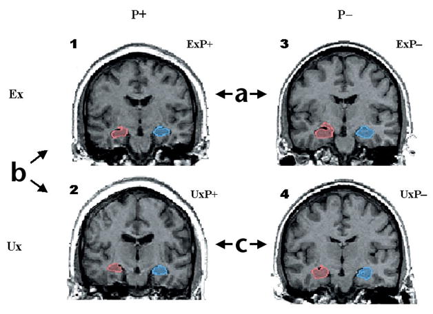
In animals, exposure to severe stress can damage the hippocampus. Recent human studies show smaller hippocampal volume in individuals with the stress-related psychiatric condition posttraumatic stress disorder (PTSD). Does this represent the neurotoxic effect of trauma, or is smaller hippocampal volume a pre-existing condition that renders the brain more vulnerable to the development of pathological stress responses? In monozygotic twins discordant for trauma exposure, we found evidence that smaller hippocampi indeed constitute a risk factor for the development of stress-related psychopathology. Disorder severity in PTSD patients who were exposed to trauma was negatively correlated with the hippocampal volume of both the patients and the patients' trauma-unexposed identical co-twin. Furthermore, severe PTSD twin pairs-both the trauma-exposed and unexposed members-had significantly smaller hippocampi than non-PTSD pairs.


Conflict of interest statement

Competing interests statement

The authors declare that they have no competing financial interests.

Figures

Fig. 1
Fig. 1
Discordant monozygotic twin paradigm for assessing MRI differences in PTSD. Sample coronal MRI images of right (red) and left (blue) hippocampi in a PTSD and a non-PTSD twin pair. Images represent four subject groups: (1) combat-exposed (Ex) subjects who developed chronic PTSD (ExP+); (2) their combat-unexposed (Ux) co-twins with no PTSD themselves (UxP+); (3) Ex subjects who never developed PTSD (ExP−) and (4) Ux co-twins also with no PTSD (UxP−). Contrast (a) provides a replication of previous work demonstrating smaller hippocampal volumes in combat veterans with versus without PTSD. Contrast (b) identifies the neurotoxicity effect—hippocampal reduction—as environmentally acquired, by contrasting hippocampal volumes in combat-exposed PTSD veterans with their unexposed co-twins. Contrast (c) examines pre-existing vulnerability by contrasting hippocampal volumes in the two groups of combat-unexposed co-twins whose combat-exposed brothers did versus did not develop PTSD. Model is tested by a diagnosis (P+ versus P−) × exposure (Ex versus Ux) ANOVA. Diagnosis refers to combat-exposed twin only. If hippocampal volume represents a vulnerability factor, the model predicts a significant main effect of diagnosis in the absence of a diagnosis × exposure interaction (that is, PTSD combat-exposed veterans and their unexposed co-twins show the same pattern). If hippocampal reduction results from neurotoxicity, the model predicts a significant main effect of exposure and/or a significant diagnosis × exposure interaction.
Fig. 2
Fig. 2
Hippocampal volume correlations with post-trauma symptoms. Scatter plots illustrate relationship of symptom severity in combat veterans with PTSD to: (a) their own hippocampal volumes and (b) the hippocampal volumes of their identical twin brothers who were not exposed to combat. Symptom severity represents the total score received on the Clinician-Administered PTSD Scale (CAPS).
Fig. 3
Fig. 3
Total hippocampal volumes for four subject groups. Scatter plot illustrates absolute hippocampal volumes (ml) for combat-exposed individuals with and without PTSD, as well as for their respective unexposed co-twins. Data are only presented for PTSD twin pairs in which the combat-exposed twin had a CAPS score >65.

Comment in

References

    1. Sapolsky RM, Uno H, Rebert CS, Finch CE. Hippocampal damage associated with prolonged glucocorticoid exposure in primates. J Neurosci. 1990;10:2897–2902. - PMC - PubMed
    1. McEwen BS. In: The Cognitive Neurosciences. Gazzaniga MS, editor. MIT Press; Cambridge, Massachusetts: 1995. pp. 1117–1135.
    1. Squire LR. Memory and the hippocampus: a synthesis from findings with rats, monkeys, and humans. Psychol Rev. 1992;99:195–231. - PubMed
    1. Zola-Morgan S, Squire LR. Neuroanatomy of memory. Annu Rev Neurosci. 1993;16:547–563. - PubMed
    1. Bremner JD, et al. MRI-based measurements of hippocampal volume in combat-related posttraumatic stress disorder. Am J Psychiatry. 1995;152:973–978. - PMC - PubMed

Publication types

LinkOut - more resources