. 2002 Apr;7(2):146-55.
doi: 10.1379/1466-1268(2002)007<0146:dpaaoh>2.0.co;2.

Distribution, phosphorylation, and activities of Hsp25 in heat-stressed H9c2 myoblasts: a functional link to cytoprotection

Affiliations

Distribution, phosphorylation, and activities of Hsp25 in heat-stressed H9c2 myoblasts: a functional link to cytoprotection

Anton L Bryantsev et al. Cell Stress Chaperones. 2002 Apr.

Abstract

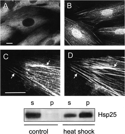
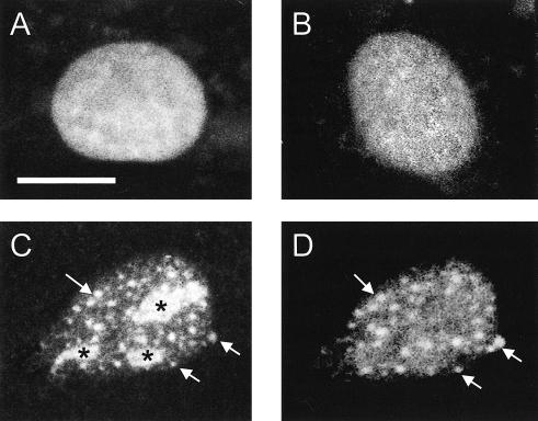
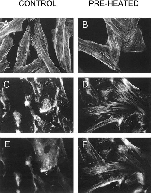
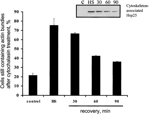
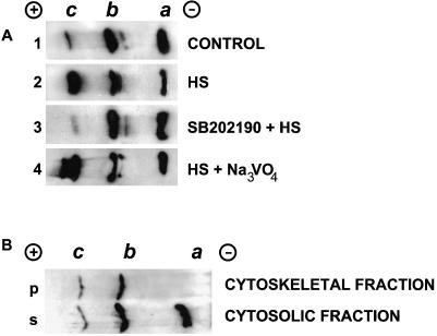
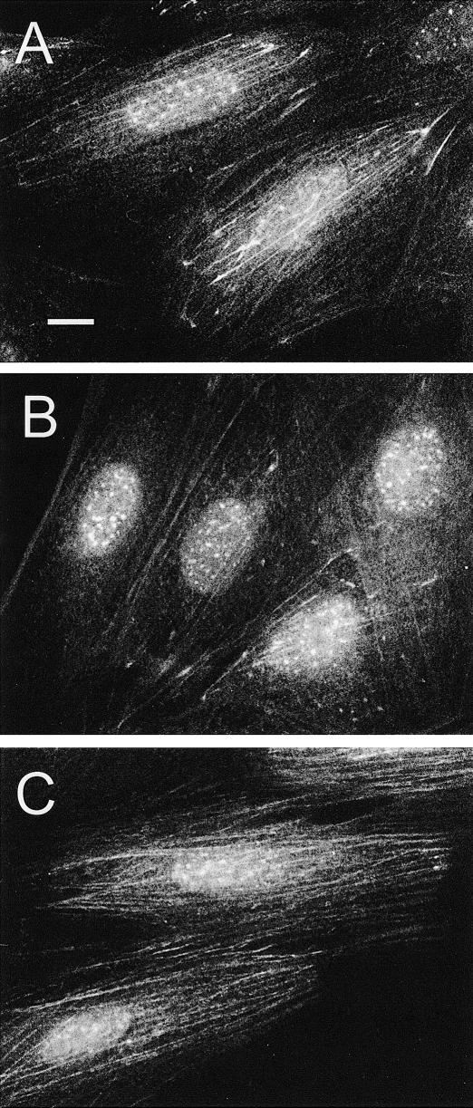
The behavior of the endogenous heat shock protein 25 (Hsp25) in heat-stressed rat H9c2 myoblasts was studied. After mild or severe heating, this protein became less extractable with Triton X-100 and displayed characteristic immunofluorescence patterns, namely (1) granules in the nucleus, and (2) association with F-actin bundles in the cytoplasm. The intranuclear granulation of Hsp25 and its association with F-actin were sensitive to drugs affecting Hsp25 phosphorylation (cantharidin, sodium orthovanadate, SB203580, SB202190). Isoform analysis of Hsp25 translocated to the nucleus-free cytoskeletal fraction revealed only mono- and biphosphorylated Hsp25 and no unphosphorylated Hsp25. Transfected luciferase with initial localization in the nucleosol became colocalized with the Hsp25-containing granules after a heat shock treatment that denatured the enzyme in the cells. The association of Hsp25 with actin filaments after a mild heat stress conferred protection from subsequent F-actin-damaging treatments with cytochalasins (D and B) or severe heat stress. We hypothesize that (1) the binding of heat-denatured nucleosolic proteins to the Hsp25 contained in specific granular structures may serve for the subsequent chaperoning or degradation of the bound proteins, and (2) the actin cytoskeleton is stabilized by the direct targeting of phosphorylated Hsp25 to microfilament bundles.


Figures

Fig. 1.
Fig. 1.
Redistribution of Hsp25 in heat-stressed H9c2 cells. (A) Intracellular localization of Hsp25 under normal conditions and (B, C) after a 30-minute recovery after heat shock (43°C, 30 minutes). (B) Note bright granules of Hsp25 in the nucleus and (C) colocalization of Hsp25 with actin fibers in the cytoplasm. The same cell preparation displays double labeling with (C) anti-Hsp25 and Texas Red conjugates and (D) fluorescein isothiocyanate–phalloidin. Arrows show examples of Hsp25 colocalization with actin fibers. Bars, 10 μm. (Lower panel) Segments of ECL–developed blots showing the heat stress–induced translocation of cytosolic Hsp25 to the cytoskeletal-nuclear fraction. Equal numbers (150 000) of the cells were either not heated (control) or heated (heat shock) for 30 minute at 43°C and fractionated into the Triton X-100–soluble fraction (s) and -insoluble (nuclear-cytoskeletal) fraction (p)
Fig. 2.
Fig. 2.
Specific relocalization of the transfected nuclear luciferase–enhanced green fluorescence protein (EGFP) fusion protein (nuc-luc-EGFP) in heat-shocked H9c2 cells. Cell nuclei expressing nuc-luc-EGFP (A) before and (C) after heat shock (43°C, 30 minutes). (C) Green fluorescence of the EGFP label in the fixed cell preparations was compared with (D) Texas Red fluorescence after the indirect immunostaining with anti-Hsp25 antibodies. (B) As a control, the same heat shock conditions were applied for the cells expressing EGFP alone. The same EGFP distribution was observed in unstressed cells. Arrows denote typical nuclear granules containing both Hsp25 and nuc-luc. Nuc-luc-EGFP entrapped into the nucleoli is marked by asterisk (*). Bar, 10 μm
Fig. 3.
Fig. 3.
Translocation of Hsp25 to actin fibers is associated with their resistance to cytochalasin D. Control, non-preheated cells (A, C, E), or cells preconditioned with a 30-minute heat shock (preheated: B, D, F) were fixed before (A, B) or after treatment with cytochalasin D (0.5 μg/mL) for 10 minutes and were stained with fluorescein isothiocyanate (FITC)-phalloidin (A–D) or were double labeled with FITC-phalloidin (C, D) and anti-Hsp25–Texas Red conjugates (E, F). Bar, 10 μm
Fig. 4.
Fig. 4.
Transient F-actin resistance to cytochalasin D in preheated H9c2 cells. The non-preheated control cells and cells recovering for indicated times after heat shock (43°C, 30 minutes) were treated with cytochalasin D (0.5 μg/mL) for 10 minute, fixed, and stained with fluorescein isothiocyanate–phalloidin. The percentage of the cells containing extended native-like actin fibers was counted. Amounts of Hsp25 translocated to actin bundles are demonstrated by Western blot of Triton-insoluble fractions (insertion) prepared from cytoplasts, as described in Materials and Methods. Each line corresponds to the bar on the diagram
Fig. 5.
Fig. 5.
Analysis of the Hsp25 isoform spectra (A) in whole H9c2 cells and (B) in the fractionated nucleus-free cytoplasts. (A) Immunoblots showing the Hsp25 isoform spectra in unstressed H9c2 cells (lane 1), in cells 30 minutes after heat shock (43°C, 30 minutes) (lane 2), after the same heating and recovery in the presence of 10 μM SB202190 (lane 3) or 50 μM sodium orthovanadate (lane 4). Each blot carries the material from 150 000 cells that were totally lysed, run by IEF, and immunoblotted, as described in Materials and Methods. The same results were obtained with 10 μM SB203580 and 20 μM cantharidinas in lanes 3 and 4, respectively (data not shown). (B) Immunoblots demonstrating Hsp25 isoforms in the Triton X-100–soluble fraction (s) and -insoluble (cytoskeletal) fraction (p) in heat-shocked cytoplasts. Cytoplasts derived from 100 000 cells were heat shocked for 30 minutes at 43°C, fractionated with Triton X-100, run by IEF, and immunoblotted, as described in Materials and Methods. Note the full absence of nonphosphorylated a isoform of Hsp25 in the cytoskeletal fraction
Fig. 6.
Fig. 6.
Effects of drugs affecting phosphorylation of Hsp25 on the intracellular distribution of Hsp25 after a mild heat shock (43°C, 30 minutes). (A) The heat-shocked control cells recovered for 1 hour. (B) The cells pretreated with the medium containing 10 μM SB202190 for 2 hours before heat shock and recovered for 1 hour in the same medium. (C) The cells recovered in the medium containing 50 μM sodium orthovanadate. Bar, 10 μm

References

    1. Alessi DR, Cuenda A, Cohen P, Dudley DT, Saltiel AR. PD098059 is a specific inhibitor of the activation of mitogen-activated protein kinase kinase in vitro and in vivo. J Biol Chem. 1995;270:27489–27494. - PubMed
    1. Arrigo AP. Small stress proteins: chaperones that act as regulators of intracellular redox state and programmed cell death. Biol Chem. 1998;379:19–26. - PubMed
    1. Arrigo AP, Landry J 1994 Expression and function of the low-molecular-weight heat shock proteins. In: The Biology of Heat Shock Proteins and Molecular Chaperones, ed Morimioto RI, Tissieres A, Georgopoulos C. Cold Spring Harbor Laboratory Press, Cold Spring Harbor, NY, 335–373.
    1. Arrigo AP, Suhan JP, Welch WJ. Dynamic changes in the structure and intracellular locate of the mammalian low-molecular-weight heat shock protein. Mol Cell Biol. 1988;8:5059–5071. - PMC - PubMed
    1. Benndorf R, Hayess K, Ryazantsev S, Wieske M, Behlke J, Lutsch G. Phosphorylation and supramolecular organization of murine small heat shock protein HSP25 abolish its actin polymerization-inhibiting activity. J Biol Chem. 1994;269:20780–20784. - PubMed

Publication types

LinkOut - more resources