Does lung diffusion impairment affect exercise capacity in patients with heart failure?
- PMID: 12381630
- PMCID: PMC1767418
- DOI: 10.1136/heart.88.5.453
Does lung diffusion impairment affect exercise capacity in patients with heart failure?
Abstract
Objective: To determine whether there is a relation between impairment of lung diffusion and reduced exercise capacity in chronic heart failure.
Design: 40 patients with heart failure in stable clinical condition and 40 controls participated in the study. All subjects underwent standard pulmonary function tests plus measurements of resting lung diffusion (carbon monoxide transfer, TLCO), pulmonary capillary volume (VC), and membrane resistance (DM), and maximal cardiopulmonary exercise testing. In 20 patients and controls, the following investigations were also done: (1) resting and constant work rate TLCO; (2) maximal cardiopulmonary exercise testing with inspiratory O2 fractions of 0.21 and 0.16; and (3) rest and peak exercise blood gases. The other subjects underwent TLCO, DM, and VC measurements during constant work rate exercise.
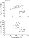
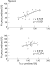
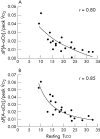
Results: In normoxia, exercise induced reductions of haemoglobin O2 saturation never occurred. With hypoxia, peak exercise uptake (peak O2) decreased from (mean (SD)) 1285 (395) to 1081 (396) ml/min (p < 0.01) in patients, and from 1861 (563) to 1771 (457) ml/min (p < 0.05) in controls. Resting TLCO correlated with peak O2 in heart failure (normoxia < hypoxia). In heart failure patients and normal subjects, TLCO and peak O2 correlated with O2 arterial content at rest and during peak exercise in both normoxia and hypoxia. TLCO, VC, and DM increased during exercise. The increase in TLCO was greater in patients who had a smaller reduction of exercise capacity with hypoxia. Alveolar-arterial O2 gradient at peak correlated with exercise capacity in heart failure during normoxia and, to a greater extent, during hypoxia.
Conclusions: Lung diffusion impairment is related to exercise capacity in heart failure.
Figures


Similar articles
-
Reduced alveolar-capillary membrane diffusing capacity in chronic heart failure. Its pathophysiological relevance and relationship to exercise performance.Circulation. 1995 Jun 1;91(11):2769-74. doi: 10.1161/01.cir.91.11.2769. Circulation. 1995. PMID: 7758183
-
Pulmonary and peripheral vascular factors are important determinants of peak exercise oxygen uptake in patients with heart failure.J Am Coll Cardiol. 1993 Mar 1;21(3):641-8. doi: 10.1016/0735-1097(93)90096-j. J Am Coll Cardiol. 1993. PMID: 8436745
-
Impaired pulmonary diffusion during exercise in patients with chronic heart failure.Circulation. 1999 Sep 28;100(13):1406-10. doi: 10.1161/01.cir.100.13.1406. Circulation. 1999. PMID: 10500041
-
Impedance to gas transfer across the alveolar-capillary membrane in chronic cardiac failure.Ital Heart J. 2000 Mar;1(3):169-73. Ital Heart J. 2000. PMID: 10806983 Review.
-
The Role of Exercise Testing in Pulmonary Vascular Disease: Diagnosis and Management.Clin Chest Med. 2021 Mar;42(1):113-123. doi: 10.1016/j.ccm.2020.11.003. Clin Chest Med. 2021. PMID: 33541605 Review.
Cited by
-
The pulmonary circulation and exercise responses in the elderly.Semin Respir Crit Care Med. 2010 Oct;31(5):528-38. doi: 10.1055/s-0030-1265894. Epub 2010 Oct 12. Semin Respir Crit Care Med. 2010. PMID: 20941654 Free PMC article. Review.
-
Brisk walking can be a maximal effort in heart failure patients: a comparison of cardiopulmonary exercise and 6 min walking test cardiorespiratory data.ESC Heart Fail. 2022 Apr;9(2):812-821. doi: 10.1002/ehf2.13781. Epub 2021 Dec 30. ESC Heart Fail. 2022. PMID: 34970846 Free PMC article.
-
Pulmonary vascular response patterns during exercise in left ventricular systolic dysfunction predict exercise capacity and outcomes.Circ Heart Fail. 2011 May;4(3):276-85. doi: 10.1161/CIRCHEARTFAILURE.110.959437. Epub 2011 Feb 3. Circ Heart Fail. 2011. PMID: 21292991 Free PMC article.
-
Effects of respiratory muscle work on blood flow distribution during exercise in heart failure.J Physiol. 2010 Jul 1;588(Pt 13):2487-501. doi: 10.1113/jphysiol.2009.186056. Epub 2010 May 10. J Physiol. 2010. PMID: 20457736 Free PMC article.
-
The cardiorenal syndrome in heart failure: cardiac? renal? syndrome?Heart Fail Rev. 2012 May;17(3):355-66. doi: 10.1007/s10741-011-9291-x. Heart Fail Rev. 2012. PMID: 22086438 Review.
References
-
- Sue DY, Oren A, Hansen JE, et al. Diffusing capacity for carbon monoxide as a predictor of gas exchange during exercise. N Engl J Med 1987;316:1301–6. - PubMed
-
- Puri S, Baker BL, Dutka DP, et al. Reduced alveolar-capillary membrane diffusing capacity in chronic heart failure. Circulation 1995;91:2769–74. - PubMed
-
- Smith AA, Cowburn PJ, Parker ME, et al. Impaired pulmonary diffusion during exercise in patients with chronic heart failure. Circulation 1999;100:1406–10. - PubMed
-
- Guazzi M, Marenzi GC, Alimento M, et al. Improvement of alveolar capillary diffusing capacity in chronic heart failure and counteracting effects of aspirin. Circulation 1997;95:1930–6. - PubMed
-
- Guazzi M, Marenzi GC, Melzi G, et al. Angiotensin-converting enzyme inhibition facilitates alveolar-capillary gas transfer and improves ventilation perfusion coupling in patients with left ventricular dysfunction. Clin Pharmacol Ther 1999;65:319–27. - PubMed
Publication types
MeSH terms
Substances
LinkOut - more resources
Full Text Sources
Medical
