Review
. 2002 Nov 5;53(2):143-56.
doi: 10.1002/neu.10098.

Development and evolution of inner ear sensory epithelia and their innervation

Affiliations
Review

Development and evolution of inner ear sensory epithelia and their innervation

B Fritzsch et al. J Neurobiol. .

Abstract

The development and evolution of the inner ear sensory patches and their innervation is reviewed. Recent molecular developmental data suggest that development of these sensory patches is a developmental recapitulation of the evolutionary history. These data suggest that the ear generates multiple, functionally diverse sensory epithelia by dividing a single sensory primordium. Those epithelia will establish distinct identities through the overlapping expression of genes of which only a few are currently known. One of these distinctions is the unique pattern of hair cell polarity. A hypothesis is presented on how the hair cell polarity may relate to the progressive segregation of the six sensory epithelia. Besides being markers for sensory epithelia development, neurotrophins are also expressed in delaminating cells that migrate toward the developing vestibular and cochlear ganglia. These delaminating cells originate from multiple sites at or near the developing sensory epithelia and some also express neuronal markers such as NeuroD. The differential origin of precursors raises the possibility that some sensory neurons acquire positional information before they delaminate the ear. Such an identity of these delaminating sensory neurons may be used both to navigate their dendrites to the area they delaminated from, as well as to help them navigate to their central target. The navigational properties of sensory neurons as well as the acquisition of discrete sensory patch phenotypes implies a much more sophisticated subdivision of the developing otocyst than the few available gene expression studies suggest.


Figures

Figure 1
Figure 1
These images show the conjecture of de Burlet (1934) about the evolutionary similarities of endorgan and innervation multiplication from two hypothetical ancestors (A,B) to the vertebrates with the largest number of sensory epithelia, Southeast Asian caecilians (C). In his accompanying text, de Burlet pointed this out explicitly, but without proposing any mechanism. Conceivably, two mechanisms are apparent. In one mechanism there is lineage relationship between hair cells and sensory neurons. Thus whenever hair cell precursors are split their innervation will split. Alternatively, there is no relationship at all between hair cells and sensory neurons, and the multiplication of innervation comes about through the selective attraction of sensory afferents to newly formed endorgans. In the latter scenario both central and peripheral specifications of the sensory projections need to be accomplished independently of the changes in hair cell specification. In the lineage relationship scenario, any alteration in the specification of hair cells would also affect the information conveyed by sensory cells, thus allowing for a rapid coevolution of hair cell and sensory neuron fate specification. The work of Norris (1892) on salamanders indicated that the above-presented evolutionary scenario might actually be recapitulated during development. He depicted that a single prosensory anlage splits over time into the different sensory organs (D,E). It is unclear at which point in development polarity of hair cells is established. We tentatively superimposed the known adult polarity distribution in salamanders (Lewis et al., 1985) onto the almost completely segregated patches of forming sensory epithelia. Note that all canal cristae have only one hair cell polarity, that the utricle has only polarity toward the striola (indicated by the dotted line), and that the saccule and sensory epithelia derived from the saccule are all polarized away from a dividing line in many salamanders. Abbreviations: AC, anterior crista; AP, amphibian papilla; BP, basilar papilla; HC, horizontal crista; PC, posterior crista; PN, papilla neglecta. Modified after Norris (1892), de Burlet (1934), and Lewis et al. (1985).
Figure 2
Figure 2
These wholemounted ears show the expression of various genes as revealed with a lac-Z marker inserted into GATA3, BDNF, NeuroD, and NT-3 genes (A,B,D,E). At embryonic day 11 (E11) distinct patterns of expression are apparent. Note that the distribution of GATA3 and BDNF has some similarities, but also show clear differences. In contrast, similarities between the NeuroD and NT-3 expression in the otocyst are obvious in the area of the future utricle and saccule. Note that each of these four genes is expressed both in restricted areas of the ear and in cells of the forming vestibulo-cochlear ganglion. LacZ-positive cells (arrows) can be seen to delaminate from the otocyst in all four cases. Delamination and cellular migration to the forming vestibulo-cochlear ganglion is even more obvious at later stages [E12 (C,F)] where lacZ- positive cells can be traced from equally lacZ-positive areas of the otocyst. Bar indicates 100 μm.
Figure 3
Figure 3
These images show the distribution of lacZ-labeled cells in histological sections (B,C,E), wholemounts (A,D), BDNF (A,B,D,E), and NeuroD (C). These images show the similarities in distribution of NeuroD and BDNF inside the utricle as well as in delaminating cells (B,C). The wholemounts show the distribution of BDNF in forming hair cells inside the saccule and utricle as well as the delaminating cells converging from specific areas of these sensory epithelia toward the forming vestibular ganglion (A). Such cells delaminate as late as E17 from the hair-cell-free hilus region of the utricle that is filled with hair cells only after completion of this delamination (D,E). Bar indicates 100 μm.
Figure 4
Figure 4
Horizontal sections show the distribution of delaminating saccular cells and various markers. Note that the NT-3lac Zneo-positive cells (A,B) emigrate from the saccular anlage (S) to aggregate near the forming cochleo-vestibular ganglion (G). The sensory neurons have a distinct morphology (C) and are positive for α-acetylated tubulin (B) and trkB (D). In contrast, the delaminating cells (labeled “primordia”) are positive for NT-3lac Zneo (A,B) but not for tubulin (B) or trkB (D). Fibers to the saccule are passing between these cells in a fascicle (F) surrounded by lacZ-positive precursors. Bar indicates 100 μm.
Figure 5
Figure 5
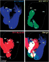
The distribution of BDNF, NT-3, and NeuroD expression in E11 ears. Each image is a computer generated color-coded image of the lacZ expression shown in Figure 2. The panel labeled “merge” shows the computer generated superposition of these expression domains to highlight the relative level of overlap of the three genes. Note that the area delineated by the delaminating sensory neuron precursors that are NeuroDlacZ positive comprises at this stage areas of what has been delineated as sensory epithelia primordia based on neurotrophin lacZ expression. The only exception from this expression of NeuroDlacZ is the posterior crista, which is already fully innervated at this stage in development, suggesting an even earlier delamination of sensory precursors. These data also demonstrate that the ear consists already at this early stage of a mosaic of variously overlapping gene expression domains.

Similar articles

Cited by

References

    1. Acampora D, Gulisano M, Simeone A. Genetic and molecular roles of Otx homeodomain proteins in head development. Gene. 2000;246:23–35. - PubMed
    1. Begbie J, Graham A. Integration between the epibranchial placodes and the hindbrain. Science. 2001;294:595– 598. - PubMed
    1. Bermingham NA, Hassan BA, Price SD, Vollrath MA, Ben-Arie N, Eatock RA, Bellen HJ, Lysakowski A, Zoghbi HY. Math1: an essential gene for the generation of inner ear hair cells. Science. 1999;284:1837–1841. - PubMed
    1. Brichta AM, Goldberg JM. The papilla neglecta of turtles: a detector of head rotations with unique sensory coding properties. J Neurosci. 1998;18:4314–4324. - PMC - PubMed
    1. Brigande JV, Kiernan AE, Gao X, Iten LE, Fekete DM. Molecular genetics of pattern formation in the inner ear: do compartment boundaries play a role? Proc Natl Acad Sci USA. 2000;97:11700–11706. - PMC - PubMed

Publication types

LinkOut - more resources