. 2002 Jun;83(3):121-32.
doi: 10.1046/j.1365-2613.2002.00221.x.

Resolution of skeletal muscle inflammation in mdx dystrophic mouse is accompanied by increased immunoglobulin and interferon-gamma production

Affiliations

Resolution of skeletal muscle inflammation in mdx dystrophic mouse is accompanied by increased immunoglobulin and interferon-gamma production

Jussara Lagrota-Candido et al. Int J Exp Pathol. 2002 Jun.

Abstract

Mdx mouse, the animal model of Duchenne muscular dystrophy, develops an X-linked recessive inflammatory myopathy with an apparent sustained capacity for muscle regeneration. We analysed whether changes in the skeletal muscle during myonecrosis and regeneration would correlate with functional alterations in peripheral lymphoid tissues. Here we show that during the height of myonecrosis, mdx mice display marked atrophy of peripheral lymph nodes and extensive muscle inflammation. In contrast, enlargement of draining lymph nodes with accumulation of CD4+ CD44+, CD4+ CD25+, CD8+ CD44+ T lymphocytes and type-2 B cells was consistently observed during amelioration of the muscle lesion. In addition, regeneration of the muscular tissue was accompanied by concomitant increase of immunoglobulin-secreting cells in regional lymph nodes and bone marrow. Double immunolabelling analysis revealed intense B cell proliferation and formation of germinal centre in the follicles of dystrophic regional lymph nodes. Furthermore, lymph node cells produced large amounts of IFN-gamma but not IL-4, IL-6 or IL-10 after in vitro mitogen stimulation with Concanavalin A. As these alterations occurred mainly during the recovery period, we suggested that local activation of the immune system could be an influence which mitigates the myonecrosis of muscular tissue in the mdx dystrophic mouse.


Figures

Figure 1
Figure 1
Haematoxylin-eosin staining of a 5 µm wax-embedded section of Triceps brachii muscle fragments of 4 week (a, d), 12 week (b, e) and 24 week (c, f) dystrophic mice. (a–d) Extensive myonecrosis with hyaline fibres (large arrow) and inflammatory cell infiltration (star) with many polymorphonuclear (arrow) and mononuclear cells (arrow head) at the height of myonecrosis; (b–e) numerous myofibres with central nuclei (arrow head), scarce inflammatory focus (star) and connective tissue (asterisk); (c–f) hypertrophied (arrow head) and necrotic (arrow) myofibres and overdeposition of connective tissue (asterisk) at early fibrosis period. Original magnification ×100 (a, b), ×200 (c), ×1000 (d, e, f).
Figure 2
Figure 2
Flow cytometry analysis of mononuclear cells present in axillary and brachial lymph nodes of mice at 4 weeks, 12 weeks and 24 weeks. One representative experiment is presented for each age. Results are expressed as mean ± SD of four animals. This experiment was performed four times. □ C57BL/ 10, formula imagemdx. *P < 0.05 and **P < 0.0001.
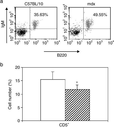
Figure 3
Figure 3
Flow cytometry analysis with anti-B220/R-PE, anti-IgM/FITC and anti-CD5 biotin/streptoavidin-quantum red of single cell suspensions from axillary and brachial lymph nodes of control and mdx mice at 24 weeks. (a) Expression of B220 and IgM; (b) percentage of CD5+ cells in the B220+IgM+ population. One representative experiment is presented. Results are expressed as mean ± SD of four animals. Similar results were seen in four additional experiments. □ C57BL/ 10,formula imagemdx. *P < 0.05.
Figure 4
Figure 4
Expression of CD25, CD44 and CD62L within CD4+ (panel a) and CD8+ (panel b) T cells from 24 week mdx lymph nodes. Results are expressed as mean ± SD of four animals. One representative experiment is presented. Similar results were seen in three additional experiments. □ C57BL/ 10, formula image
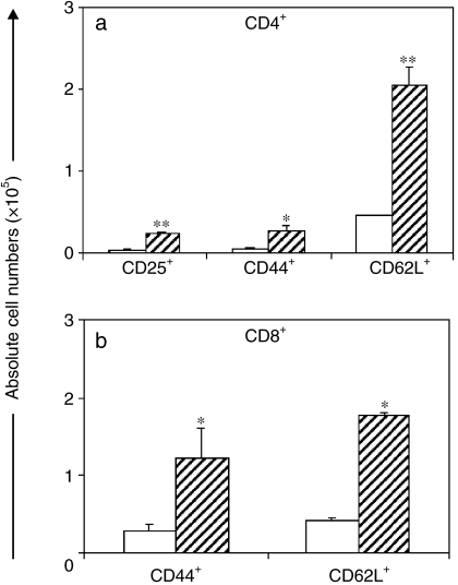
mdx. *P < 0.05, **P < 0.0001.
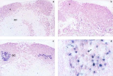
Figure 5
Figure 5
Photomicrographs showing characteristic staining of B220 (red) and PCNA (black) expression in 24 week mdx lymph node (b) compared with control C57BL/10 (a). Note the presence of germinal centre formation in mdx lymph node (c). B220+ PCNA+ lymphocytes in the germinal centre are indicated by arrow head (d). Original magnification ×200 (a, b, c) and ×1000 (d). c, cortical; PF, parafollicular; GC, germinal centre.
Figure 6
Figure 6
TCR+ and B220+ cell numbers in the bone marrow of mice at ages 4 weeks, 12 weeks and 24 weeks. One representative experiment is shown. Results are expressed as mean ± SD of four animals. Similar results were seen in three additional experiments. □ C57BL/ 10,formula imagemdx. *P < 0.05.
Figure 7
Figure 7
Numbers of Ig-secreting cells in draining lymph nodes (a, d), spleen (b, e) and bone marrow (c, f) of control and mdx mice at 12 weeks (a, b, c) and 24 weeks (d, e, f). Each symbol represents one animal. • IgM, ♦ IgG and ▴ IgA for mdx and corresponding open symbols for control mice. Bars depict mean values.
Figure 8
Figure 8
In vitro immunoglobulin secretion in the supernatants of lymphocytes derived from axillary and brachial lymph nodes of 12 week mdx mice cultivated in presence of LPS (10 µg/mL). Panel (a) shows IgM, IgG and IgA secretion in C57BL/ 10 or mdx supernatants. Panel (b) shows antigen reactivity of lymphocyte supernatants. One-hundred µL of protein antigens (10 µg/mL) were used for coating wells of microtitre plates. Supernatant reactivity was assessed with anti-IgM antibodies by direct ELISA. Representative experiments are presented. Results are expressed as mean ± SD of five animals. Similar results were obtained in three separate experiments. □ C57BL/ 10,formula imagemdx. **P < 0.001, ***P < 0.0001.
Figure 9
Figure 9
Increased production of IFN-γ by lymphocyte pool from 5 mdx or C57BL/ 10 mice at ages of 4, 12 and 24 weeks. Lymphocytes were stimulated or not with Con A (5 µg/mL) for 24 h. Results are expressed as mean ± SD of wells. One representative experiment is shown. Similar results were obtained in three separate experiments. □ C57BL/ 10,formula imagemdx. *P < 0.05, **P < 0.005.

Similar articles

Cited by

References

    1. Allamand V, Campbell KP. Animal models for muscular dystrophy: valuable tools for the development of therapies. HumMolGenet. 2000;9:2459–2467. - PubMed
    1. Amemiya K, Semino-Mora C, Granger RP, Dalakas MC. Downregulation of TGF-β1 mRNA and protein in the muscles of patients with inflammatory myopathies after treatment with high-dose intravenous immunoglobulin. ClinImmunol. 2000;94:99–104. - PubMed
    1. Bernasconi P, Torchiana E, Confalonieri P, et al. Expression of transforming growth factor-β1 in dystrophic patient muscles correlates with fibrosis. JClinInvest. 1995;96:1137–1144. - PMC - PubMed
    1. Billiau A. Interferon-γ: biology and role in pathogenesis. AdvImmunol. 1996;62:61–109. - PubMed
    1. Boland B, Himpens B, Denef JF, Gillis JM. Site-dependent pathological differences in smooth muscles and skeletal muscles of the adult mdx mouse. Muscle & Nerve. 1995;18:649–657. - PubMed

Publication types

LinkOut - more resources