Review
. 2002 Nov 1;100(9):3077-86.
doi: 10.1182/blood-2002-04-1104.

Locus control regions

Affiliations
Review

Locus control regions

Qiliang Li et al. Blood. .

Abstract

Locus control regions (LCRs) are operationally defined by their ability to enhance the expression of linked genes to physiological levels in a tissue-specific and copy number-dependent manner at ectopic chromatin sites. Although their composition and locations relative to their cognate genes are different, LCRs have been described in a broad spectrum of mammalian gene systems, suggesting that they play an important role in the control of eukaryotic gene expression. The discovery of the LCR in the beta-globin locus and the characterization of LCRs in other loci reinforces the concept that developmental and cell lineage-specific regulation of gene expression relies not on gene-proximal elements such as promoters, enhancers, and silencers exclusively, but also on long-range interactions of various cis regulatory elements and dynamic chromatin alterations.


Figures

Figure 1
Figure 1. The human and mouse β-globin loci
The human locus consists of 5 functional genes, indicated as dark boxes, arrayed in their order of developmental expression, 5′-∊-G-γ-A-γ-δ-β-3′. There are 2 developmental switches in globin chain synthesis coincident with changes in site and type of eryshropoiesis. During primitive erythropoiesis, the ∊-globin gene is expressed in the embryonic yolk sac. The first switch occurs at approximately 8 weeks' gestation; the ∊-globin gene is silenced and the Gγ- and Aγ-globin genes are expressed during definitive erythropoiesis in the fetal liver. The second switch occurs shortly after birth; the γ-globin genes are silenced and the β-globin gene and, to a lesser extent, the δ-globin gene are activated in the bone marrow. The HSs 5′HS1 through 5′HS7 are located −6, −11, −15, −18, −22, −28, and −35 kb relative to the ∊-globin gene, respectively, and are indicated by arrows. 5′HS1 through 5′HS4 are erythroid specific, but 5′HS5 through 5′HS 7 are not. Another HS (3′HS1) is locaied 20 kb downstream of the β-globin gene; 3′HS1 is found only in eryihroid cells. Boxes represent globin genes and ovals represent olfactor receptor genes; filled ones represent the productive genes and shaded ones the pseudogenes. The lines below the diagram of the locus indicate deletions of the LCR discussed in “In vivo function of LCRs.” The Hispanic deletion, which causes (γδβ)0 thalassemia in humans, extends an additional 20 kb 5′ of the LCR 5′HS5.
Figure 2
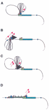
Figure 2. Models of LCR function
A globin gene is denoted by a green rectangular box with the promoter region indicated in a lighter green. Transcription factors are shown as colored ovals and circles. The 4 erythroid-specific hypersensitive site cores (HSs) are indicated by small red boxes. Blue boxes are the positions of 5′HS5 and 3′HS1, representing likely insulator elements. The flanking DNA sequences of the HSs are depicted as loops between the HS cores. Transcripts are denoted by wavy arrows. (A) Looping model. Transcription factors bind to the LCR HSs and the gene promoter. The LCR directly interacts with the gene promoter by looping out the intervening DNA, thus forming an active transcription complex at the gene promoter. (B) Tracking model. Sequence-specific transcription factors bind to the LCR, forming a complex that tracks down the DNA sequence, as depicted by the large black arrowhead, until encountering transcription factors bound to the appropriate gene promoter, initiating high-level gene expression. (C) Facilitated tracking model. Aspects of both looping and tracking models are combined. Sequence-specific transcription factors bind the LCR; looping then occurs to deliver the bound transcription factors proximal to the gene promoter, followed by tracking, until they encounter transcription factors bound to the appropriate gene promoter. (D) Linking model. Sequential binding of transcription factors along the DNA directs changes in chromatin conformation and defines the transcriptional domain. The transcription factors are linked to one another from the LCR to the gene promoter by non–DNA-binding proteins and chromatin modifiers (shown as small colored circles).

Similar articles

Cited by

References

    1. Grosveld F, van Assendelft GB, Greaves DR, Kollias G. Position-independent, high-level expression of the human beta-globin gene in transgenic mice. Cell. 1987;51:975–985. - PubMed
    1. Stamatoyannopoulos G, Grosveld F. Hemoglobin switching. In: Stamatoyannopoulos G, Majerus PW, Perlmutter RM, Varmus H, editors. Molecular Basis of Blood Diseases. 3rd ed. WB Saunders; Philadelphia, PA: 2001. pp. 135–182.
    1. Fraser P, Grosveld F. Locus control regions, chromatin activation and transcription. Curr Opin Cell Biol. 1998;10:361–365. - PubMed
    1. Li Q, Harju S, Peterson KR. Locus control regions: coming of age at a decade plus. Trends Genet. 1999;15:403–408. - PubMed
    1. Magram J, Chada K, Costantini F. Developmental regulation of a cloned adult beta-globin gene in transgenic mice. Nature. 1985;315:338–340. - PubMed

Publication types