. 2002 Oct 15;30(20):4398-405.
doi: 10.1093/nar/gkf569.

IRES-driven translation is stimulated separately by the FMDV 3'-NCR and poly(A) sequences

Affiliations

IRES-driven translation is stimulated separately by the FMDV 3'-NCR and poly(A) sequences

Sonia López de Quinto et al. Nucleic Acids Res. .

Abstract

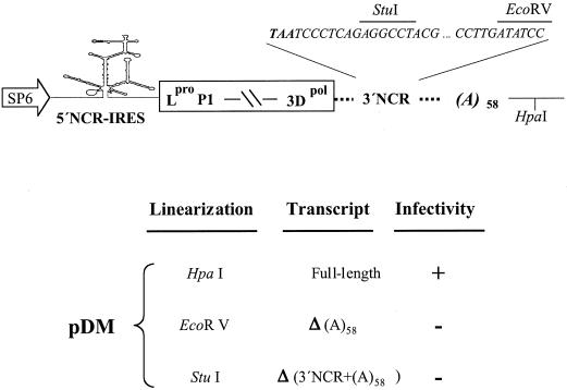
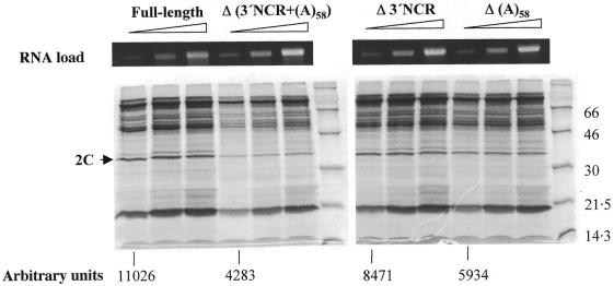
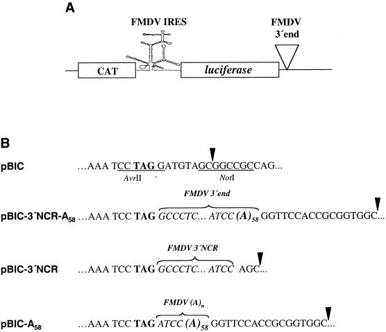
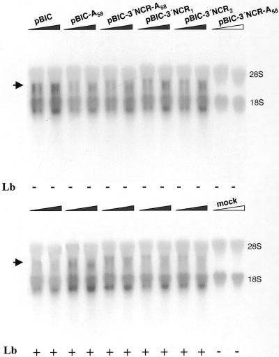
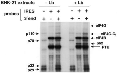
The 3' end region of foot-and-mouth disease virus (FMDV) consists of two distinct elements, a 90 nt untranslated region (3'-NCR) and a poly(A) tract. Removal of either the poly(A) tract or both the 3'-NCR and the poly(A) tract abrogated infectivity in susceptible cells in the context of a full-length cDNA clone. We have addressed the question of whether the impairment of RNA infectivity is related to defects at the translation level using a double approach. First, compared to the full-length viral RNA, removal of the 3' sequences reduced the efficiency of translation in vitro. Secondly, a stimulatory effect of the 3' end sequences on IRES-dependent translation was found in vivo using bicistronic constructs. RNAs carrying the FMDV 3' end sequences linked to the second cistron showed a significant stimulation of IRES-dependent translation, whereas cap-dependent translation was not affected. Remarkably, IRES-dependent stimulation exerted by the poly(A) tract or the 3'-NCR seems to be the result of two separate events, as the 3'-NCR alone enhanced IRES activity on its own. Under conditions of FMDV Lb protease-induced translation shut-off, the stimulation of IRES activity reached values above 6-fold in living cells. A northern blot analysis indicated that IRES stimulation was not the consequence of a change in the stability of the bicistronic RNA produced in transfected cells. Analysis of the RNA-binding proteins interacting with a mixture of 3' end and IRES probes showed an additive pattern. Altogether, our results strongly suggest that individual signals in the viral 3' end ensure stimulation of FMDV translation.


Figures

Figure 1
Figure 1
Effects of 3′ end sequences on FMDV infectivity. Schematic representation of the cDNA present in pDM (21) including relevant restriction sites (top panel). The broken line between P1 and 3Dpol is used for simplicity to represent the coding region. Infectivity of the different transcripts was measured as the ability of an equal amount (4 µg) of the indicated transcripts, transfected into 1–2 × 10⁶ BHK-21 cells, to induce cytopathic effect up to 48 h post-transfection (lower panel). The positive control pDM induced a cytopathic effect 16–20 h post-transfection.
Figure 2
Figure 2
Translation efficiency of FMDV transcripts harboring deleted 3′ end sequences. Synthetic FMDV RNAs of ∼8.5 kb, bearing the indicated deletions, were used to program in vitro translation reactions. Δ3′NCR transcripts were derived from the previously described pΔ74 construct (21). Numbers below each RNA indicate the intensity of protein 2C in arbitrary units. Mobility of molecular weight markers is shown on the right of each gel autoradiograph. The upper panel shows the ethidium bromide staining of an agarose gel loaded with aliquots of the RNAs (100, 200 and 400 ng each).
Figure 3
Figure 3
Bicistronic constructs harboring FMDV 3′ end sequences. (A) Diagram of the bicistronic RNA, CAT–FMDV IRES–luciferase. (B) Differences in the 3′ end sequences in the bicistronic constructs used in transfection assays. Sequences in pBIC generating unique AvrII and NotI restriction sites are underlined. The luciferase stop codon TAG is in bold. The 58 nt poly(A) present in some constructs is denoted as (A)58. A brace embracing residues in italic denotes the FMDV 3′ end sequences included in each bicistronic construct, pBIC-3′NCR-A58, pBIC-3′NCR and pBIC-A58. An arrowhead denotes the last residue of transcripts obtained after NotI linearization of the respective constructs.
Figure 4
Figure 4
Effects of FMDV 3′ end sequences on cap- and IRES-dependent translation efficiency in living cells. (A) IRES-dependent translation was estimated as the luciferase activity, measured in extracts from BHK-21 cells transfected with NotI-linearized plasmids, relative to the value obtained in pBIC, that was set at 100%. Plus or minus symbols indicate whether or not cells were co-transfected with pLb plasmid. (B) Cap-dependent translation was estimated by CAT activity, measured in the same extract as luciferase activity. Error bars correspond to the SEM, from three to six experiments.
Figure 5
Figure 5
Northern blot analysis of RNAs extracted from BHK-21 cells at 12 h after transfection with bicistronic constructs. A filled triangle, as opposed to an empty triangle, depicts RNA extracted from cells expressing T7 RNA polymerase. For each construct two lanes were loaded, containing in the right lane a double amount of RNA compared to the left lane. pBIC-3′NCR1 or pBIC-3′NCR2 corresponds to two independent transfections carried out with two plasmid samples of identical sequence. Below each lane is indicated whether or not co-transfection with pLb was performed. The blot was probed with a [α-32P]UTP-labeled RNA fragment complementary to nucleotides 1–462 of the FMDV IRES. The positions of 28S and 18S rRNA have been marked as migration and loading controls. The mobility of the RNAs derived from the bicistronic plasmids is indicated with an arrow.
Figure 6
Figure 6
Analysis of the interaction of the FMDV 3′ end and FMDV IRES sequences with cellular proteins. The radiolabeled transcripts (0.03 pmol) were used in UV-crosslinking assays with native proteins present in S10 extracts from BHK-21 cells transfected or not with pLb plasmid. When indicated, probes corresponding to the FMDV IRES and 3′ end were co-incubated. Following RNase A treatment, proteins were fractionated by 10% SDS–PAGE. The apparent molecular masses of the polypeptides crosslinked to the 3′ end probe are indicated on the left and those crosslinked to the IRES probe on the right side of the autoradiograph. Identification of p220 as eIF4G, p80 as eIF4B, p110 as a component of eIF3, and p57 as PTB is described in López de Quinto (11) and references therein.

References

    1. Belsham G.J. and Jackson,R.J. (2000) Translation initiation on picornavirus RNA. In Sonenberg,N., Hershey,J.W.B. and Mathews,M.B. (eds), Translational Control of Gene Expression. Cold Spring Harbor Laboratory Press, Cold Spring Harbor, NY, pp. 869–900.
    1. Martínez-Salas E., Ramos,R., Lafuente,E. and López de Quinto,S. (2001) Functional interactions in internal translation initiation directed by viral and cellular IRES. J. Gen. Virol., 82, 973–984. - PubMed
    1. Hellen C.U. and Sarnow,P. (2001) Internal ribosome entry sites in eukaryotic mRNA molecules. Genes Dev., 15, 1593–1612. - PubMed
    1. Bergamini G., Preiss,T. and Hentze,M.W. (2000) Picornavirus IRESes and the poly(A) tail jointly promote cap-independent translation in a mammalian cell-free system. RNA, 6, 1781–1790. - PMC - PubMed
    1. Michel Y.M., Poncet,D., Piron,M., Kean,K.M. and Borman,A.M. (2000) Cap-poly(A) synergy in mammalian cell-free extracts. Investigation of the requirements for poly(A)-mediated stimulation of translation initiation. J. Biol. Chem., 275, 32268–32276. - PubMed

Publication types