. 2002 Oct 15;30(20):4500-8.
doi: 10.1093/nar/gkf583.

La autoantigen is required for the internal ribosome entry site-mediated translation of Coxsackievirus B3 RNA

Affiliations

La autoantigen is required for the internal ribosome entry site-mediated translation of Coxsackievirus B3 RNA

Partho Sarothi Ray et al. Nucleic Acids Res. .

Abstract

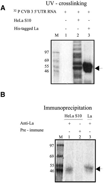
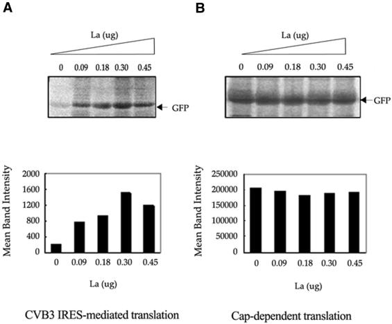
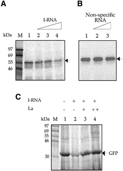
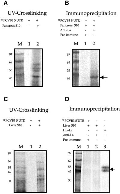
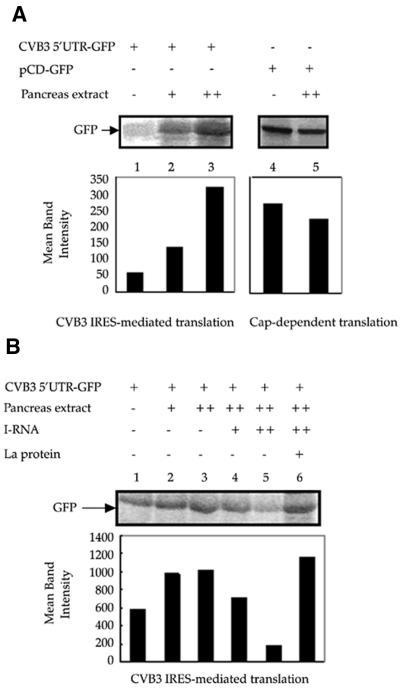
Translation initiation in Coxsackievirus B3 (CVB3) occurs via ribosome binding to an internal ribosome entry site (IRES) located in the 5'-untranslated region (UTR) of the viral RNA. This unique mechanism of translation initiation requires various trans-acting factors from the host. We show that human La autoantigen (La) binds to the CVB3 5'-UTR and also demonstrate the dose-dependent effect of exogenously added La protein in stimulating CVB3 IRES-mediated translation. The requirement of La for CVB3 IRES mediated translation has been further demonstrated by inhibition of translation as a result of sequestering La and its restoration by exogenous addition of recombinant La protein. The abundance of La protein in various mouse tissue extracts has been probed using anti-La antibody. Pancreatic tissue, a target organ for CVB3 infection, was found to have a large abundance of La protein which was demonstrated to interact with the CVB3 5'-UTR. Furthermore, exogenous addition of pancreas extract to in vitro translation reactions resulted in a dose dependent stimulation of CVB3 IRES-mediated translation. These observations indicate the role of La in CVB3 IRES-mediated translation, and suggest its possible involvement in the efficient translation of the viral RNA in the pancreas.


Figures

Figure 1
Figure 1
Specific interaction of La protein with the CVB3 5′-UTR. (A) UV crosslinking of 32P-labeled CVB3 5′-UTR with 9 µg of HeLa S10 extract (lane 2) and 400 ng of purified His-La protein (lane 3). Lane 1 contains the probe in absence of the added proteins. (B) CVB3 5′-UTR–La UV-crosslinked complexes with HeLa S10 (lanes 1 and 2) and purified His-La protein (lane 3) were immunoprecipitated using anti-La antibodies (lanes 1 and 3) and pre-immune rabbit serum (lane 2). The samples were resolved by SDS–10% PAGE and the gels were exposed for phosphorimaging. Lane M represents 14C-labeled protein markers. (A and B) The position of the La–CVB3 RNA complex is indicated by arrows.
Figure 2
Figure 2
Stimulation of CVB3 IRES mediated translation of GFP in RRL by His-tagged La protein. (A) Representative translation of in vitro transcribed uncapped CVB3 5′-UTR–GFP RNA in RRL supplemented with increasing amounts of purified La protein (as indicated above the panel). (B) Translation of in vitro transcribed, capped GFP RNA in presence of the same increasing concentrations of La protein. The translation reactions were carried out using ∼1 µg of RNA template. The gels were treated with 1 M sodium salicylate, dried and exposed for phosphorimaging. Intensities of GFP bands in the translation reactions were quantified by Fuji Imagegauge and are graphically represented below each panel.
Figure 3
Figure 3
Competition of I-RNA with CVB3 5′-UTR for binding to La and rescue of CVB3 IRES-mediated translation by recombinant La protein. (A) [32P]UTP labeled CVB3 5′-UTR RNA probe was UV crosslinked to His-tagged La protein in the absence (lane 1) and presence (lanes 2–4) of 200-, 400- and 600-fold molar excess of unlabeled I-RNA. (B) [32P]UTP labeled CVB3 5′-UTR RNA probe was UV crosslinked to His-tagged La protein in the absence (lane 1) and presence (lanes 2 and 3) of 400- and 600-fold molar excess of a non-specific RNA. (C) CVB3 5′-UTR–GFP RNA was translated in vitro in RRL supplemented with 0.5 µg HeLa S10 (lanes 1–4), 50-fold excess of I-RNA (lanes 2–4) and two concentrations (200 and 400 ng) of La protein (lanes 3 and 4). The positions of the La–CVB3 RNA complex (A and B) and the translation product GFP (C) are indicated by arrows.
Figure 4
Figure 4
Immunoblot of mouse tissue extracts using anti-La polyclonal antibodies. Various mouse tissue extracts (as indicated) containing equivalent amounts of total protein were resolved on SDS–10% PAGE and probed with polyclonal anti-La antibody. After probing with HRP-conjugated secondary antibody, immunoreactive bands were visualized by peroxidase reaction using diaminobenzidine as substrate. A replica blot containing the same amounts of protein per lane was probed with anti-β actin monoclonal antibody and anti-mouse secondary antibody followed by ECL.
Figure 5
Figure 5
Interaction of CVB3 5′-UTR RNA with La protein from mouse pancreas. (A) 32P-labeled CVB3 5′-UTR RNA was UV crosslinked to pancreas cytoplasmic extract of Balb/c mice (lane 2). Lane 1 contains the free probe in the absence of any added proteins. (B) 32P-labeled CVB3 5′-UTR RNA–protein complexes from the pancreas extract were immunoprecipitated using anti-La antibodies (lane 2) and pre-immune serum (lane 1). (C and D) UV crosslinking and immunoprecipitation of 32P-labeled CVB3 5′-UTR RNA–protein complexes from liver cytoplasmic extract of Balb/c mice. Lanes 1 and 2 correspond to those in (A) and (B). Lane 3 in (D) contains purified His-tagged La protein immunoprecipitated with anti-La antibodies. The samples were resolved by SDS–10% PAGE, the gels were dried and exposed for phosphorimaging. Lane M represents 14C-labeled protein markers. The arrow indicates CVB3 5′-UTR RNA–La complex.
Figure 6
Figure 6
Effect of exogenously added mouse pancreas extract on CVB3 IRES-mediated translation. (A) Two increasing doses of pancreas extract from Balb/c mice were exogenously added to in vitro translation reactions of CVB3 5′-UTR–GFP RNA in RRL (lanes 2 and 3). Approximately 1 µg of RNA template was used in each reaction. The higher dose of the pancreas extract was added to in vitro translation reaction using capped GFP RNA as template (lane 5). Control lanes (lanes 1 and 4) indicate translation of RNA templates in the absence of any supplementation. (B) Two increasing doses of pancreas extract (lanes 2 and 3) were added to in vitro translation reactions of CVB3 5′-UTR–GFP in RRL. Approximately 2 µg of RNA template was used in each reaction. In the presence of the higher dose of pancreas extract, 20- and 40-fold excess of I-RNA (lanes 4 and 5) was added to the reaction medium. In the presence of the higher dose of the pancreas extract and the 40-fold excess of I-RNA, 600 ng of purified La protein was added to the reaction mixture (lane 6). The GFP bands are indicated by arrows. The GFP band intensities were quantified and are represented graphically below each panel.

References

    1. Jackson R.J. and Kaminski,A. (1995) Internal initiation of translation in eukaryotes: the picornavirus paradigm and beyond. RNA, 1, 985–1000. - PMC - PubMed
    1. Iizuka N., Chen,C., Yang,O., Johannes,G. and Sarnow,P. (1995) Cap-independent translation and internal initiation of translation in eukaryotic cellular mRNA molecules. Curr. Top. Microbiol. Immunol., 203, 155–177. - PubMed
    1. Jackson R.J., Hunt,S.L., Reynolds,J.E. and Kaminski,A. (1995) Cap-dependent and cap-independent translation: operational distinctions and mechanistic interpretations. Curr. Top. Microbiol. Immunol. 203, 1–29. - PubMed
    1. Vagner S., Galy,B. and Pyronnet,S. (2001) Irresistible IRES. Attracting the translation machinery to internal ribosome entry sites. EMBO Rep., 2, 893–898. - PMC - PubMed
    1. Pelletier J. and Sonenberg,N. (1988) Internal initiation of translation of eukaryotic mRNA directed by a sequence derived from poliovirus RNA. Nature, 334, 320–325. - PubMed

Publication types

MeSH terms