. 2002 Oct 15;30(20):4509-18.
doi: 10.1093/nar/gkf573.

Induction of sporulation in Saccharomyces cerevisiae leads to the formation of N6-methyladenosine in mRNA: a potential mechanism for the activity of the IME4 gene

Affiliations

Induction of sporulation in Saccharomyces cerevisiae leads to the formation of N6-methyladenosine in mRNA: a potential mechanism for the activity of the IME4 gene

Mary J Clancy et al. Nucleic Acids Res. .

Abstract

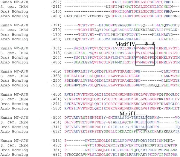
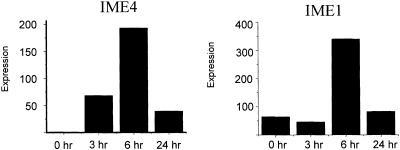
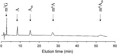
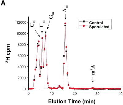
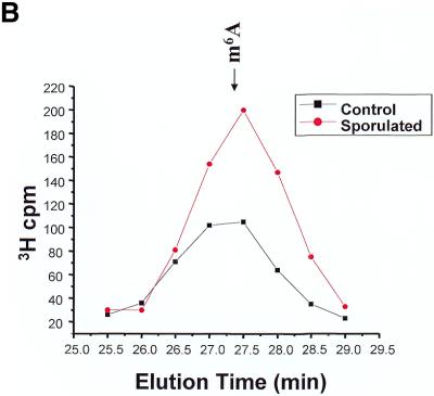
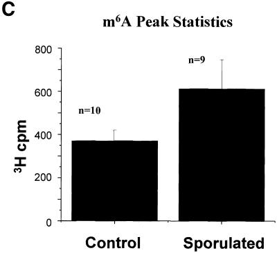
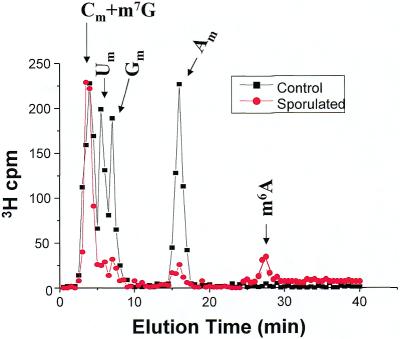
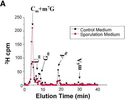
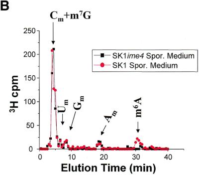
N6-methyladenosine (m6A) is present at internal sites in mRNA isolated from all higher eukaryotes, but has not previously been detected in the mRNA of the yeast Saccharomyces cerevisiae. This nucleoside modification occurs only in a sequence- specific context that appears to be conserved across diverse species. The function of this modification is not fully established, but there is some indirect evidence that m6A may play a role in the efficiency of mRNA splicing, transport or translation. The S.cerevisiae gene IME4, which is important for induction of sporulation, is very similar to the human gene MT-A70, which has been shown to be a critical subunit of the human mRNA [N6-adenosine]-methyltransferase. This observation led to the hypothesis that yeast sporulation may be dependent upon methylation of yeast mRNA, mediated by Ime4p. In this study we show that induction of sporulation leads to the appearance of low levels of m6A in yeast mRNA and that this modification requires IME4. Moreover, single amino acid substitutions in the putative catalytic residues of Ime4p lead to severe sporulation defects in a strain whose sporulation ability is completely dependent on this protein. Collectively, these data suggest very strongly that the activation of sporulation by Ime4p is the result of its proposed methyltransferase activity and provide the most direct evidence to date of a physiologic role of m6A in a gene regulatory pathway.


Figures

Figure 1
Figure 1
Conserved protein sequences of putative mRNA N6-adenosine methyltransferases. Amino acid sequences of human MT-A70, S.cerevisiae IME4 and closely related proteins from Arabidopsis and Drosophila that were identified by database searches were aligned using AlignX (a component of VectorNTI Suite 6.0 software). The subsequences shown roughly correspond to the C-terminal halves of the respective proteins, as these regions were most highly conserved. Regions of similarity were also present in alignments of the N-terminal portions, but are not shown. Methyltransferase consensus motifs are shown in boxes. The positions that correspond to Ime4p residues D348 and W351, which are predicted catalytic residues, are identified by asterisks. Red letters represent consensus residues derived from a completely conserved amino acid at a given position. Residues that are identical in two or three of the sequences are shown in blue. Residues that are highly similar to a consensus at a given position are shown in pink. Weak similarities are depicted in green. Overall sequence identity is ∼40% across all four proteins (excluding gaps). Sequence similarity is almost 95% across this region (when calculated for at least three of the four proteins at each position). GenBank accession numbers for the sequences are: AF014837 (human MT-A70), D23721 (S.cerevisiae SPO8/IME4), g15236910 (Arabidopsis thaliana homolog), AE003746 (translation of subsequence beginning at position 1654, also CG5933 gene product, Drosophila melanogaster homolog).
Figure 2
Figure 2
Induction of IME4 and IME1 mRNA levels. Yeast were grown vegetatively as described, and then switched to sporulation medium. Cells were harvested and RNA was prepared at the indicated times. Levels of IME4 and IME1 mRNA were measured by real-time RT–PCR using a LightCycler. Relative levels of expression are indicated.
Figure 3
Figure 3
Absorbance profile of an HPLC separation of adenosine and methylated adenosine nucleosides. N7-Methylguanosine and methylated adenosine analogs were separated by isocratic elution from a Supelcosil LC-18-S column (250 × 2.1 mm). The mobile phase was 7.5% methanol/30 mM sodium phosphate. Additional standards were also run, all of which were well-resolved from m6A; their elution times are indicated on subsequent figures. m7G, N7-methyl guanosine; A, adenosine; Am, 2′-O-methyladenosine; m6A, N6-methyladenosine; m6Am, (N6-,2′-O-)-dimethyladenosine.
Figure 4
Figure 4
Methylated nucleoside content of total RNA isolated from sporulating and non-sporulating yeast. (A) Yeast grown in methionine-deficient medium were labeled with [3H-methyl-]methionine. Total RNA was prepared, digested with ribonuclease P1 and nucleoside pyrophosphatase, and dephosphorylated with alkaline phosphatase. The sample was then chromatographed as described in the text. The positions of the predominant 2′-O-methylribonucleosides (Nm) are shown, as is the position of the much smaller m6A peak. The data shown represent the mean of nine separate experiments (control) and 10 experiments (sporulated). Each sample consisted of ∼10 µg of total RNA, containing ∼100 000 c.p.m. of incorporated 3H. All data were standardized to account for differences in the total counts loaded onto the column. (B) The same data are replotted on an expanded scale to better illustrate the m6A peak. (C) The radioactivity in the m6A peak is represented graphically. Error bars represent the standard deviation from the mean for each group of samples.
Figure 4
Figure 4
Methylated nucleoside content of total RNA isolated from sporulating and non-sporulating yeast. (A) Yeast grown in methionine-deficient medium were labeled with [3H-methyl-]methionine. Total RNA was prepared, digested with ribonuclease P1 and nucleoside pyrophosphatase, and dephosphorylated with alkaline phosphatase. The sample was then chromatographed as described in the text. The positions of the predominant 2′-O-methylribonucleosides (Nm) are shown, as is the position of the much smaller m6A peak. The data shown represent the mean of nine separate experiments (control) and 10 experiments (sporulated). Each sample consisted of ∼10 µg of total RNA, containing ∼100 000 c.p.m. of incorporated 3H. All data were standardized to account for differences in the total counts loaded onto the column. (B) The same data are replotted on an expanded scale to better illustrate the m6A peak. (C) The radioactivity in the m6A peak is represented graphically. Error bars represent the standard deviation from the mean for each group of samples.
Figure 5
Figure 5
Methylated nucleoside content of polyadenylated RNA isolated from sporulating and non-sporulating yeast. Total RNA was prepared as before, but the preparations were scaled up 10-fold to yield ∼100 µg of 3H- labeled RNA. From this, ∼1 µg of polyadenylated RNA was isolated, containing ∼1000 c.p.m. 3H. The samples were treated with ribonuclease, pyrophosphatase, phosphatase, and then chromatographed. All data were standardized to account for differences in the total counts loaded onto the column. The curves represent the mean values from three independent experiments.
Figure 6
Figure 6
Methylated nucleoside content of polyadenylated RNA isolated from SK1-ime4 (null mutant). (A) Comparison of methylated nucleosides from SK1-ime4 polyadenylated RNA prepared from the null-mutant yeast labeled in control medium and sporulation medium. (B) Comparison of methylated nucleosides in polyadenylated RNA prepared from from SK1-ime4 cells and wild-type SK1 cells, both labeled in sporulation medium. In this experiment, the m6A had a slightly longer retention time than in the other experiments due to regeneration of the HPLC column in between the experiments. The m6A retention time was verified with unlabeled standards.
Figure 6
Figure 6
Methylated nucleoside content of polyadenylated RNA isolated from SK1-ime4 (null mutant). (A) Comparison of methylated nucleosides from SK1-ime4 polyadenylated RNA prepared from the null-mutant yeast labeled in control medium and sporulation medium. (B) Comparison of methylated nucleosides in polyadenylated RNA prepared from from SK1-ime4 cells and wild-type SK1 cells, both labeled in sporulation medium. In this experiment, the m6A had a slightly longer retention time than in the other experiments due to regeneration of the HPLC column in between the experiments. The m6A retention time was verified with unlabeled standards.

References

    1. Rottman F.M., Shatkin,A.J. and Perry,R.P. (1974) Sequences containing methylated nucleotides at the 5′-termini of messenger RNAs: possible implications for processing. Cell, 3, 197–199. - PubMed
    1. Banerjee A.K. (1980) 5′-Terminal cap structure in eucaryotic messenger ribonucleic acids. Microbiol. Rev., 44, 175–205. - PMC - PubMed
    1. Reddy R., Singh,R. and Shimba,S. (1992) Methylated CAP structures in eukaryotic RNAs: structure, synthesis and function. Pharmacol. Ther., 54, 249–267. - PubMed
    1. Desrosiers R.C., Friderici,K.H. and Rottman,F.M. (1975) Characterization of Novikoff hepatoma mRNA methylation and heterogeneity in the methylated 5′ terminus. Biochemistry, 14, 4367–4374. - PubMed
    1. Perry R.P., Kelley,D.E., Friderici,K. and Rottman,F. (1975) The methylated constituents of L cell messenger RNA: evidence for an unusual cluster at the 5′ terminus. Cell, 4, 387–394. - PubMed

Publication types

MeSH terms