. 2002 Oct 29;99(22):14159-64.
doi: 10.1073/pnas.182539599. Epub 2002 Oct 17.

Role of the lever arm in the processive stepping of myosin V

Affiliations

Role of the lever arm in the processive stepping of myosin V

Thomas J Purcell et al. Proc Natl Acad Sci U S A. .

Abstract

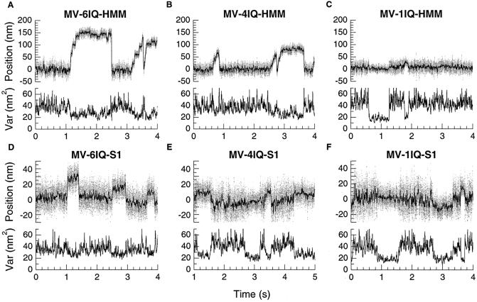
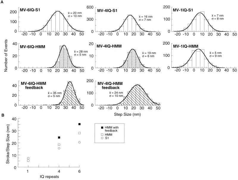
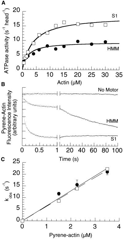
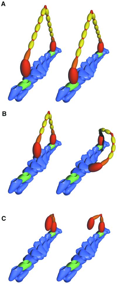
Myosin V is a two-headed molecular motor that binds six light chains per heavy chain, which creates unusually long lever arms. This motor moves processively along its actin track in discrete 36-nm steps. Our model is that one head of the two-headed myosin V tightly binds to actin and swings its long lever arm through a large angle, providing a stroke. We created single-headed constructs with different-size lever arms and show that stroke size is proportional to lever arm length. In a two-headed molecule, the stroke provides the directional bias, after which the unbound head diffuses to find its binding site, 36 nm forward. Our two-headed construct with all six light chains per head reconstitutes the 36-nm processive step seen in tissue-purified myosin V. Two-headed myosin V molecules with only four light chains per head are still processive, but their step size is reduced to 24 nm. A further reduction in the length of the lever arms to one light chain per head results in a motor that is unable to walk processively. This motor produces single small approximately 6-nm strokes, and ATPase and pyrene actin quench measurements show that only one of the heads of this dimer rapidly binds to actin for a given binding event. These data show that for myosin V with its normal proximal tail domain, both heads and a long lever arm are required for large, processive steps.


Figures

Fig 1.
Fig 1.
Diagram of constructs and experimental trap setup. (A) Diagrams of constructs. The myosin V head consists of the catalytic domain (yellow) and the light-chain binding IQ repeats (blue). The two-headed HMM constructs have a tail domain that contains native myosin V coiled coil (orange) with a short segment of GCN4 (red) to ensure dimerization. All constructs have GFP (green) and a FLAG tag (not shown) at the C terminus. (B) Experimental setup for recording single myosin molecules. An actin filament, stretched between two trapped polystyrene beads, is brought into contact with myosin molecules adsorbed onto the surface by anti-GFP antibodies.
Fig 2.
Fig 2.
Sample data from trap. Bead position is shown in the upper trace in each graph, and variance is shown below. Variance, which is inversely proportional to the net stiffness of all attachments to the bead, is calculated over a 1-ms window. Decreases in variance correspond to the increased stiffness of the system due to actin binding to a motor on the surface. One source of the apparent wide distribution of single-step sizes is caused by randomness in the starting position of the actin filament due to Brownian motion before myosin binding (16).
Fig 3.
Fig 3.
Effect of lever arm length on step size. (A) Step size histograms. Comparison of step sizes for the various constructs, pooled from several experiments. The mean (x̄) and standard deviation (σ) were obtained by a least-squares fit of each histogram to a Gaussian distribution. Molecules that produce only a single step are shown with plain bars. Histograms for MV-6IQ-HMM and MV-4IQ-HMM represent step sizes in processive staircases. Stippled histograms represent the step size of the motor stepping against a stationary trap. Striped histograms represent stepping against constant 1 pN of backward force, by using a feedback-controlled trap (8, 9). (B) Step size is proportional to lever arm length. The mean of each histogram from A is plotted against lever arm length, showing the dependence of step size on lever arm length.
Fig 4.
Fig 4.
One head is excluded in the myosin V-1IQ-HMM. (A) Actin-activated ATPase. The ATPase activity per head of the two-headed HMM (•) is approximately half that of the single-headed S1 (□). (B) Time course of pyrene quenching after mixing 2.25 μM pyrene actin with KMg50 (Top), 0.2 μM heads 1IQ-HMM (Middle), or 0.2 μM heads 1IQ-S1 (Bottom). The time axis has been split to show both the fast and slow phase of pyrene quenching in the HMM construct. (C) Actin concentration dependence of the fast rate of binding of MV-1IQ-HMM and MV-1IQ-S1. The fast phase binding rate of the myosin V constructs was measured at pyrene actin concentrations from 1.5 to 3.5 μM at 25°C in KMg50 buffer with 250 μM MgADP added. The fast phase rates of the HMM (•) and S1 (□) plotted against actin concentration was fit by using linear regressions passing through the origin with the slope describing the apparent second-order binding constant.
Fig 5.
Fig 5.
Model of myosin V stepping. A short segment of an actin filament is shown in blue, with the 36-nm pseudorepeat highlighted by green subunits. Each actin subunit has a single myosin binding site depicted as a pit. Each myosin is colored to show the heavy chain including the catalytic domain (red), the essential light chain (orange), and the calmodulin light chains (yellow). (A) The rear head of a 6IQ-HMM in a poststroke state, tightly bound to an actin subunit (Left). The near head is not yet bound, but in its diffusional search it can easily bind to the green monomer (Right). (B) The smaller stroke of the bound head of the 4IQ-HMM is shown (Left). The unbound head is not positioned so that a binding site is immediately available. The geometry of the actin filament dictates that some portion of the myosin molecule must distort to bind both heads at once. It is possible that the lever arm elastically bends to achieve this conformation (Right). (C) The 1IQ-HMM is shown bound by one head to an actin filament. The second head can bind only if considerable distortion exists in the myosin. These extreme conformations are likely to be rare, accounting for the fact that the second head binds to actin many orders of magnitude slower than the first head.

References

    1. Cheney R. E., O'Shea, M. K., Heuser, J. E., Coelho, M. V., Wolenski, J. S., Espreafico, E. M., Forscher, P., Larson, R. E. & Mooseker, M. S. (1993) Cell 75, 13-23. - PubMed
    1. Reck-Peterson S. L., Novick, P. J. & Mooseker, M. S. (1999) Mol. Biol. Cell 10, 1001-1017. - PMC - PubMed
    1. Spudich J. A. (2001) Nat. Rev. Mol. Cell. Biol. 2, 387-392. - PubMed
    1. Tyska M. J. & Warshaw, D. M. (2002) Cell Motil. Cytoskeleton 51, 1-15. - PubMed
    1. Geeves M. A. & Holmes, K. C. (1999) Annu. Rev. Biochem. 68, 687-728. - PubMed

Publication types

MeSH terms

LinkOut - more resources