. 2002 Oct 15;22(20):8808-18.
doi: 10.1523/JNEUROSCI.22-20-08808.2002.

Neuronal premotor networks involved in eyelid responses: retrograde transneuronal tracing with rabies virus from the orbicularis oculi muscle in the rat

Affiliations

Neuronal premotor networks involved in eyelid responses: retrograde transneuronal tracing with rabies virus from the orbicularis oculi muscle in the rat

Sara Morcuende et al. J Neurosci. .

Abstract

Retrograde transneuronal tracing with rabies virus from the right orbicularis oculi muscle was used to identify neural networks underlying spontaneous, reflex, and learned blinks. The kinetics of viral transfer was studied at sequential 12 hr intervals between 3 and 5 d after inoculation. Rabies virus immunolabeling was combined with the immunohistochemical detection of choline acetyltransferase expression in brainstem motoneurons or Fluoro-Ruby injections in the rubrospinal tract. Virus uptake involved exclusively orbicularis oculi motoneurons in the dorsolateral division of the facial nucleus. At 3-3.5 d, transneuronal transfer involved premotor interneurons of trigeminal, auditory, and vestibular reflex pathways (in medullary and pontine reticular formation, trigeminal nuclei, periolivary and ventral cochlear nuclei, and medial vestibular nuclei), motor pathways (dorsolateral quadrant of contralateral red nucleus and pararubral area), deep cerebellar nuclei (lateral portion of interpositus nucleus and dorsolateral hump ipsilaterally), limbic relays (parabrachial and Kölliker-Fuse nuclei), and oculomotor structures involved in eye-eyelid coordination (oculomotor nucleus, supraoculomotor area, and interstitial nucleus of Cajal). At 4 d, higher order neurons were revealed in trigeminal, auditory, vestibular, and deep cerebellar nuclei (medial, interpositus, and lateral), oculomotor and visual-related structures (Darkschewitsch, nucleus of the posterior commissure, deep layers of superior colliculus, and pretectal area), lateral hypothalamus, and cerebral cortex (particularly in parietal areas). At 4.5 and 5 d the labeling of higher order neurons occurred in hypothalamus, cerebral cortex, and blink-related areas of cerebellar cortex. These results provide a comprehensive picture of the premotor networks mediating reflex, voluntary, and limbic-related eyelid responses and highlight potential sites of motor learning in eyelid classical conditioning.


Figures
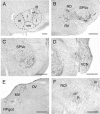
Fig. 1.
Fig. 1.
Shown are photomicrographs of rabies-immunolabeled brainstem neurons at 3 d (A), 3.5 d (B, D, F), and 4 d (C, E) after rabies virus injection into the right orbicularis oculi muscle. A, Labeled orbicularis oculi motoneurons in the dorsolateral (dl) division of the facial nucleus. Note the absence of labeling in the other divisions. B, Labeled neurons in the ipsilateral medullary reticular formation.C, Labeled neurons in the ipsilateral spinal trigeminal nucleus, pars oralis (SPVo). D, Labeled neurons in the ipsilateral ventral cochlear nucleus (VCN). E, Labeled neurons in the ipsilateral medial vestibular nucleus (MV) and in the nucleus reticularis paragigantocellularis dorsalis (RPgcd). F, Labeled neurons in the dorsolateral quadrant of the contralateral red nucleus (R) and overlying pararubral area. Scale bars:A, 100 μm; B–F, 500 μm.i, l, m,vm, Intermediate, lateral, medial, and ventromedial divisions of the facial nucleus; III, oculomotor nucleus; DV, descending vestibular nucleus;RCf, nucleus reticularis cuneiformis; RD, nucleus reticularis dorsalis; RV, nucleus reticularis ventralis, SPVc, spinal trigeminal nucleus, pars caudalis.
Fig. 2.
Fig. 2.
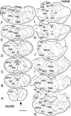
Brainstem distribution of rabies-immunolabeled neurons at 3.5 d after rabies virus injection into the right orbicularis oculi muscle. A–L, Labeled sections are arranged caudorostrally. The arrows indicate the side that is ipsilateral to the injection. Asterisks inF, G, Retrogradely labeled orbicularis oculi motoneurons in the dorsolateral division of the facial (VII) nucleus. Each dot represents one labeled neuron. Scale bar, 2 mm. CG, Central gray; CPO, caudal periolivary nucleus; D, nucleus of Darkschewitsch;DCN, dorsal cochlear nucleus; Dlh, dorsolateral hump; DH, dorsal horn; DV, descending vestibular nucleus; I, interpositus nucleus;INC, interstitial nucleus of Cajal; IO, inferior olive; KF, Kölliker–Fuse nucleus;L, lateral (dentate) nucleus; LV, lateral vestibular nucleus; M, medial (fastigial) nucleus;MV, medial vestibular nucleus; NST, nucleus of the solitary tract; PB, parabrachial nucleus;PT, pyramidal tract; PV, principal trigeminal nucleus; R, red nucleus; RCf, nucleus reticularis cuneiformis; RD, nucleus reticularis dorsalis; RGc, nucleus reticularis gigantocellularis;RMc, nucleus reticularis magnocellularis;RPc, nucleus reticularis parvocellularis;RPgcd, nucleus reticularis paragigantocellularis dorsalis; RPO, rostral periolivary region;RPoC, nucleus reticularis pontis caudalis;RPoO, nucleus reticularis pontis oralis;RSc, nucleus reticularis subceruleus; RV, nucleus reticularis ventralis; sai, stratum album intermedialis; SC, superior colliculus;sgi, stratum griseum intermedialis; sgs, stratum griseum superficialis; SNr, substantia nigra pars reticulata; so, stratum opticum; SO, superior olive; sp, strata profunda; SPO, superior paraolivary nucleus; SPVc, SPVi, SPVo, spinal trigeminal nuclei, pars caudalis, interpolaris, and oralis;tz, trapezoid body; VCN, ventral cochlear nucleus; VPO, ventral periolivary nucleus;III, oculomotor nucleus; XII, hypoglossal nucleus.
Fig. 3.
Fig. 3.
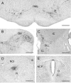
Rabies-immunolabeled neurons at 4 d after rabies virus injection into the right orbicularis oculi muscle.A, Transneuronally labeled neurons in nucleus reticularis magnocellularis (RMc). B, Labeled neurons in the ipsilateral dorsal horn (DH) and intermediate zone of the upper cervical cord. C, Labeled neurons in the contralateral dorsal nucleus of the lateral lemniscus (DLL) and inferior colliculus (IC). D, Labeled neurons in the dorsolateral quadrant of the contralateral red nucleus (R) and overlying pararubral area at 4 d (see also 3.5 d) (Fig. 1F).E, Labeled neurons in the nucleus of Darkschewitsch (D) and nucleus of the posterior commissure (NCP). Scale bars, 500 μm. III, Oculomotor nucleus; LL, lateral lemniscus.
Fig. 4.
Fig. 4.
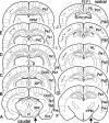
Distribution of rabies-immunolabeled neurons in the medulla and pons at 4 d after rabies virus injection into the right orbicularis oculi muscle. A–J, Labeled sections are arranged caudorostrally. Arrows indicate the side that is ipsilateral to the injected muscle. Asterisks inG, Retrogradely labeled orbicularis oculi motoneurons in the dorsolateral division of the facial (VII) nucleus. Each dot represents one labeled neuron. Scale bar, 2 mm. For abbreviations see the legend of Figure 2.
Fig. 5.
Fig. 5.
Distribution of rabies-immunolabeled neurons in the mesencephalon and caudal diencephalon at 4 d after rabies virus injection into the right orbicularis oculi muscle.A–G, Labeled sections are arranged caudorostrally.Arrows indicate the side that is ipsilateral to the injected muscle. Each dot represents one labeled neuron. Scale bar, 2 mm. For abbreviations see the legend of Figure 2.APT, Anterior pretectal nucleus; CM, central medial nucleus; CP, cerebral peduncle;Hab, habenula; IC, inferior colliculus,LH, lateral hypothalamus; LL, lateral lemniscus, MGB, medial geniculate body;MN, mamillary nuclei; NCP, nucleus of the posterior commissure; PN, pontine nuclei;PO, posterior thalamic nucleus; PR, prerubral field; RR, retrorubral nucleus;Rt, reticular thalamic nucleus; VPM, ventral posteromedial thalamic nucleus; VTA, ventral tegmental area; ZI, zona incerta.
Fig. 6.
Fig. 6.
Rabies-immunolabeled neurons in the cerebellar cortex and nuclei. A, Transneuronally labeled neurons in the medial (M, fastigial) nucleus, interpositus (I) nucleus, and dorsolateral hump (Dlh) at 4 d after the injection of rabies virus into the ipsilateral orbicularis oculi muscle. B, Transneuronal labeling of higher order neurons in the cerebellar vermis at 5 d after injection. Note the zonal distribution of labeled Purkinje cells. C, Coronal section through the paravermis, illustrating another area in which rabies-immunolabeled Purkinje cells were observed at 5 d after rabies virus injection in the ipsilateral orbicularis oculi muscle. Scale bars:A, B, 500 μm; C, 150 μm.
Fig. 7.
Fig. 7.
Distribution of rabies-immunolabeled neurons in the deep cerebellar nuclei at 4 d after rabies virus injection into the right orbicularis oculi muscle (arrow indicates ipsilateral side). Sections in A–F are arranged from caudal to rostral. The dashed line denotes the midline. Each dot represents one labeled neuron. Note transneuronally labeled neurons bilaterally, with clear ipsilateral dominance, in medial (M, fastigial) nucleus, dorsolateral hump (Dlh), lateral part of posterior interpositus (PI) and anterior interpositus (AI), and lateral (L, dentate) nuclei. Scale bar, 2 mm.
Fig. 8.
Fig. 8.
Photomicrographs illustrating transneuronally labeled pyramidal cells in the contralateral parietal cortex at 4–5 d after rabies virus injection into the right orbicularis oculi muscle. A, Coronal section of the parietal cortex, area 1 (Pa1) at 4 d.B, Parietal cortex, area 1, at 5 d. Note the increase in the number of rabies-immunolabeled pyramidal cells.C, High-power photomicrograph of parietal cortex at 4.5 d. Note Golgi-like labeling of a pyramidal cell in layer V. Scale bars: A, B, 500 μm;C, 100 μm.
Fig. 9.
Fig. 9.
Distribution of rabies-immunolabeled neurons in the cerebral cortex at 4 d after rabies virus injection into the right orbicularis oculi muscle. A–J, Labeled sections are arranged from caudal to rostral. Arrows indicate the ipsilateral side. Each dot represents one labeled neuron. Scale bar, 2 mm. At 4 d the labeled neurons were concentrated in parietal cortex, areas 1 and 2 (Pa1 andPa2) bilaterally, with a clear contralateral dominance.APT, Anterior pretectal area; CG, central gray; D, nucleus of Darkschewitsch; Ent, entorhinal cortex; F1, frontal cortex, area 1;F2, frontal cortex, area 2; HL, hindlimb area of parietal cortex; INC, interstitial nucleus of Cajal; Oc2L, occipital cortex area 2, lateral;PrC, precommissural nucleus; Prh, perirhinal cortex; R, red nucleus; SNr, substantia nigra, pars reticulata; Te1, temporal cortex, area 1; Te3, temporal cortex, area 3;VPM, ventral posteromedial thalamic nucleus.
Fig. 10.
Fig. 10.
Dual-color immunofluorescence for choline acetyltransferase (a motoneuron marker; A, C) and rabies virus (B, D) in the oculomotor nucleus at 4 d after the injection of rabies virus into the right orbicularis oculi muscle.A, C, Motoneurons in the oculomotor nucleus, identified by their expression of choline acetyltransferase (Cy3; red). B, D, Rabies-immunolabeled neurons (FITC; green) in the ipsilateral oculomotor nucleus in the same sections. They are identified here as interneurons and not motoneurons, because they are not positive for choline acetyltransferase (arrows inC point to the empty spaces corresponding to the rabies-immunolabeled neurons shown in D). Scale bars:A, B, 500 μm; C,D, 100 μm.
Fig. 11.
Fig. 11.
Rubrospinal neurons, labeled retrogradely after injections of Fluoro-Ruby into the right rubrospinal tract in the upper cervical spinal segments (A, C), and orbicularis oculi-related rubrobulbar neurons in the same section, labeled by retrograde transneuronal transfer of rabies virus from the right orbicularis oculi muscle (B, D). The photomicrographs illustrate the contralateral (left) magnocellular red nucleus at 5 d. A, Rubrospinal neurons (Fluoro-Ruby; red). Note their widespread distribution in all spinal-projecting portions of the red nucleus, including its lateral horn (left). B, Orbicularis oculi-related rubrofacial neurons (rabies immunolabeling, FITC;green). Note their exclusive location in the dorsolateral quadrant of the red nucleus. Note well that theorange spots are tissue autofluorescence, inevitably shining through the fluorescence filters. C,D, High-power view of the dorsolateral quadrant of the red nucleus from A and B. Despite their extensive overlap, rubrospinal and rubrofacial neurons are two separate populations. Arrows in C andD point to rare examples of double-labeled neurons (projecting with axon collaterals to orbicularis oculi motoneurons and to the spinal cord). Scale bars: A, B, 500 μm;C, D, 100 μm.

Similar articles

Cited by

References

    1. Aou S, Woody CD, Birt D. Changes in the activity of units of the cat motor cortex with rapid conditioning and extinction of a compound eye blink movement. J Neurosci. 1992;12:549–559. - PMC - PubMed
    1. Berger TW, Rinaldi P, Weisz DJ, Thompson RF. Single-unit analysis of different hippocampal cell types during classical conditioning of rabbit nictitating membrane response. J Neurophysiol. 1983;50:1197–1219. - PubMed
    1. Berthier NE, Moore JW. Cerebellar Purkinje cell activity related to the classically conditioned nictitating membrane response. Exp Brain Res. 1986;63:341–350. - PubMed
    1. Berthier NE, Moore JW. Activity of deep cerebellar nuclear cells during classical conditioning of nictitating membrane extension in rabbits. Exp Brain Res. 1990;83:44–54. - PubMed
    1. Bour LJ, Aramideh M, De Visser BW. Neurophysiological aspects of eye and eyelid movements during blinking in humans. J Neurophysiol. 2000;83:166–176. - PubMed

Publication types

MeSH terms

LinkOut - more resources