. 2002 Oct 15;22(20):8891-901.
doi: 10.1523/JNEUROSCI.22-20-08891.2002.

Rapsyn escorts the nicotinic acetylcholine receptor along the exocytic pathway via association with lipid rafts

Affiliations

Rapsyn escorts the nicotinic acetylcholine receptor along the exocytic pathway via association with lipid rafts

Sophie Marchand et al. J Neurosci. .

Abstract

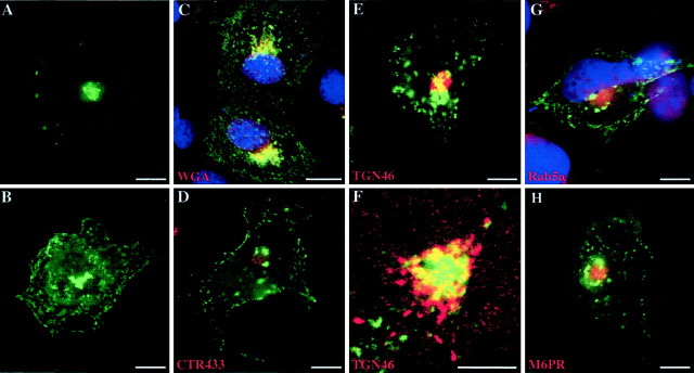
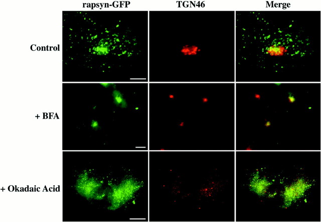
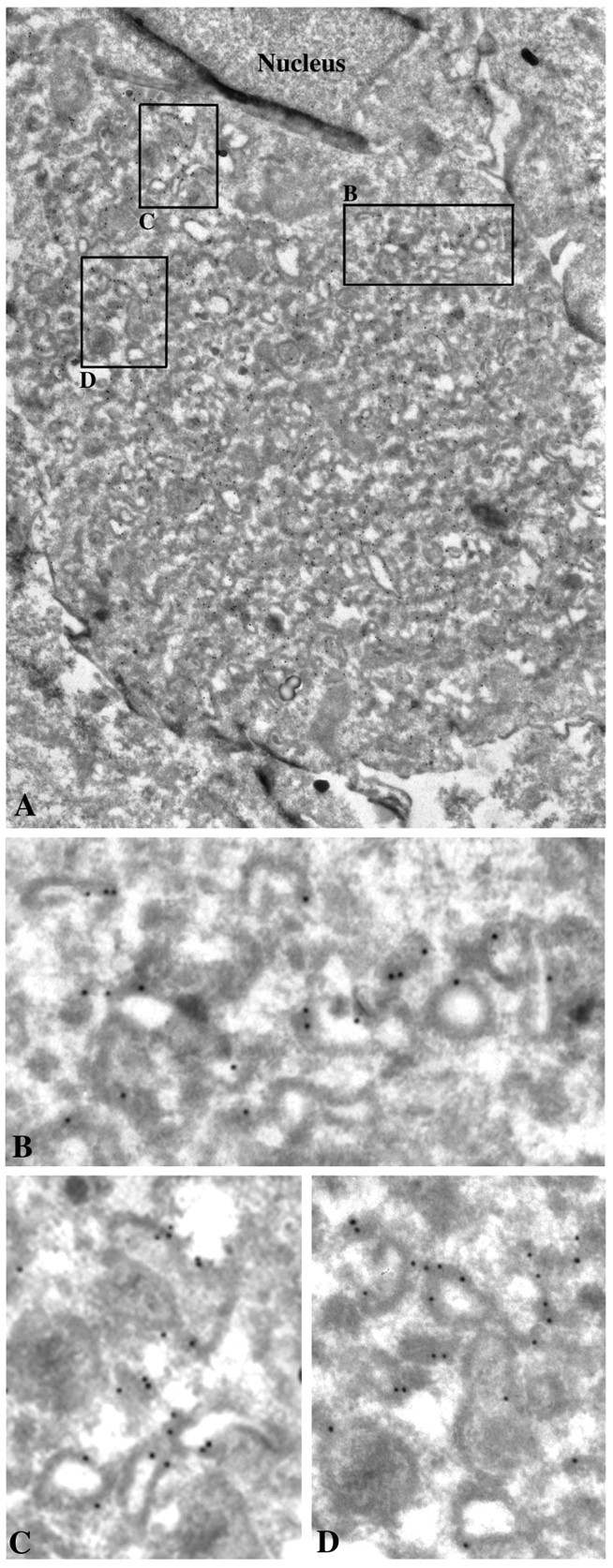
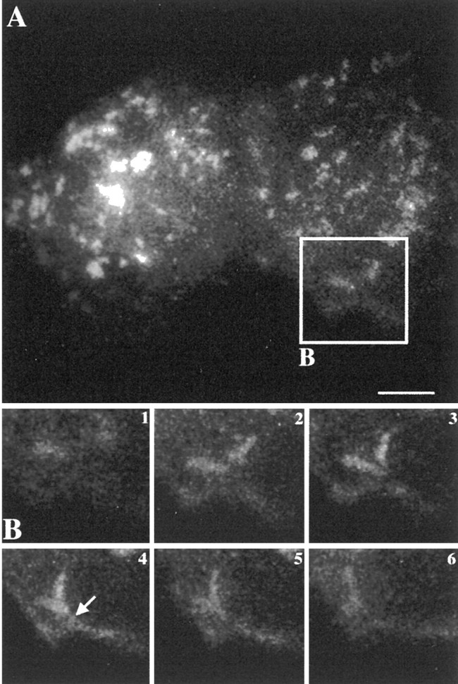
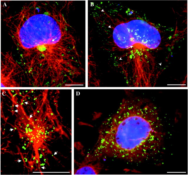
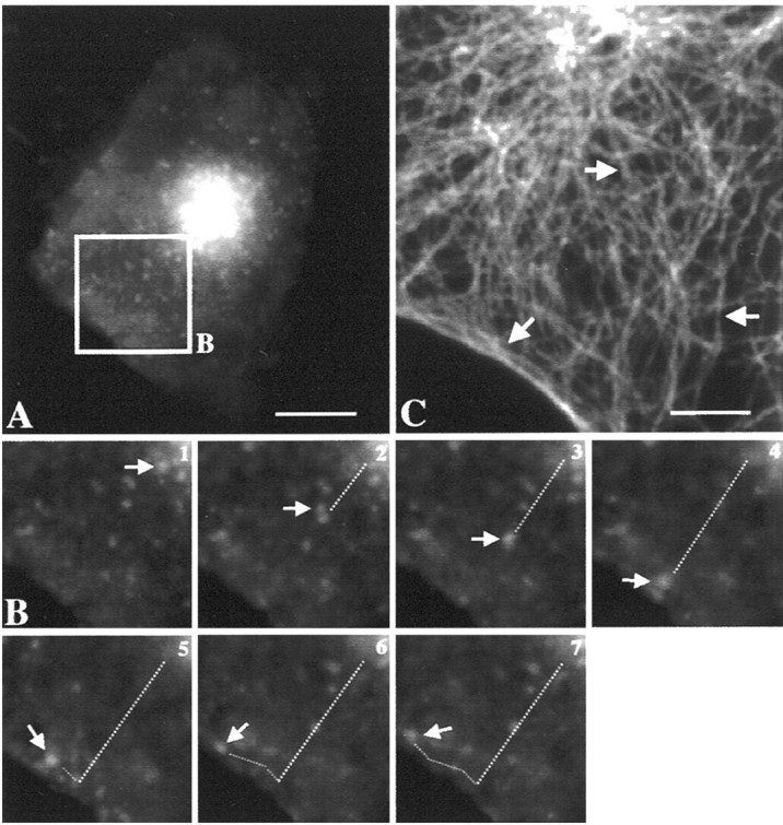
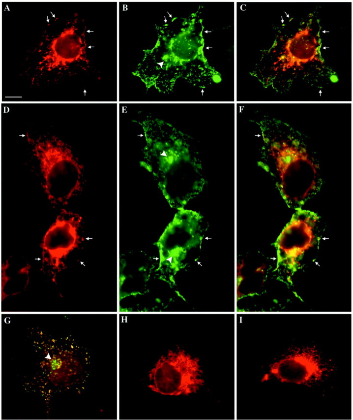
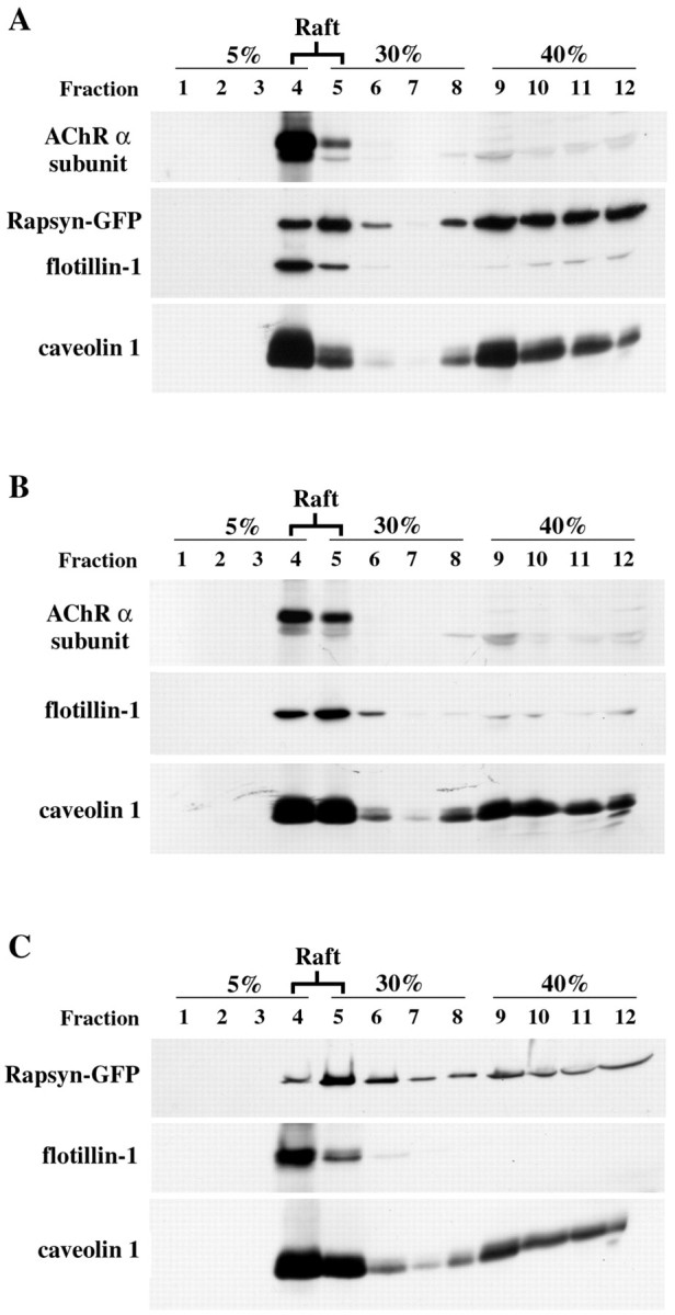
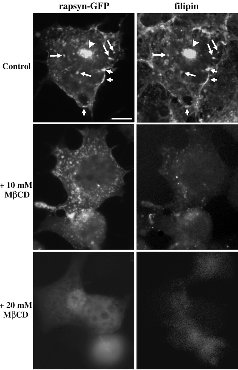
The 43 kDa receptor-associated protein rapsyn is a myristoylated peripheral protein that plays a central role in nicotinic acetylcholine receptor (AChR) clustering at the neuromuscular junction. In a previous study, we demonstrated that rapsyn is specifically cotransported with AChR via post-Golgi vesicles targeted to the innervated surface of the Torpedo electrocyte (Marchand et al., 2000). In the present study, to further elucidate the mechanisms for sorting and assembly of postsynaptic proteins, we analyzed the dynamics of the intracellular trafficking of fluorescently labeled rapsyn in the transient-expressing COS-7 cell system. Our approach was based on fluorescence, time-lapse imaging, and immunoelectron microscopies, as well as biochemical analyses. We report that newly synthesized rapsyn associates with the trans-Golgi network compartment and traffics via vesiculotubular organelles toward the cell surface of COS-7 cells. The targeting of rapsyn organelles appeared to be mediated by a microtubule-dependent transport. Using cotransfection experiments of rapsyn and AChR, we observed that these two molecules codistribute within distal exocytic routes and at the plasma membrane. Triton X-100 extraction on ice and flotation gradient centrifugation demonstrated that rapsyn and AChR are recovered in low-density fractions enriched in two rafts markers: caveolin-1 and flotillin-1. We propose that sorting and targeting of these two companion molecules are mediated by association with cholesterol-sphingolipid-enriched raft microdomains. Collectively, these data highlight rapsyn as an itinerant vesicular protein that may play a dynamic role in the sorting and targeting of its companion receptor to the postsynaptic membrane. These data also raise the interesting hypothesis of the participation of the raft machinery in the targeting of signaling molecules to synaptic sites.


Figures

Fig. 1.
Fig. 1.
Subcellular targeting of rapsyn–GFP to exocytic compartments in COS-7 cells. Cells were transfected with rapsyn fused to GFP (rapsyn–GFP) as described in Materials and Methods. Twelve to 24 hr after transfection, cells were fixed and handled for immunofluorescence using TRITC-conjugated WGA (C), monoclonal anti-CTR433 antibody (D), monoclonal anti-TGN46 antibody (E, F), polyclonal anti-Rab5a (G), or anti-M6PR antibodies (H). In some experiments, DAPI staining was performed to localize the nuclei. At early expression times, rapsyn–GFP mostly localized in a juxtanuclear region (A). At later expression times, rapsyn–GFP distributed in clusters at the plasma membrane, as well as in the juxtanuclear region, and in a dot-like pattern within the cytoplasm (B). Rapsyn–GFP fluorescence overlapped partially with WGA staining (C) and with thetrans-Golgi network marker TGN46 (E). At higher magnification, confocal analysis confirmed the codistribution of rapsyn–GFP with TGN46 (F). Rapsyn–GFP did not overlap significantly with the markers of CTR433 (D) and the early and late endosomal compartments Rab5a and M6PR, respectively (G, H). Scale bars:A–E, G, H, 10 μm;F, 5 μm.
Fig. 2.
Fig. 2.
Rapsyn–GFP localizes to thetrans-Golgi network. COS-7 cells were transfected with rapsyn–GFP. One day later, cells were incubated in either the absence (Control) or presence of 5 μg/ml brefeldin A for 2 hr (+BFA) or 0.5 μm okadaic acid for 1 hr (+Okadaic Acid). The cells were fixed and labeled with the TGN46 monoclonal antibody and observed in the fluorescence microscope. Rapsyn–GFP remained associated with condensed TGN in conditions in which the Golgi apparatus was disrupted by BFA. After okadaic acid treatment, which disrupts the entire Golgi complex, including the TGN, rapsyn–GFP-associated structures were dispersed within the cytoplasm. Scale bar, 10 μm.
Fig. 3.
Fig. 3.
Immunogold localization of rapsyn–GFP in COS-7 cells. Ultrathin cryosections of transfected COS-7 cells were immunolabeled with 10 nm of gold-conjugated monoclonal anti-GFP antibody and observed by electron microscopy. Rapsyn was associated with the cytoplasmic surface of numerous intracellular tubulovesicular organelles (details in B–D) often accumulated within the juxtanuclear region (A). Magnifications:A, 10,000×; B–D, 35,000×.
Fig. 4.
Fig. 4.
Dynamics of intracellular trafficking of rapsyn–GFP by videomicroscopy. A, The COS-7 cell transfected with rapsyn–GFP was analyzed by three-dimensional videomicroscopy after 24 hr of expression. B, Time series imaging of the selected area of the cell in A. Tubulovesicular rapsyn-enriched organelles fused together while moving toward the cell surface and with the plasma membrane (arrow). The signal then vanished as rapsyn spread out along the membrane. Time intervals between frames were 20 sec. Scale bar, 10 μm.
Fig. 5.
Fig. 5.
Rapsyn–GFP localization is dependent on the integrity of the microtubular network. COS-7 cells expressing rapsyn–GFP were fixed and handled for immunofluorescence microscopy using monoclonal anti-α-tubulin antibody. A, In cells starting to express rapsyn–GFP (24 hr), TGN-associated rapsyn–GFP appeared in close proximity with the MTOC (arrow).B, At later expression times (typically 2 d), rapsyn–GFP distributed as numerous organelles surrounding the MTOC and underlying the cell cortex (arrowheads).C, Confocal analysis suggested the association of rapsyn–GFP carriers with microtubules (arrows).D, Depolymerization of microtubules by nocodazole disrupted juxtanuclear and cortical distributions of rapsyn–GFP.A, B, D, DAPI staining was performed to localize the nuclei. Scale bars, 10 μm.
Fig. 6.
Fig. 6.
Dynamics of intracellular trafficking of rapsyn–GFP by videomicroscopy: role of microtubules. A, The COS-7 cell transfected with rapsyn–GFP was analyzed by three-dimensional videomicroscopy after 24 hr of expression.B, Time series imaging of the selected area of the cell in A. The organelle indicated by thearrow moved straight from the cell center to the periphery and then tangentially to the cell surface. Both the velocity (∼0.2 μm/sec) of the transport and the linear trajectory suggested a microtubule-guided movement. The trajectory of rapsyn–GFP transporters is consistent with the microtubular organization (C) radiating from the MTOC (arrow) toward the cell periphery and underlying the plasma membrane (arrows). Intervals between frames are 20 sec. Scale bars: A, 10 μm; C, 5 μm.
Fig. 7.
Fig. 7.
Distributions of AChR and rapsyn–GFP in the exocytic pathway in COS-7 cells. Cells transfected with AChR α, β, γ, and δ subunit constructs or cotransfected with rapsyn–GFP and AChR subunits constructs were fixed, permeabilized with 0.1% Triton X-100, and processed for immunofluorescence using anti-AChR α-subunit antibody or TRITC-conjugated α-bungarotoxin. AChR (A,D) and rapsyn (B, E) colocalized at the cell surface and partially within intracellular compartments (C–F). Intracellularly, AChR distributed within the entire exocytic pathway, whereas rapsyn–GFP fluorescence was observed only in distal compartments, including the TGN (B, E, arrowheads; also see Figs. 1, 2) and cytoplasmic organelles (G). In cells transfected only with AChR subunits, a similar intracellular distribution was observed (H, I). Note that no AChR clusters were present at the cell surface (H,I) at variance with cells cotransfected with rapsyn–GFP (A, D,arrows). A, D,H, I, AChR labeling with anti-AChR α-subunit antibody; B, E, rapsyn–GFP;C, F, G, double-labeling AChR/rapsyn–GFP. G, AChR labeling was achieved by α-bungarotoxin. Scale bar, 10 μm.
Fig. 8.
Fig. 8.
Raft fractionation of rapsyn–GFP and AChR in COS-7 cells. Cells transiently cotransfected with rapsyn–GFP and AChR subunits (A) or separately with AChR subunits (B) or rapsyn (C) were subjected to subcellular fractionation by extraction with 1% (w/v) Triton X-100 on ice. Cell lysates were separated by ultracentrifugation on discontinuous sucrose density gradients. Fractions collected from the top of the gradient were separated by SDS-PAGE (12% acrylamide) and analyzed by immunoblotting (see Materials and Methods). The distributions of rapsyn–GFP and AChR α subunit along the gradient were compared with those of caveolin-1 and flotillin-1, two endogenous markers of lipid rafts. AChR α subunit (49 kDa) and rapsyn–GFP (70 kDa) fractionated mostly in the low-density caveolin-1(26 kDa)/flotillin-1(52 kDa)-enriched raft fractions (lanes 4, 5).
Fig. 9.
Fig. 9.
Role of cholesterol in the distribution of rapsyn-GFP in COS-7 cells. Cells transfected with plasmid expressing rapsyn–GFP were treated in the absence (Control) or presence of 10 or 20 mm MβCD for 1 hr at 37°C and then fixed, permeabilized, and incubated with filipin to detect cholesterol (see Materials and Methods). In control experiments, rapsyn–GFP- and filipin-derived free cholesterol signals showed significant colocalization at the plasma membrane (shortarrows), in vesicular structures (long arrows), and in the juxtanuclear region (arrowheads). Cholesterol depletion by MβCD perturbed the subcellular localization of rapsyn–GFP. In cells treated with 10 mm MβCD, rapsyn–GFP signal appeared dispersed within the cytoplasm. On higher MβCD concentrations (20 mm) providing acute cholesterol depletion, rapsyn–GFP was dramatically redistributed and appeared diffuse in the cytoplasm. Scale bar, 10 μm.

Similar articles

Cited by

References

    1. Arni S, Keilbaugh SA, Ostermeyer AG, Brown DA. Association of GAP-43 with detergent-resistant membranes requires two palmitoylated cysteine residues. J Biol Chem. 1998;273:28478–28485. - PubMed
    1. Banting G, Ponnambalam S. TGN38 and its orthologues: roles in post-TGN vesicle formation and maintenance of TGN morphology. Biochim Biophys Acta. 1997;1355:209–217. - PubMed
    1. Barrantes FJ. The acetylcholine receptor ligand-gated channel as a molecular target of disease and therapeutic agents. Neurochem Res. 1997;22:391–400. - PubMed
    1. Bignami F, Camus G, Marchand S, Bailly L, Stetzkowski-Marden F, Cartaud J. Targeting of acetylcholine receptor and 43 kDa rapsyn to the postsynaptic membrane in Torpedo marmorata electrocyte. J Physiol (Paris) 1998;92:177–181. - PubMed
    1. Bijlmakers MJJE, Marsh M. Trafficking of an acylated cytosolic protein: newly synthetized p56lck travels to the plasma membrane via the exocytic pathway. J Cell Biol. 1999;145:457–468. - PMC - PubMed

Publication types

MeSH terms