. 2002 Oct 15;22(20):8911-21.
doi: 10.1523/JNEUROSCI.22-20-08911.2002.

Dopamine induces a PI3-kinase-independent activation of Akt in striatal neurons: a new route to cAMP response element-binding protein phosphorylation

Affiliations

Dopamine induces a PI3-kinase-independent activation of Akt in striatal neurons: a new route to cAMP response element-binding protein phosphorylation

Karen Brami-Cherrier et al. J Neurosci. .

Abstract

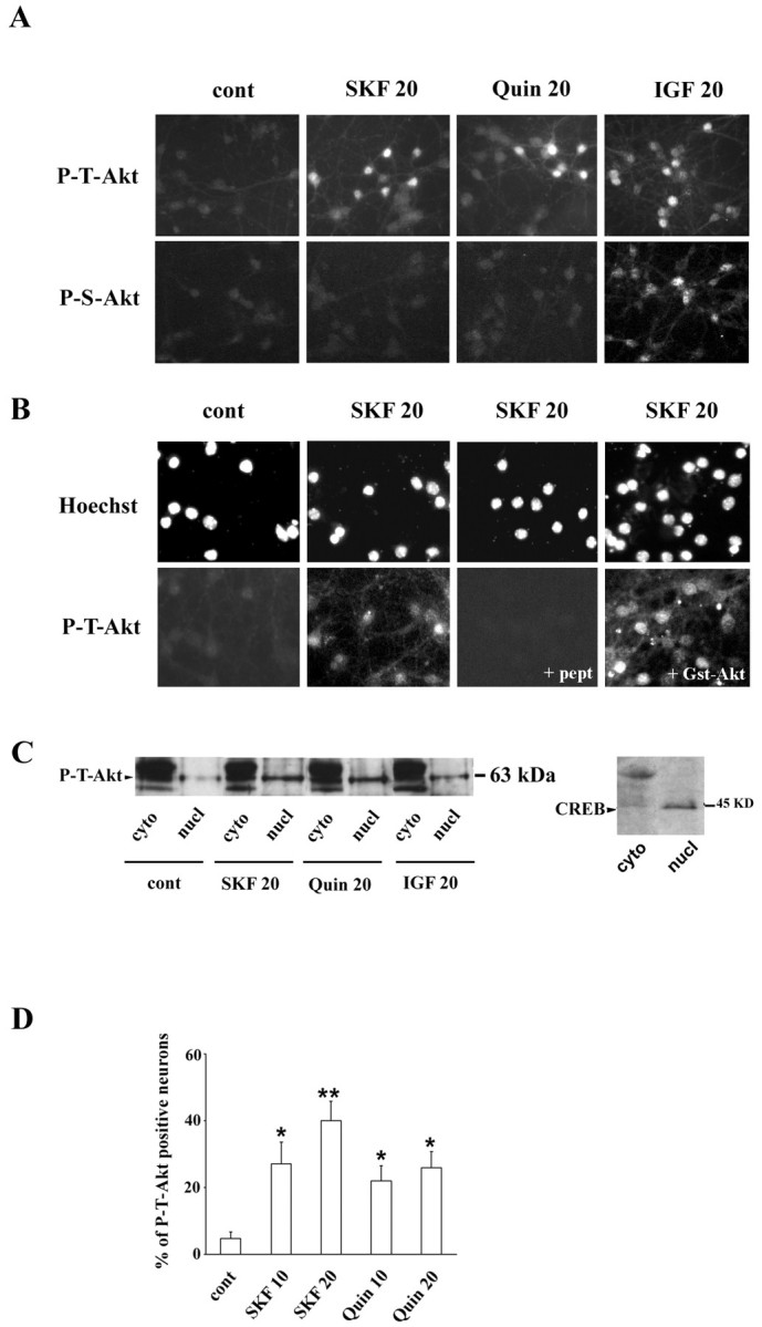
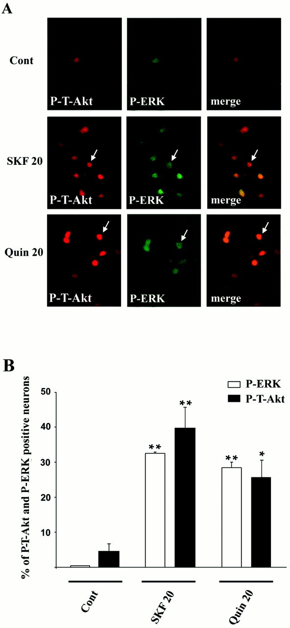
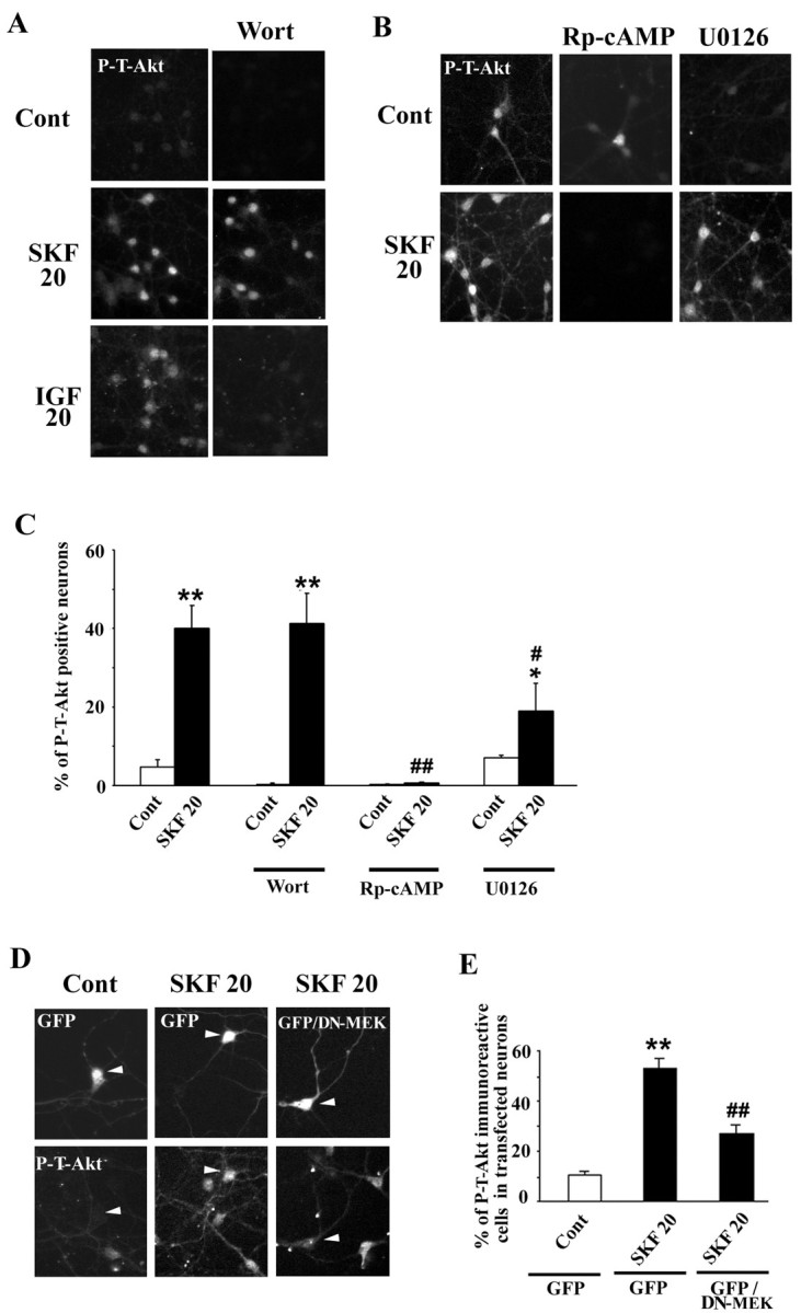
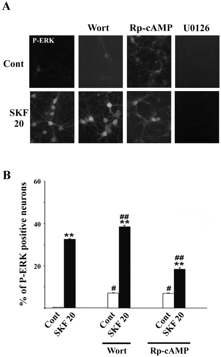
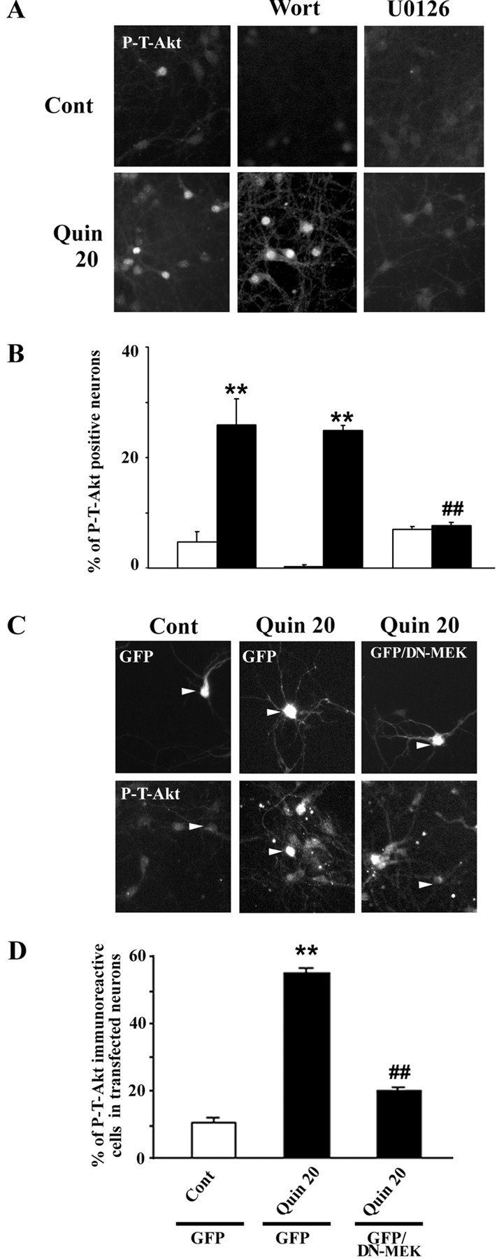
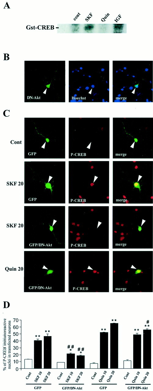
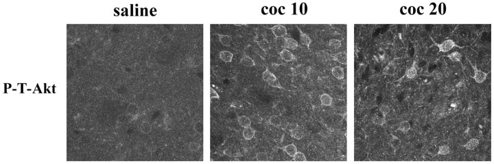
Akt is classically described as a prosurvival serine/threonine kinase activated in response to trophic factors. After activation by phosphoinositide 3-kinase (PI3-kinase), it can translocate to the nucleus where it promotes specific genetic programs by catalyzing phosphorylation of transcription factors. We report here that both dopamine (DA) D1 (SKF38393) and D2 (quinpirole) agonist treatments rapidly increase, in primary striatal neurons in culture, phosphorylation levels of Akt on Thr(308), a residue that is critically involved in its kinase activity. These treatments also activate the extracellular signal-regulated kinase (ERK) pathway in the same population of striatal neurons. Induction of active, phospho-Thr(308) Akt by dopamine D1 and D2 agonists is insensitive to wortmannin and thus PI3-kinase independent, in contrast to growth factor-induced Akt activity. D1- and D2-induced phospho-Thr(308) Akt is decreased by the mitogen-activated protein kinase kinase (MEK) inhibitor, U0126, as well as by overexpression of a dominant-negative version of MEK, thus implicating the Ras/ERK signaling cascade in this process. Furthermore, overexpression of a mutant form of Akt that cannot be activated impaired cAMP response element-binding protein (CREB) phosphorylation induced by SKF38393 and quinpirole treatments. Activation of Akt on Thr(308) was also found in vivo in striatal neurons after acute administration of cocaine, a psychostimulant that strongly increases DA transmission. Thus, multiple intracellular pathways can transduce signals from dopamine receptors to CREB in striatal neurons, one of these being Akt. We propose that this signaling pathway plays a pivotal role in DA-induced regulation of gene expression and long-term neuronal adaptation in the striatum.


Figures

Fig. 1.
Fig. 1.
Activation of Akt occurs in response to D1 and D2 receptor agonists in striatal neurons.A, Phosphorylation levels of Akt were detected by immunocytochemistry using anti-active anti-phospho-Thr308 (P-T-Akt) and anti-phospho-Ser473 (P-S-Akt) antibodies in control conditions (cont) and in striatal neurons treated for 20 min with the selective D1 agonist SKF38393 (SKF 20) (100 μm), the D2 agonist quinpirole (Quin 20) (10 μm), or IGF (IGF 20) (200 nm). B, Control of specificity of P-T-Akt in the presence of an excess of the immunogen peptide: KDGATMK[pT] FCGT (+pept) or unphosphorylated GST–Akt1 (+Gst-Akt) (bottom panel). C, Western blot analysis of P-T-Akt from subcellular fractionation of striatal neurons. Specific labeling corresponding to the expected molecular weight for Akt was detected in the nuclear compartment during SKF38393, quinpirole, and IGF treatment. As a control of fractionation, CREB immunoblotting was performed showing a specific nuclear immunostaining. D, P-T-Akt-immunoreactive nuclei were quantified during treatment with the D1 and D2 agonists for 10 min (SKF 10 and Quin 10, respectively) and 20 min (SKF 20 andQuin 20, respectively). Data report the percentage of P-T-Akt-positive nuclei when compared with the total number of Hoechst nuclei and are representative of three independent experiments for each treatment. For each experiment, cells were counted from five independent fields (∼100 cells for each field). Statistical comparisons were performed using one-way ANOVA followed by post hoc comparison (Newman–Keuls test). *p < 0.05 and **p < 0.01 when compared with the control group.
Fig. 2.
Fig. 2.
Akt and ERK activation occur in the same striatal subpopulation. A, Double immunostaining of active-Akt [polyclonal phospho-Thr308 Akt (P-T-Akt)] and active-ERK [monoclonal diphospho-Thr202-Tyr204 p44/p42 MAPK (P-ERK)] was performed using anti-rabbit Cy3- and anti-mouse FITC-coupled secondary antibodies, respectively. Note the colocalization of P-T-Akt and P-ERK immunostaining after 20 min of treatment with the D1 (SKF 20) and D2 (Quin 20) agonists (arrows).B, Single immunostaining of P-T-Akt and P-ERK was performed using polyclonal phospho-Thr308 Akt (P-T-Akt) and polyclonal diphospho-Thr202-Tyr204 p44/p42 MAPK (P-ERK), respectively. Immunoreactive cells were quantified 20 min after SKF38393 (SKF 20) and quinpirole (Quin 20) treatments (3 independent experiments each). For each experiment and each antibody, cells were counted from five independent fields (∼100 cells for each field). Statistical comparisons: *p < 0.05 and **p < 0.01 when compared with the control group (Newman–Keuls test).
Fig. 3.
Fig. 3.
Signaling pathways underlying Akt phosphorylation during D1 receptor stimulation. A, Immunocytochemical detection of P-T-Akt was analyzed as described in Figure 1 in the presence of wortmannin (Wort) (100 nm) applied 15 min before D1 agonist or IGF treatment (20 min each) (SKF 20 and IGF 20, respectively). B, Immunostaining of P-T-Akt in the presence of the selective inhibitor of PKA, Rp-cAMP (50 μm), or the selective MEK inhibitor, U0126(10 μm). C, Quantification of P-T-Akt immunolabeling was performed as described in Figure 1. Statistical analysis: *p < 0.05, **p < 0.01 when compared with the corresponding control group;#p < 0.05 and##p < 0.01 when compared with SKF38393 treatment alone (Newman–Keuls test). D, P-T-Akt (bottom panel) was analyzed as described in Figure 1 in neurons transfected with GFP (top panel) alone (GFP) or in neurons cotransfected with GFP and DN-MEK (GFP/DN-MEK).Arrowheads indicate the same neuron analyzed with filters corresponding to FITC (for GFP-positive neuron) (top panel) or Cy3 (for P-T-Akt) (bottom panel). Note the disappearance of P-T-Akt immunostaining in cells coexpressing GFP and DN-MEK. E, Quantification of P-T-Akt-immuno-reactive neurons was performed from GFP- or GFP/DN-MEK-transfected neurons (C,arrowheads) in control conditions (Cont) and after D1 (SKF 20) agonist treatments. For each experiment, P-T-Akt immunostaining was quantified from 100 transfected cells. Data are representative of three independent experiments for each treatment. **p < 0.01 when compared with the control group; ##p < 0.01 when compared with neurons transfected with GFP alone (Newman–Keuls test).
Fig. 4.
Fig. 4.
Signaling pathways underlying ERK phosphorylation during D1 receptor stimulation. A, Immunocytochemical detection of SKF38393-induced P-ERK was analyzed as described in Figure 2C, in the presence of wortmannin (Wort) (100 nm), Rp-cAMP (50 μm), or U0126 (10 μm).B, Quantification of P-ERK immunolabeling was performed as described in Figure 2C. Statistical analysis: *p < 0.05, **p < 0.01 when compared with the corresponding control group;#p < 0.05,##p < 0.01 when compared with groups in the presence or not of the inhibitors (Newman–Keuls test).
Fig. 5.
Fig. 5.
Signaling pathways underlying Akt phosphorylation during D2 receptor stimulation. A, Immunocytochemical detection of quinpirole-induced P-T-Akt was analyzed as described in Figure 1, in the presence of wortmannin (Wort) (100 nm) or U0126 (10 μm).B, Quantification of P-T-Akt immunolabeling was performed as described in Figure 1. Statistical analysis: **p < 0.01 when compared with the corresponding control group; ##p < 0.01 when compared with SKF38393 treatment alone (Newman–Keuls test). C, During quinpirole treatment, P-T-Akt (bottom panel) was analyzed as described in Figure 3 in neurons transfected (arrowheads) with GFP alone (GFP) or with GFP and DN-MEK (GFP/DN-MEK) (bottom panel). D, Quantification of P-T-Akt-immunoreactive neurons in transfected neurons as described in Figure 3E. Statistical analysis: **p< 0.01 when compared with the control group;##p < 0.01 when compared with neurons transfected with GFP alone (Newman–Keuls test).
Fig. 6.
Fig. 6.
Activated Akt controls CREB phosphorylation induced by D1 agonist treatment. A, Striatal neurons were treated for 20 min with SKF, quinpirole, and IGF as indicated. Akt was immunoprecipitated from neuronal lysates using an antibody corresponding to the PH domain, and
Fig. 7.
Fig. 7.
In vivo activation of Akt in striatal neurons after cocaine administration. P-T-Akt immunostaining was performed in striatal sections of mice treated with saline or cocaine. P-T-Akt immunoreactivity was increased 10 and 20 min after cocaine (20 mg/kg, i.p.) administration (coc 10and coc 20, respectively). Shown is fluorescent confocal analysis of P-T-Akt immunolabeling. Note its nuclear labeling during cocaine treatment (coc 20). Data are representative of at least four independent mice for each treatment. its kinase activity was determined with GST-CREB1–166 as a substrate. After washing to eliminate unincorporated radioactivity, samples were subjected to 10% SDS-PAGE (data are representative of 3 independent experiments).B, Immunolocalization of HA-tagged-DN-Akt (DN-Akt) was analyzed with an anti-HA antibody. Note the localization of DN-Akt in both cytosolic (including neuritic extension) and nuclear compartments. Note also the nuclear integrity (Hoechst) of neuronal cells overexpressing DN-Akt.C, CREB phosphorylation was analyzed by immunocytochemical detection of the anti-P-CREB antibody. Note thatP-CREB immunostaining is strongly induced during SKF38393 treatment (SKF 20), including in a GFP-transfected neuron (arrowhead). During SKF (arrowhead) but not quinpirole (Quin 20) (arrowhead) treatment, note the disappearance of P-CREB immunostaining in cells cotransfected with GFP and DN-Akt (GFP/DN-Akt). D, Quantification of P-CREB-immunoreactive neurons was performed from GFP or GFP + DN-Akt-transfected neurons in control conditions (Cont), after D1 (SKF 10 and SKF 20) or D2 (Quin 10 and Quin 20) agonist treatments. For each experiment, P-CREB immunostaining was quantified from 100 transfected cells. Data are representative of three independent experiments for each group. *p < 0.05 and **p < 0.01 when compared with the corresponding control group; #p < 0.05 and##p < 0.01 when compared with neurons transfected with GFP alone (Newman–Keuls test).

References

    1. Aizman O, Brismar H, Uhlen P, Zettergren E, Levey AI, Forssberg H, Greengard P, Aperia A. Anatomical and physiological evidence for D1 and D2 dopamine receptor colocalization in neostriatal neurons. Nat Neurosci. 2000;3:226–230. - PubMed
    1. Alessi DR, Cohen P. Mechanism of activation and function of protein kinase B. Curr Opin Genet Dev. 1998;8:55–62. - PubMed
    1. Alessi DR, Andjelkovic M, Caudwell B, Cron P, Morrice N, Cohen P, Hemmings BA. Mechanism of activation of protein kinase B by insulin and IGF-1. EMBO J. 1996;15:6541–6551. - PMC - PubMed
    1. Alessi DR, James SR, Downes CP, Holmes AB, Gaffney PR, Reese CB, Cohen P. Characterization of a 3-phosphoinositide-dependent protein kinase which phosphorylates and activates protein kinase Balpha. Curr Biol. 1997;7:261–269. - PubMed
    1. Andjelkovic M, Alessi DR, Meier R, Fernandez A, Lamb NJ, Frech M, Cron P, Cohen P, Lucocq JM, Hemmings BA. Role of translocation in the activation and function of protein kinase B. J Biol Chem. 1997;272:31515–31524. - PubMed

Publication types

MeSH terms

Substances