

In situ GABAergic modulation of synchronous gonadotropin releasing hormone-1 neuronal activity
- PMID: 12388600
- PMCID: PMC6757671
- DOI: 10.1523/JNEUROSCI.22-20-08932.2002
In situ GABAergic modulation of synchronous gonadotropin releasing hormone-1 neuronal activity
Abstract
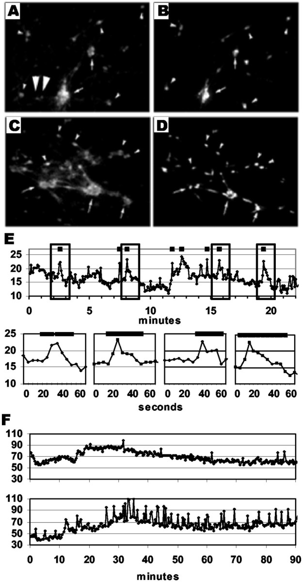
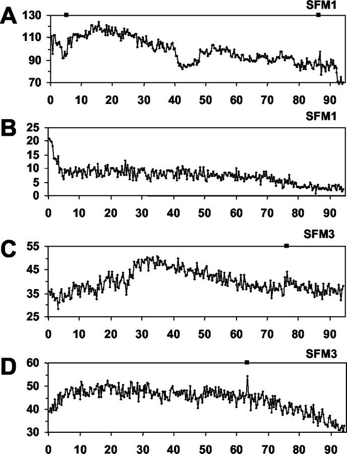
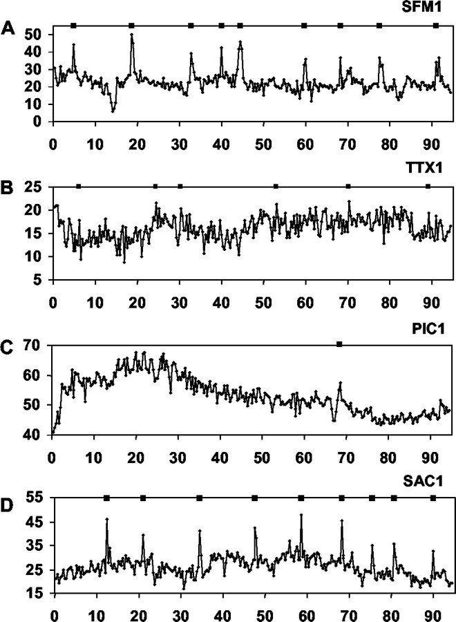
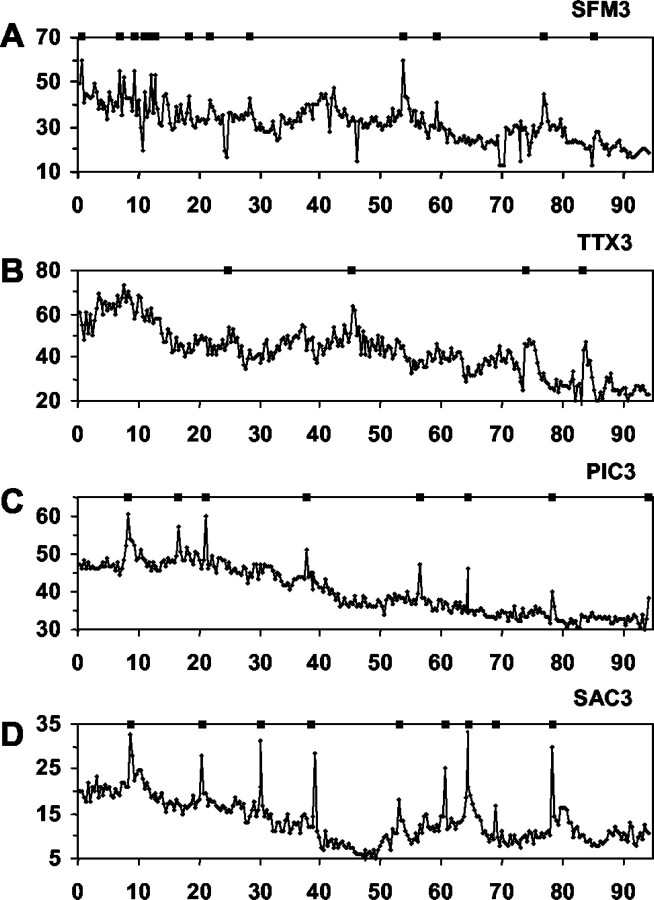
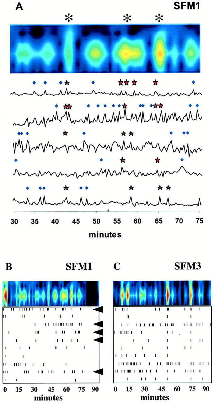
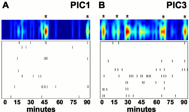
Evidence indicates that gonadotropin releasing hormone-1 [GnRH-1, also known as luteinizing hormone releasing hormone (LHRH)] neurons can exhibit synchronized neuroendocrine secretory activity before entrance into the CNS. In this study, we used calcium imaging to evaluate patterns of activity in individual, embryonic, GnRH-1 neurons as well as population dynamics of GnRH-1 neurons in mouse nasal explants maintained for 1 versus 3 weeks. Independent of age, GnRH-1 neurons displayed significant calcium peaks that synchronized at an interval of approximately 20 min across multiple GnRH-1 cells within an explant. Acute tetrodotoxin treatment decreased the amplitude of calcium peaks in individual GnRH-1 neurons and the duration but not the frequency of synchronized activity in the population of GnRH-1 neurons. Acute GABA(B) receptor antagonism increased the frequency of synchronized neuronal activity at both ages, whereas acute GABA(A) receptor antagonism decreased calcium oscillations in individual GNRH-1 cells as well as synchronization of the calcium pulses within the GnRH-1 population at the 1 week time point to background non-GNRH-1 cell levels. These results indicate that developing GnRH-1 neurons rely heavily on GABAergic signaling to initiate synchronized bouts of activity but thereafter, possess an innate capacity for synchronized activity patterns that are modulated by, but not completely dependent on GABAergic signaling.
Figures
Similar articles
-
Direct action of estradiol on gonadotropin-releasing hormone-1 neuronal activity via a transcription-dependent mechanism.J Neurosci. 2004 Jul 14;24(28):6326-33. doi: 10.1523/JNEUROSCI.1006-04.2004. J Neurosci. 2004. PMID: 15254088 Free PMC article.
-
Luteinizing hormone releasing hormone (LHRH) neurons maintained in nasal explants decrease LHRH messenger ribonucleic acid levels after activation of GABA(A) receptors.Endocrinology. 1998 Jun;139(6):2734-40. doi: 10.1210/endo.139.6.6034. Endocrinology. 1998. PMID: 9607779
-
Endogenous gamma-aminobutyric acid can excite gonadotropin-releasing hormone neurons.Endocrinology. 2005 Dec;146(12):5374-9. doi: 10.1210/en.2005-0788. Epub 2005 Aug 25. Endocrinology. 2005. PMID: 16123153
-
Understanding calcium homeostasis in postnatal gonadotropin-releasing hormone neurons using cell-specific Pericam transgenics.Cell Calcium. 2012 Mar-Apr;51(3-4):267-76. doi: 10.1016/j.ceca.2011.11.005. Epub 2011 Dec 15. Cell Calcium. 2012. PMID: 22177387 Review.
-
Calcium dynamics in gonadotropin-releasing hormone neurons.Front Neuroendocrinol. 2010 Jul;31(3):259-69. doi: 10.1016/j.yfrne.2010.05.005. Epub 2010 Jun 2. Front Neuroendocrinol. 2010. PMID: 20594958 Review.
Cited by
-
Neuropeptide Y directly inhibits neuronal activity in a subpopulation of gonadotropin-releasing hormone-1 neurons via Y1 receptors.Endocrinology. 2010 Jun;151(6):2736-46. doi: 10.1210/en.2009-1198. Epub 2010 Mar 29. Endocrinology. 2010. PMID: 20351316 Free PMC article.
-
Epigenetic changes coincide with in vitro primate GnRH neuronal maturation.Endocrinology. 2010 Nov;151(11):5359-68. doi: 10.1210/en.2010-0555. Epub 2010 Sep 22. Endocrinology. 2010. PMID: 20861233 Free PMC article.
-
Differential regulation of gonadotropin-releasing hormone neuron activity and membrane properties by acutely applied estradiol: dependence on dose and estrogen receptor subtype.J Neurosci. 2009 Apr 29;29(17):5616-27. doi: 10.1523/JNEUROSCI.0352-09.2009. J Neurosci. 2009. PMID: 19403828 Free PMC article.
-
Cell type-specific expression of a genetically encoded calcium indicator reveals intrinsic calcium oscillations in adult gonadotropin-releasing hormone neurons.J Neurosci. 2007 Jan 24;27(4):860-7. doi: 10.1523/JNEUROSCI.3579-06.2007. J Neurosci. 2007. PMID: 17251427 Free PMC article.
-
Activation of hypothalamic gono-like neurons in female rats during estrus.Neural Regen Res. 2012 Nov 5;7(31):2413-23. doi: 10.3969/j.issn.1673-5374.2012.31.002. Neural Regen Res. 2012. PMID: 25337091 Free PMC article.
References
-
- Belchetz PP, Plant TM, Nakai Y, Keogh EJ, Knobil E. Hypophysial responses to continuous and intermittent delivery of hypothalamic gonadotropin-releasing hormone. Science. 1978;202:631–633. - PubMed
MeSH terms
Substances
LinkOut - more resources
Full Text Sources
Other Literature Sources