. 2002 Nov;76(22):11273-82.
doi: 10.1128/jvi.76.22.11273-11282.2002.

Human immunodeficiency virus type 1 recombination: rate, fidelity, and putative hot spots

Affiliations

Human immunodeficiency virus type 1 recombination: rate, fidelity, and putative hot spots

Jianling Zhuang et al. J Virol. 2002 Nov.

Abstract

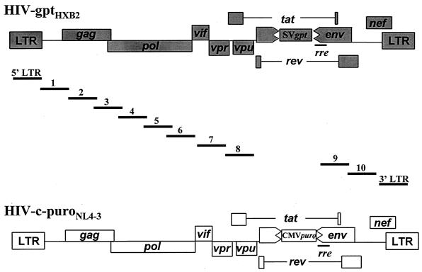
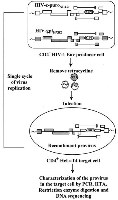
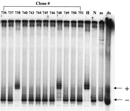
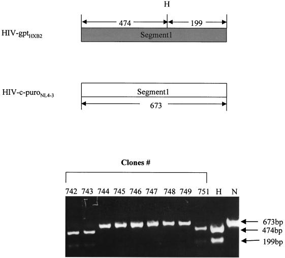
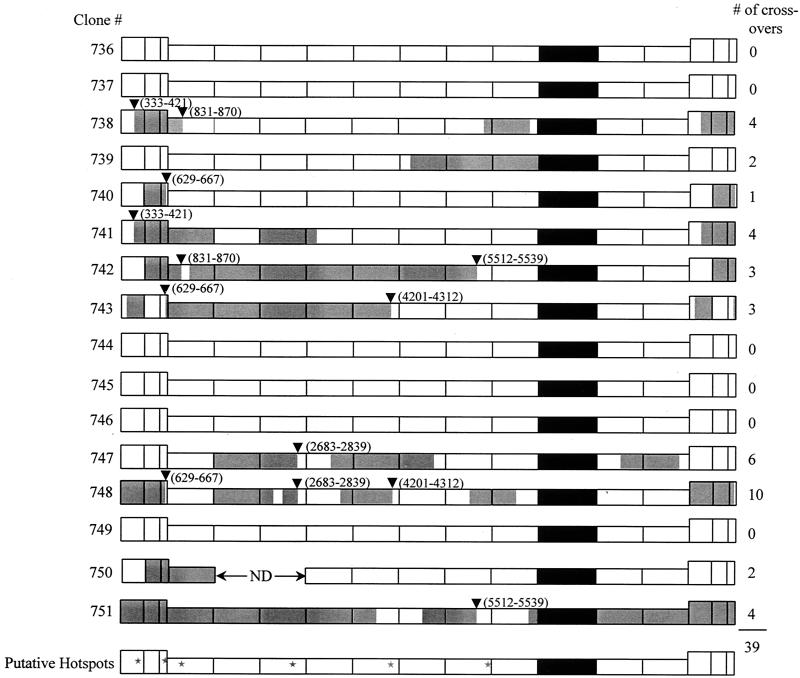
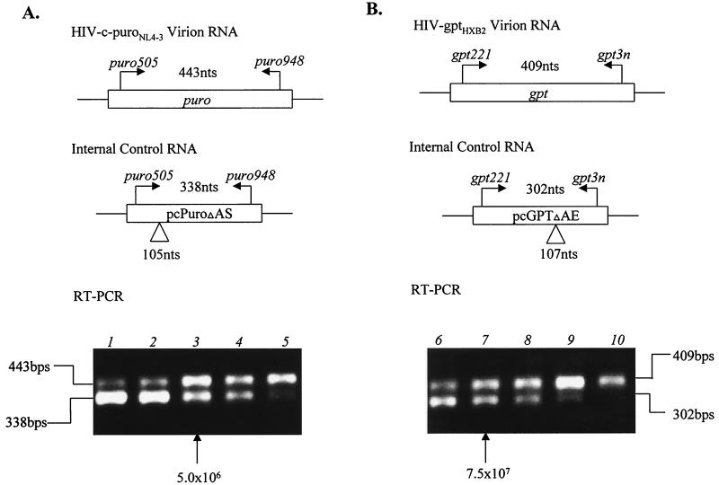
Previously, we reported that human immunodeficiency virus type 1 (HIV-1) recombines approximately two to three times per genome per replication cycle, an extremely high rate of recombination given the relatively small genome size of HIV-1. However, a recombination hot spot involving sequence of nonretroviral origin was identified in the vector system utilized, raising the possibility that this hot spot skewed the rate of recombination, and the rate of recombination observed was an overestimation. To address this issue, an HIV-1-derived vector system was used to examine the rate of recombination between autologous HIV-1 sequences after restricting replication to a single cycle in the absence of this hot spot. Viral DNA and RNA were analyzed by a combination of the heteroduplex tracking assay, restriction enzyme analysis, DNA sequencing, and reverse transcription-PCR. The results indicate that HIV-1 undergoes recombination at a minimum rate of 2.8 crossovers per genome per cycle. Again, this is a very high rate given the small size of the HIV-1 genome. The results also suggested that there might be local hot spots of recombination at different locations throughout the genome since 13 of the 33 strand transfers identified by DNA sequencing shared the same site of recombination with one or two other clones. Furthermore, identification of crossover segments also allowed examination of mutations at the point of recombination, since it has been predicted from some studies of cell-free systems that mutations may occur with a frequency of 30 to 50% at crossover junctions. However, DNA sequence analysis of crossover junctions indicated that homologous recombination during viral replication was not particularly mutagenic, indicating that there are other factors or conditions not yet reproduced in cell-free systems which contribute to fidelity during retroviral recombination.


Figures

FIG. 1.
FIG. 1.
HIV-1 vectors and segments of provirus DNA amplified to study HIV-1 recombination rate. HIV-c-puroNL4-3 is based upon HIV-1 strain NL4-3. HIV-gptHXB2 is based upon HIV-1 strain HXB2. CMVpuro represents the puro gene expressed from the cytomegalovirus immediate-early promoter. SVgpt represents the gpt gene under control of the simian virus 40 early gene promoter. The 12 different overlapping segments amplified are depicted by the bars with numbers or LTR designations above them. The coordinates of the amplified segments are as follows: 5′LTR, 53 to 799; 1, 611 to 1283; 2, 1261 to 2049; 3, 2028 to 2826; 4, 2807 to 3663; 5, 3547 to 4398; 6, 4271 to 5066; 7, 4932 to 5797; 8, 5778 to 6390; 9, 7643 to 8454; 10, 8336 to 9156; 3′LTR, 8966 to 9716 (sequence coordinates are according to the HXB2 provirus sequence).
FIG. 2.
FIG. 2.
Protocol for study of HIV-1 recombination. Individual cell clones containing a single copy of both HIV-gptHXB2 and HIV-c-puroNL4-3 were established. Upon induction by removal of tetracycline, vector virus was harvested and used to inoculate CD4-positive HeLaT4 target cells, which was followed by selection and isolation of target cell clones. Going from a provirus in a producer cell to a provirus in a target cell comprised a single cycle of virus replication. Proviruses in each target cell clone were further analyzed by PCR, the HTA, restriction enzyme digestion, and DNA sequencing in order to identify recombinants and crossover points.
FIG. 3.
FIG. 3.
Representative example of an HTA. Radioactively labeled single-stranded probe was prepared from HIV-c-puroNL4-3 plasmid DNA by asymmetric PCR using the segment 8 primer pair (lane ss). Radioactively labeled double-stranded segment was amplified at the same time as a control (lane ds). Segment 8 is 612 bp in length. The sequence difference between the two strains within this segment is approximately 1%. The probe was annealed with PCR products amplified from NL-c-puroNL4-3 (lane N, homoduplex control), HIV-gptHXB2 (lane H, heteroduplex control), or genomic DNA from different target cell clones (clones 736 to 738, 740, 743 to 746, and 748 to 751). Clones 738 and 748 are shifted compared to the band for HIV-c-puroNL4-3 DNA and were therefore scored as recombinants. Symbols: +, band shifted due to heteroduplex formation; −, nonshift homoduplex band.
FIG. 4.
FIG. 4.
Representative example of a restriction enzyme digestion to identify crossover points. At the top of the figure, the diagram depicts the anticipated sizes of amplified segment 1 from HIV-gptHXB2 and HIV-c-puroNL4-3, respectively, as well as the predicted HindIII digestion patterns. Purified proviral and plasmid DNA segments were digested with HindIII. Bands of 474 and 199 indicate this segment of the clone is from HIV-gptHXB2 (lane H) while a 673-bp band indicates it is from HIV-c-puroNL4-3 (lane N). The bottom of the figure illustrates the digestion pattern of amplified segment 1 from nine progeny target cell clones, 742 to 749 and 751. H indicates control DNA amplified from HIV-gptHXB2 plasmid, and N indicates control DNA amplified from HIV-c-puroNL4-3 plasmid. Clones 742, 743, and 751 display 474- and 199-bp bands, indicating they are of HIV-gptHXB2 origin. Clones 745 to 749 display a 673-bp band, indicating they are of HIV-c-puroNL4-3 origin.
FIG. 5.
FIG. 5.
Recombination distribution throughout the full-length proviral genomic DNA. Ten out of 16 clones had undergone a total of 39 recombination events. Thirty-three out of the 39 recombinant sites were sequenced. Six putative hot spots are indicated by inverted triangles and summarized with stars. The coordinates of the putative hot spots are as follows: 333 to 421, 629 to 667, 831 to 870, 2683 to 2839, 4201 to 4312, and 5512 to 5539 (sequence coordinates are according to the HXB2 provirus sequence). White is of HIV-c-puroNL4-3 origin and grey is of HIV-gptHXB2 origin. The marker gene cassette is depicted in black.
FIG. 6.
FIG. 6.
RT-PCR to determine relative levels of HIV-c-puroNL4-3 and HIV-gptHXB2 RNAs. (A) Quantitation of HIV-c-puroNL4-3 virion RNA. The primers used to amplify the puro-specific sequence are indicated in Materials and Methods. The primers are also specific for the puro control RNA, which has an internal deletion of 105 bases to distinguish it from the signal obtained from the viral RNA. Viral RNA (625 ng) was mixed with different dilutions of the puro control RNA ranging from 107 to 106 molecules (1.0 × 10⁷, 7.5 × 10⁶, 5 × 10⁶, 2.5 × 10⁶, and 1.0 × 10⁶ in lanes 1 to 5, respectively). As can be seen, the signal from the viral RNA is about the same as the signal obtained with 5 × 10⁶ molecules of control RNA. (B) RT-PCR for quantitation of HIV-gptHXB2 RNA. The gpt-specific segment to be amplified and the control RNA with an internal deletion are indicated. Again, 625 ng of virion RNA was added to each sample with the amount of control RNA ranging from 108 to 107 molecules (1.0 × 10⁸, 7.5 × 10⁷, 5 × 10⁷, 2.5 × 10⁷, and 1.0 × 10⁶ in lanes 6 to 10, respectively). As is evident, equivalent signals are obtained when 7.5 × 10⁷ molecules of control RNA are added.

References

    1. Adachi, A., H. E. Gendelman, S. Koenig, T. Folks, R. Willey, A. Rabson, and M. A. Martin. 1986. Production of acquired immunodeficiency syndrome-associated retrovirus in human and nonhuman cells transfected with an infectious molecular clone. J. Virol. 59:284-291. - PMC - PubMed
    1. Anderson, J. A., R. J. Teufel II, P. D. Yin, and W. S. Hu. 1998. Correlated template-switching events during minus-strand DNA synthesis: a mechanism for high negative interference during retroviral recombination. J. Virol. 72:1186-1194. - PMC - PubMed
    1. Balakrishnan, M., P. J. Fay, and R. A. Bambara. 2001. The kissing hairpin sequence promotes recombination within the HIV-I 5′ leader region. J. Biol. Chem. 276:36482-36492. - PubMed
    1. Battula, N., and L. A. Loeb. 1976. On the fidelity of DNA replication. Lack of exodeoxyribonuclease activity and error-correcting function in avian myeloblastosis virus DNA polymerase. J. Biol. Chem. 251:982-986. - PubMed
    1. Bebenek, K., J. Abbotts, J. D. Roberts, S. H. Wilson, and T. A. Kunkel. 1989. Specificity and mechanism of error-prone replication by human immunodeficiency virus-1 reverse transcriptase. J. Biol. Chem. 264:16948-16956. - PubMed

Publication types

LinkOut - more resources