. 2002 Nov;76(22):11425-33.
doi: 10.1128/jvi.76.22.11425-11433.2002.

Tropism of varicella-zoster virus for human tonsillar CD4(+) T lymphocytes that express activation, memory, and skin homing markers

Affiliations

Tropism of varicella-zoster virus for human tonsillar CD4(+) T lymphocytes that express activation, memory, and skin homing markers

Chia-Chi Ku et al. J Virol. 2002 Nov.

Abstract

Varicella-zoster virus (VZV) is an alphaherpesvirus with the characteristic neurotropism of this group, but VZV also infects T cells productively and downregulates major histocompatibility complex (MHC) class I expression on infected T cells, as shown in the SCID-hu mouse model. T-cell tropism is likely to be critical for the cell-associated viremia associated with primary VZV infection. In these experiments, we found that VZV infects human tonsillar CD4(+) T cells in culture, with 15 to 25% being positive for VZV proteins as detected by polyclonal anti-VZV immunoglobulin G (IgG) staining and monitored by flow cytometry analysis. RNA transcripts for VZV gE, a late gene product, were detected in T-cell populations that expressed VZV surface proteins, but not in the VZV-negative cell fraction. Exposure to phorbol myristate acetate resulted in an increase in VZV-positive T cells, indicating that viral DNA was present within these cells and that VZV gene expression could be induced by T-cell activation. By immune scanning electron microscopy, VZV virions were detected in abundance on the surfaces of infected tonsillar T cells. The predominant CD4(+) T-lymphocyte subpopulations that became infected were activated CD69(+) T cells with the CD45RA(-) memory phenotype. Subsets of CD4(+) T cells that expressed skin homing markers, cutaneous leukocyte antigen, and chemokine receptor 4 were also infected with VZV. By chemotaxis assay, VZV-infected T cells migrated to SDF-1, demonstrating that skin migratory function was intact despite VZV infection. The susceptibility of tonsil T cells to VZV suggests that these cells may be important targets during the initial VZV infection of upper respiratory tract sites. Viral transfer to migrating T cells in the tonsils may facilitate cell-associated viremia, and preferential infection of CD4 T cells that express skin homing markers may enhance VZV transport to cutaneous sites of replication.


Figures

FIG. 1.
FIG. 1.
FACS analysis of VZV-infected CD3+ T cells. Column-purified T cells isolated from tonsils were cocultured with VZV-infected HEL monolayers. The T cells were analyzed at days 2 to 3 postinfection and stained with anti-CD3 MAb and VZV IgG-negative human serum (A) as background staining or with anti-VZV IgG polyclonal immune serum (B) to detect VZV glycoproteins, which provide the indication of infection. The FACS plots show infection of tonsil T cells with VZV, and the frequency of CD3+ T cells that are VZV positive is as indicated.
FIG. 2.
FIG. 2.
Detection of VZV gE transcripts by RT-PCR. Ten nanograms of template RNA isolated from VZV-infected fibroblasts (lanes 1 and 2), VZV-infected and FACS-sorted CD3+ T cells (lanes 3 to 6), or uninfected T cells (lane 7) was amplified by RT-PCR in the presence (lanes 1, 3, 5, 7, and 8) or absence (lanes 2, 4, and 6) of reverse transcriptase. The RNA was treated (lanes 1, 2, 5, 6, and 7) or untreated (lanes 3 and 4) with DNase I prior to cycling amplification. A 1,070-bp fragment for VZV gE was detected as indicated. Lane 8, no-template control (NC).
FIG. 3.
FIG. 3.
PMA stimulation of VZV transcription in virally infected T cells. VZV-infected T cells at 2 days postinfection were preincubated either with 0.1% DMSO alone (A and B) or with U0126 at 10 μM in 0.1%DMSO (C) at 37°C for 30 min followed by PMA stimulation at 100 nM at 37°C for 24 h. The T cells were washed and stained with antihuman CD3 MAb and anti-VZV IgG immune serum for FACS analysis. The FACS plots show that VZV replication indicated by expression of VZV glycoproteins was enhanced by PMA treatment (B), but was inhibited by U0126 (C) compared to the unstimulated control (A).
FIG. 4.
FIG. 4.
Expression of VZV particles on infected T cells by scanning electron microscopy. (A) Two uninfected CD3+ T cells were visualized by scanning electron microscopy at a magnification of ×10,000. (B) VZV-infected T cells had a more irregular surface than was seen in uninfected T cells. Both ruffles and microvilli, as well as darkened crypts, were abundant on the surface of a representative VZV-infected T cell. In addition, viral particles were also distinguishable on the surface of the cell at magnification of ×15,000. (C) Pleomorphic virus particles ranging in size from 120 to 200 nm were distributed between microvilli and sometimes formed small clusters on the cell surface. A representative high-magnification (×50,000) scanning electron micrograph shows two viral particles in the center of panel C, to the right of microvilli; arrows indicate that the envelop of the larger particle was labeled with VZV gE immunogold. Micrographs in reference illustrate comparable VZV infection of human melanoma cells.
FIG. 5.
FIG. 5.
Phenotypic analysis of CD4+ T cells from tonsils and peripheral blood. T cells isolated from peripheral blood (PBL [upper panels]) and tonsils (lower panels) were stained with anti-CD4, -CD45RA, -CD69, or -CD71 MAbs for FACS analysis. The cells were gated on CD45RA+ CD4+ naive (shaded area) and CD45RA CD4+ memory (thick line) cells and analyzed for expression of CD69 (A and B) and CD71 (C and D) in these subpopulations. T cells from the same preparation stained with mouse IgG2a were treated as an isotype control (dashed line).
FIG. 6.
FIG. 6.
Expression of VZV glycoproteins on CD45RA+ or CD45RA CD4+ T cells. VZV infection of T cells was achieved, and the cells were then stained as described in the legend to Fig. 1. The cells were gated on CD4+ T cells and analyzed by FACS dot plots. The relationship between CD45RA expression and VZV infection in CD4+ T cells shows that more memory than naive CD4+ T cells are infected with VZV. The numbers indicated in the upper right corner are the percentages of cells in each of the corresponding quadrants.
FIG. 7.
FIG. 7.
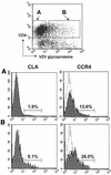
Expression of CLA and CCR4 on VZV-infected CD4+ T cells. Column-purified T cells from tonsils were infected with VZV as described in the legend to Fig. 1. The T cells were analyzed on day 2 and stained with anti-CD4 MAb, anti-VZV IgG polyclonal immune serum, and anti-CLA or anti-CCR4 antibody for FACS analysis. The cells were gated on uninfected (A) and VZV-infected (B) CD4+ T cells and analyzed for CLA and CCR4 expression. The fluorescent profiles of CLA and CCR4 in uninfected (upper panels) and VZV-infected (lower panels) CD4+ T cells show more CLA and CCR4 are expressed on VZV-infected than on uninfected CD4+ T cells. T cells from the same preparation were stained with mouse IgG1 as an isotype control for CCR4.
FIG. 8.
FIG. 8.
Chemotaxis of VZV-infected T cells in response to SDF-1α. Tonsillar T cells infected with VZV were tested for their chemotaxic response to human SDF-1α (100 nM) with 24-well plate tissue culture inserts with 5-μm-pore-size filters. All samples were performed in duplicative wells. Cells that were incubated with chemotaxis medium without containing chemokine were treated as negative controls. After 2 h of incubation at 37°C, cells that were in the starting population and that had migrated were harvested and stained with anti-CD4, anti-CD45RA, and anti-VZV IgG immune serum. The cells were gated on uninfected (VZV CD4+) and infected (VZV+ CD4+) T cells. The percentages of total CD4+ T cells (black bars), CD45RA+ naive (dotted bars), and CD45RA memory (hatched bars) CD4+ T cells that had migrated to SDF-1α from starting cell populations (maximal migration) in uninfected and infected cells were thus calculated and subtracted from background migration (range, 2.8 and 5.2%). The results shown are the averages of duplicate samples from three independent experiments.

Similar articles

Cited by

References

    1. Abendroth, A., G. Morrow, A. L. Cunningham, and B. Slobedman. 2001. Varicella-zoster virus infection of human dendritic cells and transmission to T cells: implications for virus dissemination in the host. J. Virol. 75:6183-6192. - PMC - PubMed
    1. Arvin, A. 2001. Varicella-zoster virus, p. 2731-2767. In D. Knipe and P. Howley (ed.), Fields virology, 4th ed., vol. 2. Lippincott Williams & Wilkins, Philadelphia, Pa.
    1. Arvin, A. M., J. F. Moffat, and R. Redman. 1996. Varicella-zoster virus: aspects of pathogenesis and host response to natural infection and varicella vaccine. Adv. Virus Res. 46:263-309. - PubMed
    1. Asano, Y., N. Itakura, Y. Hiroishi, S. Hirose, T. Nagai, T. Ozaki, T. Yazaki, K. Yamanishi, and M. Takahashi. 1985. Viremia is present in incubation period in nonimmunocompromised children with varicella. J. Pediatr. 106:69-71. - PubMed
    1. Berg, E. L., T. Yoshino, L. S. Rott, M. K. Robinson, R. A. Warnock, T. K. Kishimoto, L. J. Picker, and E. C. Butcher. 1991. The cutaneous lymphocyte antigen is a skin lymphocyte homing receptor for the vascular lectin endothelial cell-leukocyte adhesion molecule 1. J. Exp. Med. 174:1461-1466. - PMC - PubMed

Publication types

Substances