Comparative Study
. 2002 Nov;76(22):11447-59.
doi: 10.1128/jvi.76.22.11447-11459.2002.

Comparison of the complete DNA sequences of the Oka varicella vaccine and its parental virus

Affiliations
Comparative Study

Comparison of the complete DNA sequences of the Oka varicella vaccine and its parental virus

Yasuyuki Gomi et al. J Virol. 2002 Nov.

Erratum in

  • J Virol. 2003 Mar;77(5):3351.

Abstract

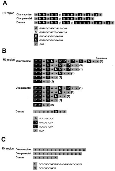
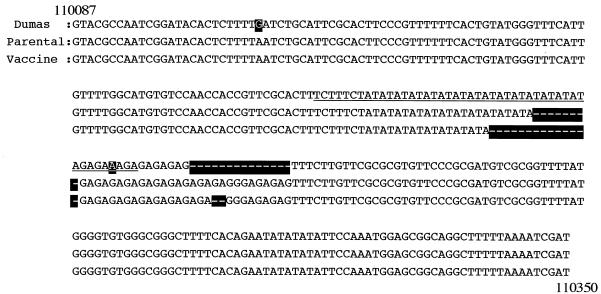
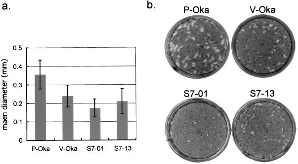
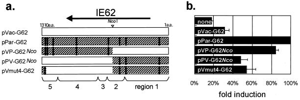
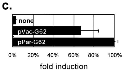
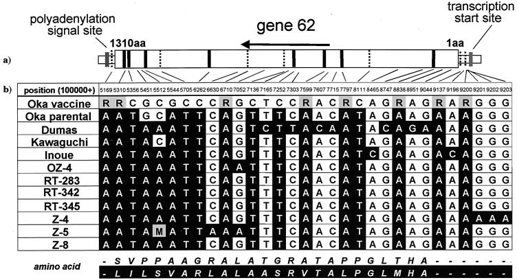
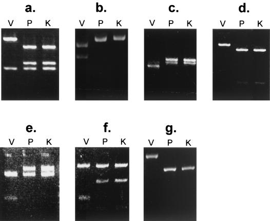
The DNA sequences of the Oka varicella vaccine virus (V-Oka) and its parental virus (P-Oka) were completed. Comparison of the sequences revealed 42 base substitutions, which led to 20 amino acid conversions and length differences in tandem repeat regions (R1, R3, and R4) and in an origin of DNA replication. Amino acid substitutions existed in open reading frames (ORFs) 6, 9A, 10, 21, 31, 39, 50, 52, 55, 59, 62, and 64. Of these, 15 base substitutions, leading to eight amino acid substitutions, were in the gene 62 region alone. Further DNA sequence analysis showed that these substitutions were specific for V-Oka and were not present in nine clinical isolates. The immediate-early gene 62 product (IE62) of P-Oka had stronger transactivational activity than the mutant IE62 contained in V-Oka in 293 and CV-1 cells. An infectious center assay of a plaque-purified clone (S7-01) from the V-Oka with 8 amino acid substitutions in ORF 62 showed smaller plaque formation and less-efficient virus-spreading activity than did P-Oka in human embryonic lung cells. Another clone (S-13) with only five substitutions in ORF 62 spread slightly faster than S7-01 but not as effectively as P-Oka. Moreover, transient luciferase assay in 293 cells showed that transactivational activities of IE62s of S7-01 and S7-13 were lower than that of P-Oka. Based on these results, it appears that amino acid substitutions in ORF 62 are responsible for virus growth and spreading from infected to uninfected cells. Furthermore, the Oka vaccine virus was completely distinguishable from P-Oka and 54 clinical isolates by seven restriction-enzyme fragment length polymorphisms that detected differences in the DNA sequence.


Figures

FIG. 1.
FIG. 1.
Comparisons of complete DNA sequences. The genome is presented in sections; horizontal lines indicate unique regions, and open boxes indicate inverted repeat regions. ORFs 1 to 71 are illustrated as pentagons. ORFs 62 through 64 are duplications of ORFs 69 through 71. Solid, shaded, and open symbols show the base substitutions causing amino acid conversions, base substitutions in noncoding regions, and silent mutations, respectively. (A) Forty-two base substitutions between P-Oka and V-Oka. (B) One hundred seventy-six base substitutions between the Dumas virus and P-Oka. All 176 substitutions between the Dumas virus and P-Oka are contained within 218 substitutions between the Dumas virus and V-Oka; the 218 substitutions between the Dumas virus and V-Oka were the total of 176 plus 42 substitutions.
FIG. 2.
FIG. 2.
Comparisons of repeat regions. V-Oka has different structures in R1 (A), R4 (B), and R3 (C) compared to P-Oka. Both V-Oka and P-Oka contain multiple clones that have different patterns and combinations of repeat elements in R3.
FIG. 3.
FIG. 3.
Comparisons of DNA sequences in the origin region. An almost palindromic sequence (underline) is located in the middle of this region. P-Oka and V-Oka have deletions, (TA)4 and (TA)7, in the palindromic sequence, respectively. The other deletion site is in the (GA) repeat just after the palindromic sequence.
FIG. 4.
FIG. 4.
(a) Plaque sizes after infection with cell-free P-Oka, V-Oka, S7-01, and S7-13. (b) HEL cells were infected with each cell-free virus and cultured for 10 days. The cells were stained with methylene blue.
FIG. 5.
FIG. 5.
Infectious center assay of P-Oka, V-Oka, S7-01, and S7-13. HEL cells (a) and GPE cells (b) in 35-mm dishes were infected independently with similar titers of these four kinds of cell-free viruses, and then the cells were washed and treated with trypsin from 1 to 5 days postinfection. The trypsin-treated cells were diluted and transferred onto monolayers of uninfected HEL cells in 35-mm dishes, and the numbers of infected cells were assessed by counting the number of VZV plaques appearing after 7 days. The number of infected cells was normalized to the initial viral titer per dish; the fold increase indicates the number of infected cells spread from one initial infected cell at day 0.
FIG. 6.
FIG. 6.
(a) Structure of IE62s. Amino acid residues are numbered from 1 to 1310 from the N to the C terminus. Vertical lines indicate the positions of amino acid changes compared with the IE62 from P-Oka. (b and c) Transactivational activity of parental and mutant IE62s on the DNA polymerase promoter in 293 cells (b) and GPE cells (c). Each of the five effector plasmids was transfected together with pLuc-Pol, and each luciferase activity was determined after 24 h. The basal luciferase activity, induced by no effector, was set equal to 1.0, and the other activities relative to that were given.
FIG. 6.
FIG. 6.
(a) Structure of IE62s. Amino acid residues are numbered from 1 to 1310 from the N to the C terminus. Vertical lines indicate the positions of amino acid changes compared with the IE62 from P-Oka. (b and c) Transactivational activity of parental and mutant IE62s on the DNA polymerase promoter in 293 cells (b) and GPE cells (c). Each of the five effector plasmids was transfected together with pLuc-Pol, and each luciferase activity was determined after 24 h. The basal luciferase activity, induced by no effector, was set equal to 1.0, and the other activities relative to that were given.
FIG. 7.
FIG. 7.
(a) Structure of gene 62. Amino acid residues are numbered from 1 to 1310 from the N to the C terminus. Fifteen vertical lines indicate the positions of base changes between Oka vaccine and its parental virus. Bold or broken lines show base mutations with or without amino acid conversions, respectively. (b) Sequence analysis of gene 62 from the V-Oka vaccine, Oka parental, Dumas strains, and nine clonal isolates. Filled cells indicate base differences compared with V-Oka. All nine viruses have identical bases at these 15 positions, but V-Oka has different bases. R, mixture of G and A; M, mixture of A and C
FIG. 8.
FIG. 8.
RFLP analysis of the PCR products from V-Oka, P-Oka, and 54 wild-type viruses. V-Oka (V) can be distinguished not only from P-Oka (P) but also from all wild-type viruses. The Kawaguchi strain (K) was used to represent the wild-type viruses by using AluI (a), BstXI (b), AccII (c), SfaNI (d), SacII (e), SmaI (f), and BsrI (g).

Similar articles

Cited by

References

    1. Abe, T., M. Sato, and M. Tamai. 2000. Variable R1 region in varicella zoster virus in fulminant type of acute retinal necrosis syndrome. Br. J. Ophthalmol. 84:193-198. - PMC - PubMed
    1. Argaw, T., J. I. Cohen, M. Klutch, K. Lekstrom, T. Yoshikawa, Y. Asano, and P. R. Krause. 2000. Nucleotide sequences that distinguish Oka vaccine from parental Oka and other varicella-zoster virus isolates. J. Infect. Dis. 181:1153-1157. - PubMed
    1. Asano, Y., N. Itakura, Y. Hiroishi, S. Hirose, T. Ozaki, K. Kuno, T. Nagai, T. Yazaki, K. Yamanishi, and M. Takahashi. 1985. Viral replication and immunologic responses in children naturally infected with varicella-zoster virus and in varicella vaccine recipients. J. Infect. Dis. 152:863-868. - PubMed
    1. Barker, D. E., and B. Roizman. 1992. The unique sequence of the herpes simplex virus 1 L component contains an additional translated open reading frame designated UL49.5. J. Virol. 66:562-566. - PMC - PubMed
    1. Baudoux, L., P. Defechereux, S. Schoonbroodt, M. P. Merville, B. Rentier, and J. Piette. 1995. Mutational analysis of varicella-zoster virus major immediate-early protein IE62. Nucleic Acids Res. 23:1341-1349. - PMC - PubMed

MeSH terms

Associated data