. 2002 Nov;76(22):11505-17.
doi: 10.1128/jvi.76.22.11505-11517.2002.

Adenovirus-facilitated nuclear translocation of adeno-associated virus type 2

Affiliations

Adenovirus-facilitated nuclear translocation of adeno-associated virus type 2

Wu Xiao et al. J Virol. 2002 Nov.

Abstract

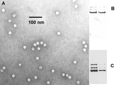
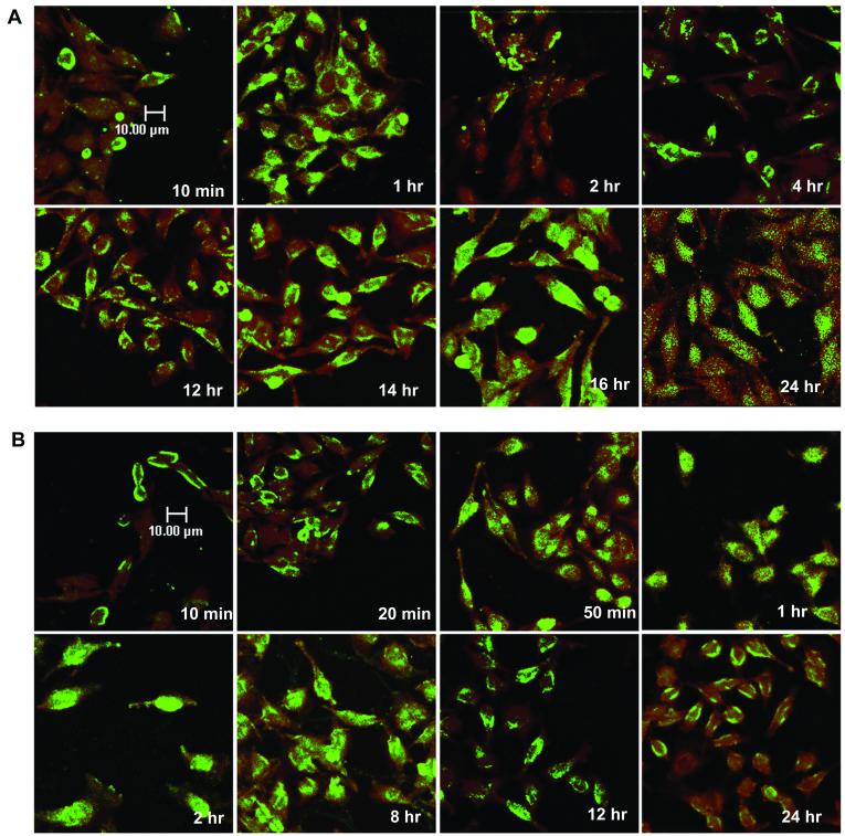
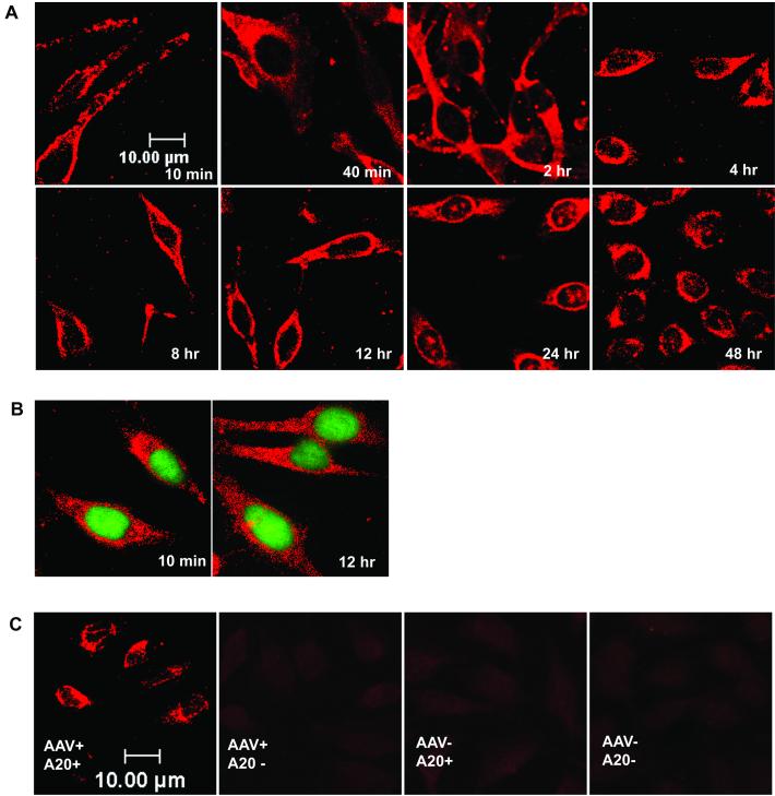
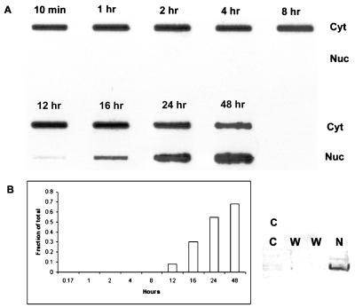
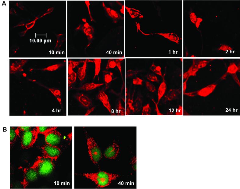
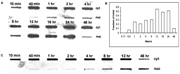
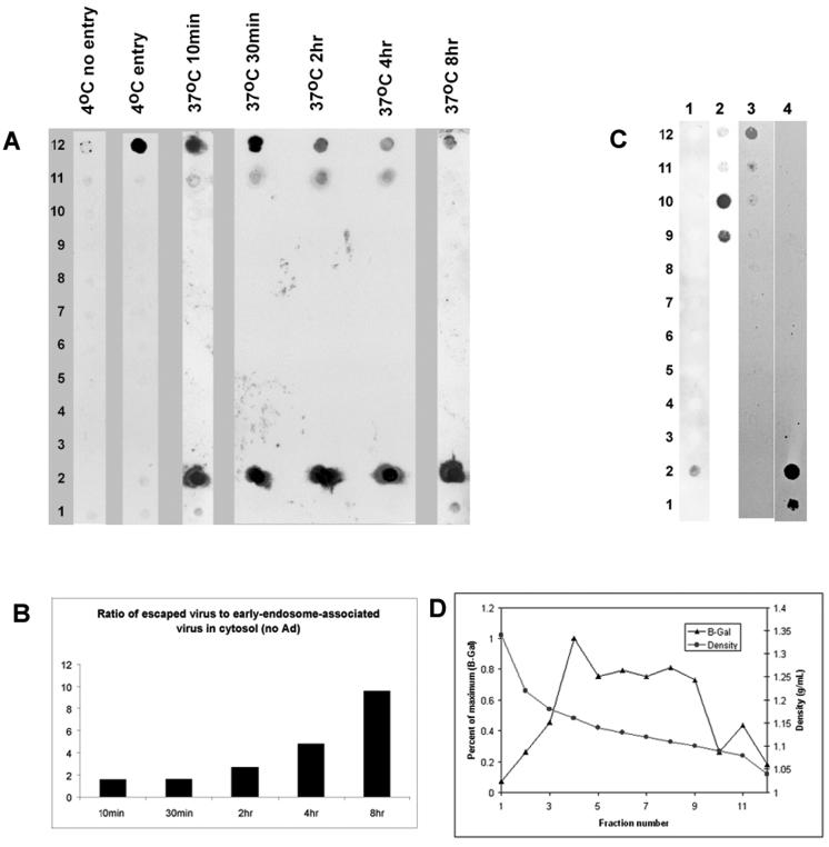
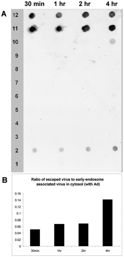
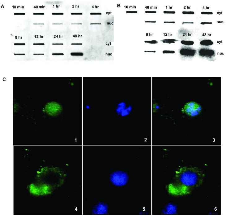
We examined cytoplasmic trafficking and nuclear translocation of adeno-associated virus type 2 (AAV) by using Alexa Fluor 488-conjugated wild-type AAV, A20 monoclonal antibody immunocytochemistry, and subcellular fractionation techniques followed by DNA hybridization. Our results indicated that in the absence of adenovirus (Ad), AAV enters the cell rapidly and escapes from early endosomes with a t(1/2) of about 10 min postinfection. Cytoplasmically distributed AAV accumulated around the nucleus and persisted perinuclearly for 16 to 24 h. Viral uncoating occurred before or during nuclear entry beginning about 12 h postinfection, when viral protein and DNA were readily detected in the nucleus. Few, if any, intact AAV capsids were found in the nucleus. In the presence of Ad, however, cytoplasmic AAV quickly translocated into the nucleus as intact particles as early as 40 min after coinfection, and this facilitated nuclear translocation of AAV was not blocked by the nuclear pore complex inhibitor thapsigargan. The rapid nuclear translocation of intact AAV capsids in the presence of Ad suggested that one or more Ad capsid proteins might be altering trafficking. Indeed, coinfection with empty Ad capsids also resulted in the appearance of AAV DNA in nuclei within 40 min. Escape from early endosomes did not seem to be affected by Ad coinfection.


Figures

FIG. 1.
FIG. 1.
Characterization of AAV preparations. (A) Electron micrograph of wild-type AAV used in this study. Particles with a black dot in the center are likely to be empty capsids. (B and C). Coomassie blue staining (B) and Western blotting (C) of wild-type AAV capsid preparations that had been fractionated on SDS-acrylamide gels. In each gel, the left lane contained 1011 particles and the right lane contained 1010 particles. The Western blot was detected with anti-capsid B1 monoclonal antibody (31, 47).
FIG. 2.
FIG. 2.
Time course of AAV infection using fluorescently labeled virus. (A) HeLa cells were infected with Alexa Fluor 488-labeled AAV at a MOI of 10,000 particles per cell for 10 min at 37°C, washed to remove unadsorbed virus, and incubated at 37°C. Cell samples were taken at the indicated time points, stained with Syto 64 red fluorescent dye, and observed on a confocal microscope as described in Materials and Methods. (B) HeLa cells were incubated with Alexa Fluor 488 fluorescent dye only, and cell samples were taken at the indicated time points.
FIG. 3.
FIG. 3.
Time course of AAV infection using A20 immunocytochemistry. (A) HeLa cells were infected with wild-type AAV at a MOI of 10,000 particles per cell for 10 min at 37°C, washed to remove unadsorbed virus, and incubated at 37°C. Samples were taken at the indicated time points, and intact viral particles were detected with A20 monoclonal antibody as described in Materials and Methods. (B) HeLa cells were infected with wild-type AAV as in panel A and incubated at 37°C for the indicated times. Samples were then stained with A20 antibody as in panel A and Syto X green, which stains nucleic acid in the nucleus. (C) Control A20 immunocytochemistry experiments. HeLa cells were infected with wild-type AAV or mock infected and then stained with A20 monoclonal antibody and Cy3-labeled goat anti-mouse Immunoglobulin G or just the secondary antibody.
FIG. 4.
FIG. 4.
Time course of AAV infection detected by DNA hybridization. (A) HeLa cells were infected with AAV as described in the legend to Fig. 3 and fractionated at the indicated time points into cytoplasmic (Cyt) and nuclear (Nuc) fractions. Viral DNA in each fraction was isolated and detected by slot blot hybridization. (B) The density of the DNA signals in the nucleus and cytoplasm was measured as described in Materials and Methods, and the fraction of the total DNA in the nucleus at each time point was calculated. (C) Western blotting of the cytoplasmic and nuclear fractions using anti-histone H3 antibody. C, cytoplasmic fraction; W, nuclear washes; N, nuclear fraction.
FIG. 5.
FIG. 5.
Time course of AAV infection in the presence of Ad coinfection. HeLa cells were infected with wild-type AAV (MOI = 10,000 DNA-containing particles) and Ad5 (MOI = 10 PFU) for 10 min at 37°C and then washed to remove unabsorbed virus. The cells were then incubated with Ad5 (MOI = 10 PFU) for the duration of the experiment. Cell samples were taken at the indicated times postinfection and stained with A20 monoclonal antibody to visualize intact AAV particles as described in Materials and Methods. (B) HeLa cells were infected with wild-type AAV as in panel A and incubated at 37°C for the indicated time. Samples were then stained with A20 antibody as in panel A and Syto X green, which stains nucleic acid in the nucleus.
FIG. 6.
FIG. 6.
Time course of viral DNA distribution in the presence of Ad coinfection. (A) HeLa cells were infected with AAV and Ad as described in the legend to Fig. 5. At the indicated times, cell samples were fractionated into nuclear (nuc) and cytoplasmic (cyt) fractions and viral DNA from each fraction was detected by slot blot hybridization. (B) The density of the DNA signals in the nucleus and cytoplasm was measured as described in Materials and Methods, and the fraction of the total DNA in the nucleus at each time point was calculated. (C) HeLa cells were infected with AAV and Ad as described in the Legend to Fig. 5, except that empty Ad5 ts369 particles (MOI = 1,000 particles) were used in place of wild-type Ad5. Cell samples were fractionated at the indicated times into nuclear and cytoplasmic fractions, and viral DNA was detected by slot blot hybridization.
FIG. 7.
FIG. 7.
Subcellular fractionation of AAV-infected cells. (A) HeLa cells were infected with AAV as described below, and the nucleus-free cytoplasmic fraction was fractionated on a continuous iodixanol gradient as described in Materials and Methods. Viral DNA was isolated from each fraction and detected by dot blot hybridization as described in Materials and Methods. Lanes: 4°C no entry, cells and virus were precooled to 4°C, and then cell samples were incubated at 4°C for 1 h; 4°C entry, AAV was added directly to warm cells, and then cells were incubated at 4°C for 1 h; 37°C 10min to 37°C 8hr; Cells were infected with AAV at 37°C and harvested at the indicated time. (B) The intensities of the free cytoplasmic virus (fraction 2) and early-endosome-associated virus signals (fractions 11 and 12) were scanned and plotted as the ratio of escaped to endosome-associated virus. (C) Continuous iodixanol gradients were loaded with AAV (lane 1), viral DNA isolated from purified AAV (lane 2), endosomally trapped biotinylated human transferrin (see Materials and Methods) (lane 3), or free biotinylated transferrin (lane 4). (D) The distribution profile of β-galactosidase (β-Gal) activity, a marker for dense endocytic vesicles, was determined on a continuous iodixanol density gradient. Density was determined by weight.
FIG. 8.
FIG. 8.
Subcellular fractionation of cells coinfected with AAV and Ad. (A) Nucleus-free cytoplasmic extracts of AAV- plus Ad5-infected HeLa cells were taken at the indicated time points postinfection and subjected to continuous iodixanol gradient centrifugation as in Fig. 7. Viral DNA was detected by dot blot hybridization. (B) The intensities of the free cytoplasmic virus (fraction 2) and early-endosome-associated virus signals (fractions 11 and 12) were scanned and plotted as the ratio of escaped to endosome-associated virus.
FIG. 9.
FIG. 9.
Effect of the NPC inhibitor on AAV nuclear translocation in the presence of Ad. (A) Thapsigargin-treated cells were infected with AAV and Ad in Ca2+-free S-MEM. Cell samples were taken at the indicated time points and fractionated into cytoplasmic (cyt) and nuclear (nuc) fractions. Viral DNA from each fraction was detected by slot blot hybridization. (B) Cells were treated the same way as in panel A but maintained in normal calcium-containing DMEM. (C) Thapsigargin inhibition of the nuclear movement of 10-kDa dextran. Alexa Fluor 488-conjugated 10-kDa dextran was introduced into HeLa cells as described in Materials and Methods. Nuclei were stained with DAPI, and samples were observed under deconvolution microscope. Panels: 1 and 4, labeled dextran; 2 and 5, DAPI stain; 3 and 6, merged. Panels 1 to 3 were not treated with thapsigargin; panels 4 to 6 were treated with thapsigargin.

Similar articles

Cited by

References

    1. Agbandje, M., T. C. Jenkins, R. McKenna, A. P. Reszka, and S. Neidle. 1992. Anthracene-9,10-diones as potential anticancer agents. Synthesis, DNA-binding, and biological studies on a series of 2,6-disubstituted derivatives. J. Med. Chem. 35:1418-1429. - PubMed
    1. Bantel-Schaal, U., B. Hub, and J. Kartenbeck. 2002. Endocytosis of adeno-associated virus type 5 leads to accumulation of virus particles in the Golgi compartment. J. Virol. 76:2340-2349. - PMC - PubMed
    1. Bartlett, J. S., and R. J. Samulski. 1998. Fluorescent viral vectors: a new technique for the pharmacological analysis of gene therapy. Nat. Med. 4:635-637. - PubMed
    1. Bartlett, J. S., R. Wilcher, and R. J. Samulski. 2000. Infectious entry pathway of adeno-associated virus and adeno-associated virus vectors. J. Virol. 74:2777-2785. - PMC - PubMed
    1. Bruder, J. T., and I. Kovesdi. 1997. Adenovirus infection stimulates the Raf/MAPK signaling pathway and induces interleukin-8 expression. J. Virol. 71:398-404. - PMC - PubMed

Publication types

LinkOut - more resources