. 2002 Nov;76(22):11518-29.
doi: 10.1128/jvi.76.22.11518-11529.2002.

Generation of a replication-competent, propagation-deficient virus vector based on the transmissible gastroenteritis coronavirus genome

Affiliations

Generation of a replication-competent, propagation-deficient virus vector based on the transmissible gastroenteritis coronavirus genome

Javier Ortego et al. J Virol. 2002 Nov.

Abstract

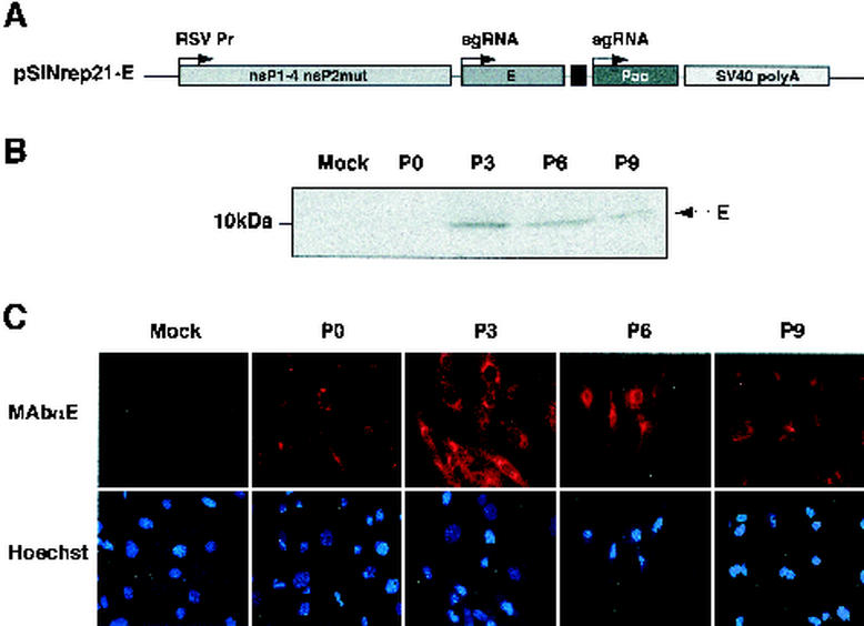
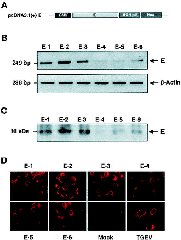
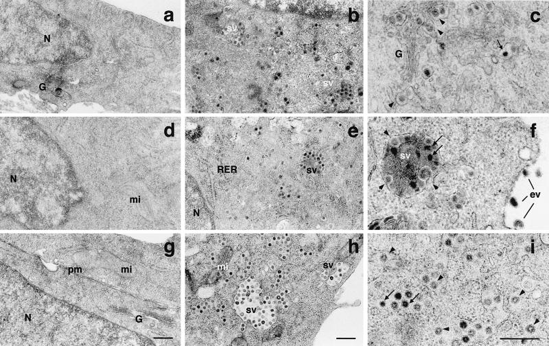
Replication-competent propagation-deficient virus vectors based on the transmissible gastroenteritis coronavirus (TGEV) genome that are deficient in the essential E gene have been developed by complementation within E(+) packaging cell lines. Cell lines expressing the TGEV E protein were established using the noncytopathic Sindbis virus replicon pSINrep21. In addition, cell lines stably expressing the E gene under the CMV promoter have been developed. The Sindbis replicon vector and the ectopic TGEV E protein did not interfere with the rescue of infectious TGEV from full-length cDNA. Recombinant TGEV deficient in the nonessential 3a and 3b genes and the essential E gene (rTGEV-Delta3abDeltaE) was successfully rescued in these cell lines. rTGEV-Delta3abDeltaE reached high titers (10(7) PFU/ml) in baby hamster kidney cells expressing porcine aminopeptidase N (BHK-pAPN), the cellular receptor for TGEV, using Sindbis replicon and reached titers up to 5 x 10(5) PFU/ml in cells stably expressing E protein under the control of the CMV promoter. The virus titers were proportional to the E protein expression level. The rTGEV-Delta3abDeltaE virions produced in the packaging cell line showed the same morphology and stability under different pHs and temperatures as virus derived from the full-length rTGEV genome, although a delay in virus assembly was observed by electron microscopy and virus titration in the complementation system in relation to the wild-type virus. These viruses were stably grown for >10 passages in the E(+) packaging cell lines. The availability of packaging cell lines will significantly facilitate the production of safe TGEV-derived vectors for vaccination and possibly gene therapy.


Figures

FIG. 1.
FIG. 1.
Expression of the E protein by a Sindbis virus replicon vector in BHK-pAPN cell lines. (A) Scheme of the Sindbis virus replicon construct encoding the TGEV E gene. RSV Pr, Rous sarcoma virus promoter; sgRNA, subgenomic RNA; Pac, puromycin resistance gene; SV40 poly A, transcription-termination polyadenylation signal from simian virus 40. nsP1-4, nonstructural proteins 1 to 4; nsP2mut, mutant nonstructural protein 2. (B) Western blot analysis of E protein expression at different cell passages (P) of BHK-pAPN transformed with the Sindbis virus replicon expressing the E protein. The position of the E protein is indicated by an arrow. Mock, untransfected. (C) Immunofluorescence microscopy using an E protein-specific MAb at different cell passages of BHK-pAPN transformed with the Sindbis virus replicon expressing the E protein.
FIG. 2.
FIG. 2.
TGEV growth kinetics and rescue from cDNA in BHK-pAPN cells expressing E protein using Sindbis replicon. (A) Growth kinetics of TGEV in BHK-pAPN cells expressing TGEV E protein. Virus titers were determined by plaque assay. (B) Titration of infectious TGEV recovered from a transfected cDNA in BHK-pAPN cells expressing TGEV E protein by Sindbis virus replicon. E, BHK-pAPN cells expressing the TGEV E protein. SIN, cells transfected with pSINrep21 plasmid. C, mock-transfected cells. Mean values from four experiments are represented, and standard deviations are shown as error bars.
FIG. 3.
FIG. 3.
Rescue of rTGEV-Δ3abΔE from cDNA in cells expressing E protein using Sindbis replicon. (A) Scheme of the pBAC-TGEV-(MluI-FseI)-FL plasmid, containing MluI and FseI restriction sites as indicated above the bar, and the pBAC-TGEV-Δ3abΔE plasmid, with 3ab and E genes deleted as indicated above the bar. CMV, CMV promoter; HDV, hepatitis delta virus ribozyme; BGH, bovine growth hormone termination and polyadenylation (An) sequence. (B) Rescue of recombinant TGEV containing the MluI and FseI restriction sites (Vwt) by transfecting BHK-pAPN cells (CE) and BHK-pAPN cells expressing the TGEV E protein (CE+). VΔE, rTGEV-Δ3abΔE. (C) Detection of virus mRNAs for the E and S genes by RT-PCR analysis in BHK-pAPN cells expressing the TGEV E protein and infected with recombinant viruses rTGEV-(MluI-FseI)-FL (Vwt) and rTGEV-Δ3abΔE (VΔE). V, uninfected cells; P1, P2, and P3, passages 1, 2, and 3, respectively. (D) Growth kinetics of recombinant viruses rTGEV-(MluI-FseI)-FL (Vwt) and rTGEV-Δ3abΔE (VΔE) in BHK-pAPN cells expressing TGEV E protein (CE+). Mean values from four experiments are represented, and standard deviations are shown as error bars.
FIG. 4.
FIG. 4.
Characterization of virus mRNAs and structural proteins from recombinant viruses rTGEV-(MluI-FseI)-FL and rTGEV-Δ3abΔE. (A) Northern blot analysis of cytoplasmic RNAs from BHK-pAPN cells (BHK-pAPN-E) and BHK-pAPN cells expressing the E protein (BHK-pAPN-E+) infected with rTGEV-(MluI-FseI)-FL (wt) or with rTGEV-Δ3abΔE (Δ3abΔE). Δ3abΔE × 10 was exposed 10-fold longer than Δ3abΔE. C, uninfected BHK-pAPN cells. The subgenomic RNAs are indicated by arrows and named according to the coded gene. (B) Western blot analysis using S, N, M, or E protein-specific MAb (see Materials and Methods) of cell lysates from BHK-pAPN cells (E) and BHK-pAPN-E+ cells (E+) infected with rTGEV-(MluI-FseI)-FL (wt) or rTGEV- Δ3abΔE (ΔE). The analyses were carried out at 12 and 24 h postinfection, as indicated. V, uninfected cells.
FIG. 5.
FIG. 5.
Expression of the TGEV E protein in LLC-PK1 cell lines stably transformed with E protein expression plasmids. (A) Scheme of the E gene expression cassette in the pcDNA3.1(+) plasmid. CMV, CMV promoter; BGH pA, bovine growth hormone transcription stop signal and polyadenylation sequence; Neo, gene coding for neomycin resistance. (B) Detection of the mRNA for the E protein and β-actin by semiquantitative RT-PCR analysis in six cell lines (E-1, E-2, E-3, E-4, E-5, and E-6) stably transformed with the E gene. DNA amplification fragments were resolved by agarose gel electrophoresis and stained with ethidium bromide. (C) Western blot analysis of cell lysates from the six LLC-PK1 cell lines stably expressing the E protein, using an E-protein specific MAb. The position of the E protein band is indicated by an arrow. (D) Detection of E protein expression by immunofluorescence microscopy, using an E protein-specific MAb, in the six LLC-PK1 cell lines stably expressing the TGEV E protein. Mock, untransfected LLC-PK1 cells; TGEV, TGEV-infected LLC-PK1 cells.
FIG. 6.
FIG. 6.
Effects of E protein expression levels in rescue of recombinant viruses rTGEV-(MluI-FseI)-FL and rTGEV- Δ3abΔE. (A and B) Growth kinetics of rTGEV-(MluI-FseI)-FL (A) and rTGEV-Δ3abΔE (B) virus in LLC-PK1 cell lines expressing different levels of E protein (Fig. 5). C, untransformed LLC-PK1 cells; E-1 to E-6, cell lines expressing different amounts of E protein. (C) Relation between virus (rTGEV-Δ3abΔE) titers and E protein expression levels in transformed LLC-PK1 cells. Mean values from four experiments are represented, with standard deviations shown as error bars.
FIG. 7.
FIG. 7.
Effects of temperature and pH changes on rTGEV-(MluI-FseI)-FL and rTGEV-Δ3abΔE virus stabilities. Titrations of virus supernatants from cells infected with recombinant viruses rTGEV-(MluI-FseI)-FL and rTGEV-Δ3abΔE after incubation for 30 min at the indicated temperature (A) or pH (B) are shown. Mean values from four experiments are represented, with standard deviations shown as error bars. The abbreviations are defined in the legend to Fig. 3B.
FIG. 8.
FIG. 8.
Characterization of the assembly of recombinant viruses rTGEV-(MluI-FseI)-FL and rTGEV-Δ3abΔE in BHK-pAPN cells by electron microscopy. Electron microscopy sections of BHK-pAPN cells that do not express (a to c) or express (d to i) the TGEV E protein, infected with rTGEV-(MluI-FseI)-FL (b, c, e, and f) or rTGEV-Δ3abΔE (h and i). Large annular viruses are indicated with arrowheads, and small and dense viruses are indicated with arrows. sv, secretory vesicle; ev, extracellular virus; N, nucleus; G, Golgi complex; mi, mitochondria; RER, rough endoplasmic reticulum; pm, plasma membrane. Bars, 400 nm.
FIG. 9.
FIG. 9.
Quantification of large and small virions in BHK-pAPN cells expressing the TGEV E protein and infected with recombinant virus rTGEV-(MluI-FseI)-FL or rTGEV-Δ3abΔE. Shown are the percentages of intracellular virions with a large (68- to 80-nm diameter) (top) or small (50- to 65-nm) (bottom) core present at 18 and 24 h postinfection. A total of 500 virions were included in the measurements.

Similar articles

Cited by

References

    1. Almazán, F., J. M. González, Z. Pénzes, A. Izeta, E. Calvo, J. Plana-Durán, and L. Enjuanes. 2000. Engineering the largest RNA virus genome as an infectious bacterial artificial chromosome. Proc. Natl. Acad. Sci. USA 97:5516-5521. - PMC - PubMed
    1. Alonso, S., A. Izeta, I. Sola, and L. Enjuanes. 2002. Transcription regulatory sequences and mRNA expression levels in the coronavirus transmissible gastroenteritis virus. J. Virol. 76:1293-1308. - PMC - PubMed
    1. An, S., C. J. Chen, X. Yu, J. L. Leibowitz, and S. Makino. 1999. Induction of apoptosis in murine coronavirus-infected cultured cells and demonstration of E protein as an apoptosis inducer. J. Virol. 73:7853-7859. - PMC - PubMed
    1. Baric, R. S., K. S. Fu, M. C. Schaad, and S. A. Stohlman. 1990. Establishing a genetic recombination map for MHV-A59 complementation groups. Virology 177:646. - PMC - PubMed
    1. Baudoux, P., C. Carrat, L. Besnardeau, B. Charley, and H. Laude. 1998. Coronavirus pseudoparticles formed with recombinant M and E proteins induce alpha interferon synthesis by leukocytes. J. Virol. 72:8636-8643. - PMC - PubMed

Publication types

Substances

LinkOut - more resources