. 2002 Nov;76(22):11570-83.
doi: 10.1128/jvi.76.22.11570-11583.2002.

Interleukin-8 and growth-regulated oncogene alpha mediate angiogenesis in Kaposi's sarcoma

Affiliations

Interleukin-8 and growth-regulated oncogene alpha mediate angiogenesis in Kaposi's sarcoma

Brian R Lane et al. J Virol. 2002 Nov.

Abstract

The development of the complex neoplasm Kaposi's sarcoma is dependent on infection with the Kaposi's sarcoma-associated herpesvirus (KSHV) and appears to be greatly enhanced by cytokines and human immunodeficiency virus type 1 (HIV-1) Tat. Interleukin-8 (IL-8) and growth-regulated oncogene alpha (GRO-alpha) are chemokines involved in chemoattraction, neovascularization, and stimulation of HIV-1 replication. We have previously demonstrated that production of GRO-alpha is stimulated by exposure of monocyte-derived macrophages (MDM) to HIV-1. Here we show that exposure of MDM to HIV-1, viral Tat, or viral gp120 leads to a substantial increase in IL-8 production. We also demonstrate that IL-8 and GRO-alpha are induced by KSHV infection of endothelial cells and are crucial to the angiogenic phenotype developed by KSHV-infected endothelial cells in cell culture and upon implantation into SCID mice. Thus, the three known etiological factors in Kaposi's sarcoma pathogenesis-KSHV, HIV-1 Tat, and cellular growth factors-might be linked, in part, through induction of IL-8 and GRO-alpha.


Figures
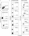
FIG. 1.
FIG. 1.
Intracellular IL-8 is present in the monocytic subpopulation of PBMC exposed to HIV-1. PBMC were treated with monensin (control) along with HIV-1BaL, HIV-1BRU, or TNF-α (100 ng/ml). PBMC were harvested after 6 h and analyzed for intracellular IL-8 protein by flow cytometry. (A) Lymphocyte and monocyte subpopulations were gated according to the pattern of forward scatter and side scatter. (B) Background staining in the lymphocytes and monocytes was determined by incubation with phycoerythrin-mouse IgG1 isotype control (mouse IgG-PE) and FITC-mouse IgG2a isotype control (mouse IgG-FITC). The histograms show fluorescence intensity on logarithmic scales along the x axis (FITC) and y axis (phycoerythrin). The percentage of FITC-positive cells is indicated for both the double-positive and single-positive quadrants. (C) Lymphocytes and monocytes were stained with an FITC-conjugated mouse anti-human IL-8 antibody (IL-8-FITC) and either phycoerythrin-conjugated mouse anti-human CD4 (CD4-PE) or phycoerythrin-conjugated mouse anti-human CD14 (CD14-PE), respectively. The percentage of CD4+ and CD14+ cells staining positive for IL-8 in each condition is indicated. Data shown are representative of four independent experiments.
FIG. 2.
FIG. 2.
HIV-1 and the viral proteins Tat and gp120 stimulate IL-8 production by MDM. (A) MDM were left untreated (control) or treated with either HIV-1 Tat (10 ng/ml; from HIV-1HXB2), R5 gp120 (1 μg/ml; from HIV-1CM235), HIV-1BaL, X4 gp120 (1 μg/ml; from HIV-1MN), or HIV-1BRU. Supernatants were collected from cultures in triplicate after 24 to 48 h and tested for IL-8 by ELISA. A box plot of the amounts of IL-8 for each treatment group is shown. Each point represents a single experiment, with the center horizontal line marking the median of the sample. The length of each box shows the range within which the central 50% of the values fell (Hspread), with the box edges (hinges) at the first and third quartiles. The whiskers show the range of values that fell within 1.5 Hspreads of the hinges. Values outside the whiskers are plotted with asterisks. Also indicated for each treatment condition are the mean amount of IL-8 produced, the number of experiments performed (n), and a statistical measure of the difference in the amount of IL-8 relative to the untreated controls with the Wilcoxon signed ranks test for nonparametric data (P value). (B) Total cellular RNA was extracted from control MDM (−) and MDM exposed to HIV-1BRU (+) after 2 days. RNA from two different donors was then analyzed for IL-8 gene expression by Northern (RNA) blot analysis with an IL-8-specific probe.
FIG. 3.
FIG. 3.
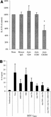
HIV-1 gp120 and Tat both induce IL-8 production by MDM exposed to X4 HIV. (A) MDM were treated with mouse IgG, anti-CD4, anti-CCR5, or anti-CXCR4 (each at 20 μg/ml) and then infected with HIV-1BRU. Supernatants were collected 1 or 2 days after infection and analyzed for IL-8 by ELISA. Data shown are the means ± standard errors from five independent experiments. The reduction in IL-8 production due to anti-CXCR4 was significant (P = 0.04) relative to that of the mouse IgG control by Student's t test. (B) MDM were treated with the CXCR4 inhibitor AMD3100 (5 μg/ml), normal rabbit serum (NRS, 1:200), anti-Tat (1:200), or anti-TNF-α (1:200) as indicated, alone or in combination, just prior to exposure to HIV-1BRU and every 3 days thereafter. Supernatants were collected 8 days after infection and analyzed for IL-8 by ELISA. Data shown are the means ± standard errors from three independent experiments.
FIG. 4.
FIG. 4.
Heat inactivation inhibits the stimulation of IL-8 production by HIV-1 in MDM. HIV-1BaL or HIV-1BRU was either left untreated, heated at 56°C for 30 min, or heated at 100°C for 10 min and then used to infect MDM from two different donors. Supernatants were collected 40 h after infection and analyzed for IL-8 by ELISA. IL-8 production is presented as a percentage of the amount produced by MDM infected with active HIV-1 by the following formula: [(IL-8 in heat-inactivated HIV-infected MDM − IL-8 in uninfected MDM)/(IL-8 in active HIV-infected MDM − IL-8 in uninfected MDM)] × 100. In these two experiments, the mean amount of IL-8 induced by HIV-1BaL was 206 ng/ml, and that induced by HIV-1BRU was 422 ng/ml.
FIG. 5.
FIG. 5.
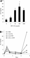
HIV-1 Tat and TNF-α stimulate IL-8 production by MDM. (A) Dose-response curve for treatment of MDM with recombinant HIV-1 Tat (1 to 1,000 ng/ml). ELISA data are plotted as the mean amount of IL-8 present in 24-h supernatants from multiple wells from two experiments (± standard deviation). Statistical significance (*, P < 0.05) was determined with a paired-sample t test. (B) MDM were treated with normal rabbit serum (NRS, 1:200), anti-Tat (1:200), or anti-TNF-α (1:200) as indicated, alone or in combination, just prior to exposure to HIV-1BaL and every 3 days thereafter. Supernatants were collected 1, 2, 4, 6, and 8 days after infection and analyzed for IL-8 by ELISA. Data shown are representative of three independent experiments.
FIG. 6.
FIG. 6.
The KSHV cyclin D gene is detected in KSHV-infected HDMEC by PCR after multiple passages in culture. After 12 passages, total DNA present in the cellular lysates was purified from uninfected HDMEC (lane 2), HDMEC treated with unstimulated BCBL-1 cell lysate (mock-infected, lane 3), and HDMEC treated with 48-h PMA-stimulated BCBL-1 cell lysate (KSHV-infected, lane 4). DNA from PMA-stimulated BCBL-1 cells (lane 5) served as a positive control. PCR analysis demonstrated the presence of the viral cyclin D gene in the BCBL-1 cells and in KSHV-infected HDMEC. HindIII-digested DNA markers (lane 1) are shown for size comparison.
FIG. 7.
FIG. 7.
KSHV infection of HDMEC induces the release of the angiogenic factors IL-8, GRO-α, and VEGF. HDMEC (80 to 95% confluent) were left uninfected (control) or infected with KSHV and maintained in culture for one to four passages. Fresh medium was added to the KSHV-infected HDMEC and collected 24 h later and analyzed by ELISA for IL-8 (A), GRO-α (B), or VEGF (C). Data shown in panels A and B are the means from four experiments (± standard deviation), and those in panel C are from a representative experiment.
FIG. 8.
FIG. 8.
IL-8 and GRO-α are major components of the angiogenic activity present in the conditioned medium of KHSV-infected HDMEC. Conditioned medium derived from uninfected or KSHV-infected HDMEC, with or without neutralizing antibodies to IL-8, GRO-α, or VEGF, was assayed for the ability to stimulate the chemotaxis of endothelial cells. Specific migration was determined after subtracting background migration (unstimulated control). Values are reported as a percentage (± standard error of the mean) of the migration induced by recombinant IL-8.
FIG. 9.
FIG. 9.
KSHV-infected HDMEC stimulate IL-8-dependent angiogenesis in SCID mice. Cells (106) were passaged five times (to avoid confounding effects from contaminants in the original inoculum of KSHV) before being seeded into a PLA sponge. (A) HDMEC (left panel) and KSHV-infected HDMEC (right panel) were seeded into a PLA sponge and implanted in duplicate into the flanks of SCID mice (n = 4). After 14 days, implanted sponges were removed from the mice, and photographs were taken with a Nikon anatomy scope (magnification, ×1.5 to ×2). (B) The sponges were stained with a rat anti-mouse CD31 antibody at high power (400×). (C) Cells were seeded in a PLA sponge with or without neutralizing antibodies to VEGF or IL-8 and implanted into the flanks of SCID mice in duplicate. After 14 days, the PLA sponges were removed, and CD31-positive blood vessels were counted in 10 random fields from three independent sponges for each condition.
FIG. 9.
FIG. 9.
KSHV-infected HDMEC stimulate IL-8-dependent angiogenesis in SCID mice. Cells (106) were passaged five times (to avoid confounding effects from contaminants in the original inoculum of KSHV) before being seeded into a PLA sponge. (A) HDMEC (left panel) and KSHV-infected HDMEC (right panel) were seeded into a PLA sponge and implanted in duplicate into the flanks of SCID mice (n = 4). After 14 days, implanted sponges were removed from the mice, and photographs were taken with a Nikon anatomy scope (magnification, ×1.5 to ×2). (B) The sponges were stained with a rat anti-mouse CD31 antibody at high power (400×). (C) Cells were seeded in a PLA sponge with or without neutralizing antibodies to VEGF or IL-8 and implanted into the flanks of SCID mice in duplicate. After 14 days, the PLA sponges were removed, and CD31-positive blood vessels were counted in 10 random fields from three independent sponges for each condition.

Similar articles

Cited by

References

    1. Alkhatib, G., C. Combadiere, C. C. Broder, Y. Feng, P. E. Kennedy, P. M. Murphy, and E. A. Berger. 1996. CC CKR5: a RANTES, MIP-1alpha, MIP-1beta receptor as a fusion cofactor for macrophage-tropic HIV-1. Science 272:1955-1958. - PubMed
    1. Ariyoshi, K., M. Schim van der Loeff, P. Cook, D. Whitby, T. Corrah, S. Jaffar, F. Cham, S. Sabally, D. O'Donovan, R. A. Weiss, T. F. Schulz, and H. Whittle. 1998. Kaposi's sarcoma in the Gambia, West Africa, is less frequent in human immunodeficiency virus type 2 than in human immunodeficiency virus type 1 infection despite a high prevalence of human herpesvirus 8. J. Hum. Virol. 1:193-199. - PubMed
    1. Baggiolini, M., B. Dewald, and B. Moser. 1994. Interleukin-8 and related chemotactic cytokines—CXC and CC chemokines. Adv. Immunol. 55:97-179. - PubMed
    1. Barillari, G., R. Gendelman, R. C. Gallo, and B. Ensoli. 1993. The Tat protein of human immunodeficiency virus type 1, a growth factor for AIDS Kaposi sarcoma and cytokine-activated vascular cells, induces adhesion of the same cell types by using integrin receptors recognizing the RGD amino acid sequence. Proc. Natl. Acad. Sci. USA 90:7941-7945. - PMC - PubMed
    1. Beral, V., T. A. Peterman, R. L. Berkelman, and H. W. Jaffe. 1990. Kaposi's sarcoma among persons with AIDS: a sexually transmitted infection? Lancet 335:123-128. - PubMed

Publication types

MeSH terms

LinkOut - more resources