. 2002 Nov;76(22):11729-37.
doi: 10.1128/jvi.76.22.11729-11737.2002.

Characterization of a putative alpha-helix across the capsid-SP1 boundary that is critical for the multimerization of human immunodeficiency virus type 1 gag

Affiliations

Characterization of a putative alpha-helix across the capsid-SP1 boundary that is critical for the multimerization of human immunodeficiency virus type 1 gag

Chen Liang et al. J Virol. 2002 Nov.

Abstract

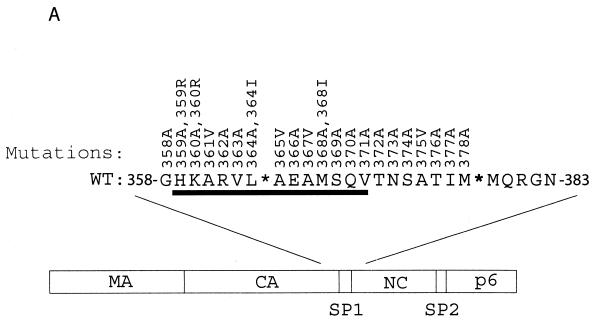
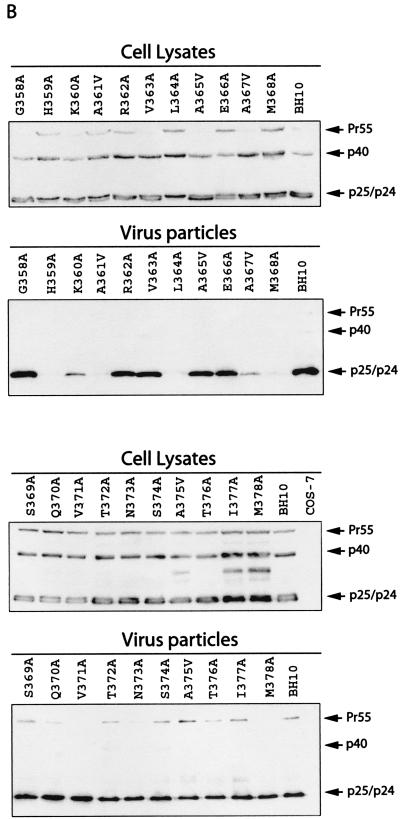
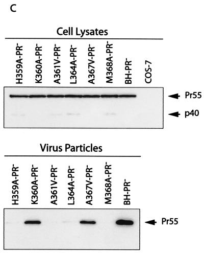
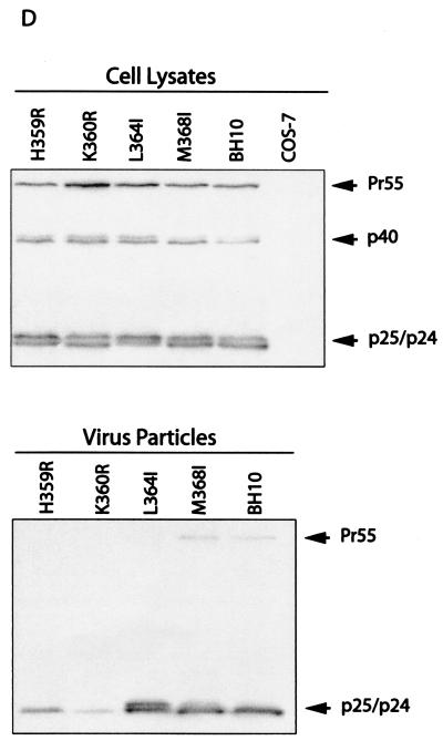
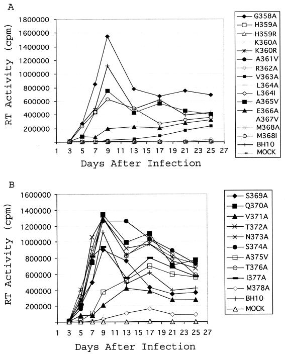
A 14-amino-acid spacer peptide termed SP1 that separates the capsid (CA) and nucleocapsid (NC) sequences plays an active role in the assembly of human immunodeficiency virus type 1. This activity of SP1 involves its amino-terminal residues that, together with adjacent CA residues, constitute a putative alpha-helical structure spanning Gag residues from positions 359 to 371. In this study, we have determined that the virus assembly determinants within this putative alpha-helix were residues H359, K360, A361, L364, A367, and M368, of which K360 and A367 contribute to virus production to lesser extents. Notably, changes of the two basic amino acids H359 and K360 to arginine (R) impaired virus production, whereas mutations L364I and M368I, in contrast to L364A and M368A, generated near-wild-type levels of virus particles. This suggests that within Gag complexes, amino acids H359 and K360 are involved in stricter steric interactions than L364 and M368. Since L364 and M368 are separated by four residues and thus presumably located on the same side of the helical surface, they may initiate synergistic hydrophobic interactions to stabilize Gag association. Further analysis in the context of the protease-negative mutation D185H confirmed the key roles of amino acids H359, A361, L364, and M368 in virus assembly. Importantly, when transfected cells were subjected to Dounce homogenization and the cell lysates were treated by ultracentrifugation at 100,000 x g, Gag molecules containing each of the H359A, A361V, L364A, and M368A mutations were found mainly in the supernatant fraction (S100), whereas approximately 80% of wild-type Gag proteins were found in the pellet. Therefore, these four mutations must have prevented Gag from generating large complexes.


Figures

FIG. 1.
FIG. 1.
Effects of the mutations in SP1 and the carboxy terminus of CA on virus production. (A) Amino acids from positions 358 to 378 were substituted with alanine except that A361, A365, A367, and A375 were changed to valine. Amino acids H359 and K360 were further changed to arginine, and L364 and M368 were changed to isoleucine. These mutations were generated through use of a PCR strategy as described previously (40). The primers employed are listed in Table 1. The underlined residues from positions 359 to 371 may form an α-helix as predicted by the PHD program (1, 41-44). The asterisks denote viral protease cleavage sites. Numbering of amino acids starts from the first residue of Gag. WT, wild type. (B) Analysis of viral proteins expressed in cells or associated with virus particles. Either COS-7 or HeLa cells were transfected with mutated or wild-type DNA constructs. Forty-eight hours after transfection, cells were lysed in NP-40 lysis buffer containing 50 mM Tris-Cl (pH 7.4), 150 mM NaCl, 1% NP-40, 0.02% sodium azide, and protease inhibitor cocktails (Roche, Laval, Quebec, Canada). The lysates were clarified in a Beckman GR-6S centrifuge at 3,000 rpm for 30 min at 4°C and then assessed by Western blotting using monoclonal antibodies (MAbs) against CA (p24) (ID Lab, Inc., London, Ontario, Canada). Mock-transfected cells were also used in Western blots as negative controls. Virus particles in culture fluids were concentrated by ultracentrifugation at 210,000 × g for 1 h and then analyzed by Western blotting using the same antibodies. The positions of Gag precursor Pr55 and its derivatives are shown to the right of the gels. Similar results from Western blots were obtained from transfection of COS-7 and HeLa cells, and results from COS-7 are shown. The quantities of Gag proteins that were associated with either the cell lysates or virus particles were further determined by quantitative ELISA (Vironostika HIV-1 Antigen Microelisa System; Organon Teknika Corporation, Durham, N.C.). The data were used to calculate the efficiency of virus production for each of the mutated and wild-type DNA constructs. The results are summarized in Table 2. (C) Effects of mutations H359A, K360A, A361V, L364A, A367V, and M368A on virus production in the context of a protease-negative virus. These mutated DNA constructs were tested in both COS-7 and HeLa cells. Similar results from Western blots were obtained in both cases, and results from COS-7 cells are shown. The Pr55 precursor is the major form of Gag protein that was detected by Western blotting using MAbs against CA (p24). The quantities of Pr55 associated with cell lysates and virus particles were measured by quantitative ELISA, and the data were used to calculate the efficiency of virus production. The results are summarized in Table 3. (D) Effects of mutations H359R, K360R, L364I, and M368I on virus production. Either COS-7 or HeLa cells were transfected with the mutated or wild-type constructs. Gag proteins associated with the cell lysates or virus particles were assessed by Western blotting using MAbs against CA (p24). Similar results were obtained for both cell lines, and results from COS-7 cells are shown. The efficiency of virus production by the mutated or wild-type constructs was calculated on the basis of quantitative ELISA. The results are summarized in Table 2 FIG. 1—Continued..
FIG. 1.
FIG. 1.
Effects of the mutations in SP1 and the carboxy terminus of CA on virus production. (A) Amino acids from positions 358 to 378 were substituted with alanine except that A361, A365, A367, and A375 were changed to valine. Amino acids H359 and K360 were further changed to arginine, and L364 and M368 were changed to isoleucine. These mutations were generated through use of a PCR strategy as described previously (40). The primers employed are listed in Table 1. The underlined residues from positions 359 to 371 may form an α-helix as predicted by the PHD program (1, 41-44). The asterisks denote viral protease cleavage sites. Numbering of amino acids starts from the first residue of Gag. WT, wild type. (B) Analysis of viral proteins expressed in cells or associated with virus particles. Either COS-7 or HeLa cells were transfected with mutated or wild-type DNA constructs. Forty-eight hours after transfection, cells were lysed in NP-40 lysis buffer containing 50 mM Tris-Cl (pH 7.4), 150 mM NaCl, 1% NP-40, 0.02% sodium azide, and protease inhibitor cocktails (Roche, Laval, Quebec, Canada). The lysates were clarified in a Beckman GR-6S centrifuge at 3,000 rpm for 30 min at 4°C and then assessed by Western blotting using monoclonal antibodies (MAbs) against CA (p24) (ID Lab, Inc., London, Ontario, Canada). Mock-transfected cells were also used in Western blots as negative controls. Virus particles in culture fluids were concentrated by ultracentrifugation at 210,000 × g for 1 h and then analyzed by Western blotting using the same antibodies. The positions of Gag precursor Pr55 and its derivatives are shown to the right of the gels. Similar results from Western blots were obtained from transfection of COS-7 and HeLa cells, and results from COS-7 are shown. The quantities of Gag proteins that were associated with either the cell lysates or virus particles were further determined by quantitative ELISA (Vironostika HIV-1 Antigen Microelisa System; Organon Teknika Corporation, Durham, N.C.). The data were used to calculate the efficiency of virus production for each of the mutated and wild-type DNA constructs. The results are summarized in Table 2. (C) Effects of mutations H359A, K360A, A361V, L364A, A367V, and M368A on virus production in the context of a protease-negative virus. These mutated DNA constructs were tested in both COS-7 and HeLa cells. Similar results from Western blots were obtained in both cases, and results from COS-7 cells are shown. The Pr55 precursor is the major form of Gag protein that was detected by Western blotting using MAbs against CA (p24). The quantities of Pr55 associated with cell lysates and virus particles were measured by quantitative ELISA, and the data were used to calculate the efficiency of virus production. The results are summarized in Table 3. (D) Effects of mutations H359R, K360R, L364I, and M368I on virus production. Either COS-7 or HeLa cells were transfected with the mutated or wild-type constructs. Gag proteins associated with the cell lysates or virus particles were assessed by Western blotting using MAbs against CA (p24). Similar results were obtained for both cell lines, and results from COS-7 cells are shown. The efficiency of virus production by the mutated or wild-type constructs was calculated on the basis of quantitative ELISA. The results are summarized in Table 2 FIG. 1—Continued..
FIG. 2.
FIG. 2.
Analysis of Gag complexes within the cytoplasm by velocity sedimentation. COS-7 cells were transfected with mutated or wild-type DNA constructs containing the protease-negative mutation D185H. Cells were then treated by Dounce homogenization in ice-cold TNE buffer containing protease inhibitor cocktails (Roche). After the supernatants were clarified in a Beckman GR-6S centrifuge at 3,000 rpm for 30 min at 4°C, they were subjected to ultracentrifugation at 100,000 × g for 1 h at 4°C through a 20% sucrose cushion. The supernatants (termed S100) and pelleted materials (termed P100) were analyzed for Gag proteins either by anti-p24 Western blotting or by quantitative anti-p24 ELISA. The relative quantities of Gag proteins in the S100 and P100 fractions for each construct are shown in the bar graph at the bottom of the figure.
FIG. 3.
FIG. 3.
Effects of the various mutations on viral replication. Jurkat cells were transfected with mutated or wild-type viral cDNA. Viral growth was monitored by measuring levels of RT activity in culture fluids at various times. For clarity, the growth curves are presented in two separate graphs (A and B).

Similar articles

Cited by

References

    1. Accola, M. A., S. Hoglund, and H. G. Gottlinger. 1998. A putative alpha-helical structure which overlaps the capsid-p2 boundary in the human immunodeficiency virus type 1 Gag precursor is crucial for viral particle assembly. J. Virol. 72:2072-2078. - PMC - PubMed
    1. Accola, M. A., B. Strack, and H. G. Gottlinger. 2000. Efficient particle production by minimal Gag constructs which retain the carboxy-terminal domain of human immunodeficiency virus type 1 capsid-p2 and a late assembly domain. J. Virol. 74:5395-5402. - PMC - PubMed
    1. Berthet-Colominas, C., S. Monaco, A. Novelli, G. Sibai, F. Mallet, and S. Cusack. 1999. Head-to-tail dimers and interdomain flexibility revealed by the crystal structure of HIV-1 capsid protein (p24) complexed with a monoclonal antibody Fab. EMBO J. 18:1124-1136. - PMC - PubMed
    1. Borsetti, A., A. Ohagen, and H. G. Gottlinger. 1998. The C-terminal half of the human immunodeficiency virus type 1 Gag precursor is sufficient for efficient particle assembly. J. Virol. 72:9313-9317. - PMC - PubMed
    1. Burniston, M. T., A. Cimarelli, J. Colgan, S. P. Curtis, and J. Luban. 1999. Human immunodeficiency virus type 1 Gag polyprotein multimerization requires the nucleocapsid domain and RNA and is promoted by the capsid-dimer interface and the basic region of matrix protein. J. Virol. 73:8527-8540. - PMC - PubMed

Publication types

LinkOut - more resources