. 2002 Oct;13(10):3466-76.
doi: 10.1091/mbc.e02-05-0281.

RNA polymerase III in Cajal bodies and lampbrush chromosomes of the Xenopus oocyte nucleus

Affiliations

RNA polymerase III in Cajal bodies and lampbrush chromosomes of the Xenopus oocyte nucleus

Christine Murphy et al. Mol Biol Cell. 2002 Oct.

Abstract

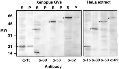
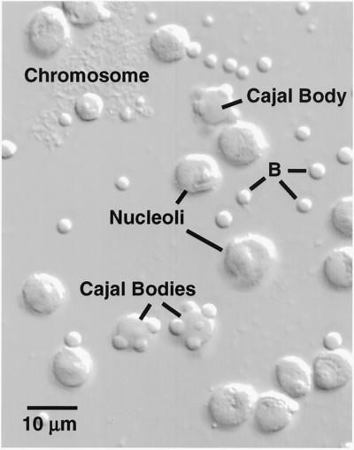
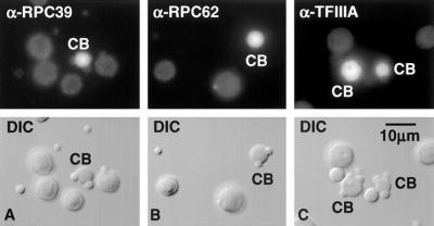
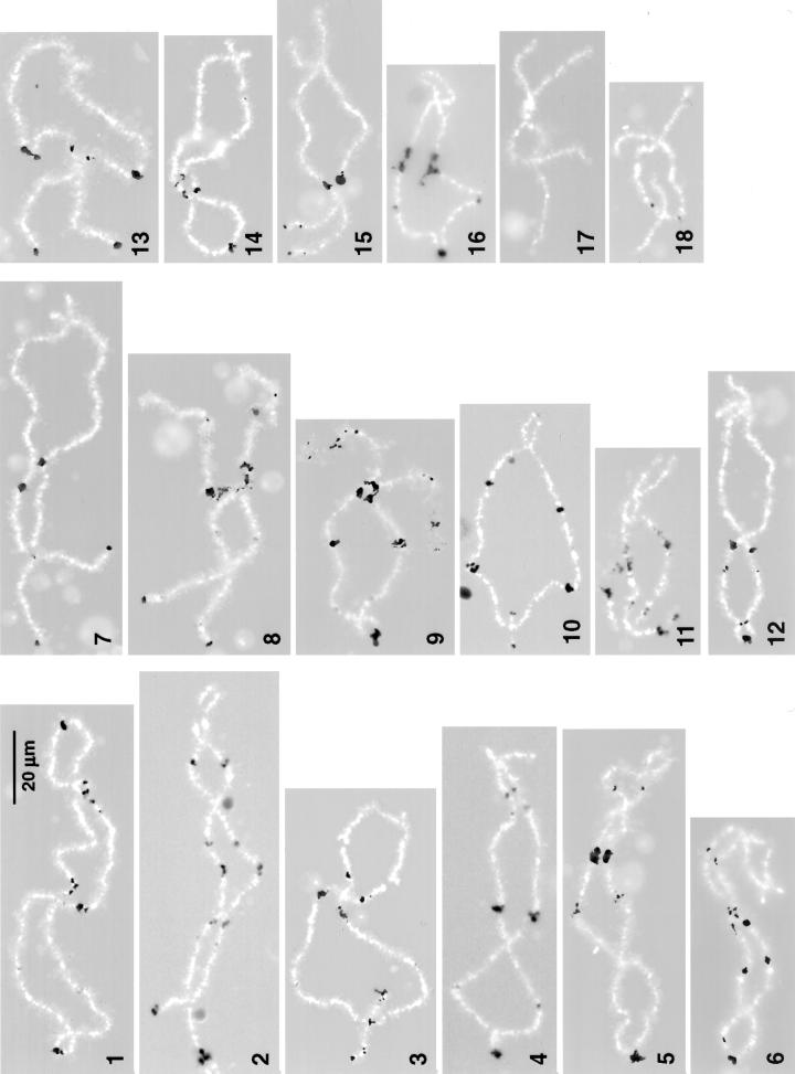
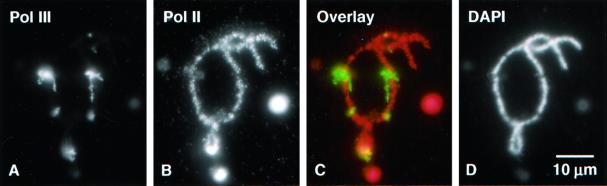
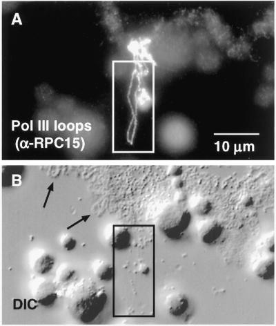
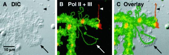
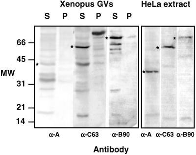
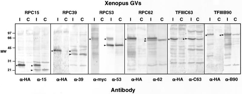
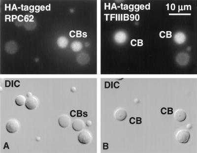
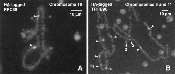
We used immunofluorescence to study the distribution and targeting of RNA polymerase (pol) III subunits and pol III transcription factors in the Xenopus laevis oocyte nucleus. Antibodies against several of these proteins stained Cajal bodies and approximately 90 specific sites on the lampbrush chromosomes. Some of the chromosomal sites had been identified previously by in situ hybridization as the genes for 5S rRNA. The remaining sites presumably encode tRNAs and other pol III transcripts. Pol III sites were often resolvable as loops similar to the much more abundant pol II loops, but without a matrix detectable by phase contrast or differential interference contrast. This morphology is consistent with the transcription of short repeated sequences. Hemagglutinin-tagged transcripts encoding core subunits and transcription factors were injected into the oocyte cytoplasm, and the distribution of newly translated proteins inside the nucleus was monitored by immunostaining. Cajal bodies were preferentially targeted by these proteins, and in some cases the chromosomal sites were also weakly stained. The existence of pol III subunits and pol III transcription factors in Cajal bodies and their targeting to these organelles are consistent with a model of Cajal bodies as sites for preassembly of the nuclear transcription machinery.


Figures

Figure 1
Figure 1
Western blots of proteins from Xenopus GVs and HeLa nuclear extract probed with antibodies against human pol III core subunits RPC15, RPC39, RPC53, and RPC62. GVs were separated into supernatant (S) and pellet (P) fractions before solubilization and electrophoresis. In the HeLa extract each antibody recognizes a single major band of the expected size. Similar bands (*) in the Xenopus GVs presumably correspond to Xenopus pol III subunits. Anti-RPC39 recognizes an additional protein or breakdown product of ∼15 kDa in the GV. In the GVs most pol III is in the supernatant (nucleoplasm), not in the pellet (organelles). Because this is a montage of gels run at different times under different conditions, the molecular weight markers at the left should be considered approximate only.
Figure 2
Figure 2
A small portion of the contents of a single Xenopus GV centrifuged onto a microscope slide and viewed by DIC. The major nonchromosomal structures are nucleoli, Cajal bodies, and B-snurposomes (B). Cajal bodies often have one or more B-snurposomes embedded in them or attached to their surface.
Figure 3
Figure 3
CBs are stained by antibodies against subunits of the core pol III enzyme and pol III transcription factors. Shown herein is staining by α-RPC39, α-RPC62, and α-TFIIIA. Nucleoli and B-snurposomes are near the background level of staining.
Figure 4
Figure 4
Pol III sites in the lampbrush chromosomes of Xenopus. This figure consists of a montage of chromosomes from several different nuclei stained with antibodies against RPC15 (chromosomes 2, 4, 5, 9–11, and 16–18), RPC53 (chromosomes 1, 3, 7, 8, and 12–15), or RPC62 (chromosome 6). Pol III loops are shown black against the white chromosome axis (4′,6 diamidino-2-phenylindole stain). Stain at the left end of all chromosomes except 17 and 18 corresponds to previously identified oocyte-type 5S gene loci. Each chromosome of the set can be recognized solely on the basis of its pattern of pol III loops. The 18 chromosomes are numbered according to their relative lengths and are displayed with their long arms to the left. The relative lengths shown herein differ somewhat from those published previously (Callan et al., 1987). Updated cytological maps of X. laevis lampbrush chromosomes can be obtained from us.
Figure 5
Figure 5
Pol III loops are resistant to α-amanitin. Oocytes were injected with 0.9 ng of α-amanitin, an amount that inhibits pol II but not pol III transcription. Spread GV contents were double stained with mAb H14 against the phosphorylated C-terminal domain of RPB1, which stains pol II loop axes (red), and α-RPC15, which stains pol III loop axes (green). Amanitin causes a striking overall shortening of the chromosome (B). After amanitin treatment pol II loops contract and lose most of their matrix (compare control pol II loops in Figure 7). At the same time, pol III loops remain extended (A) and presumably continue to transcribe.
Figure 6
Figure 6
Pol III sites on the chromosomes consist of irregular patches of stain from which distinct loops may extend laterally. (A) After staining with α-RPC15, loops appear as a more-or-less uniform line ∼0.4 μm in diameter, suggesting that they are diffraction-limited images of polymerase attached to the underlying DNA loop axis. (B) Pol III loops (box) are barely detectable by DIC microscopy, even after digital image processing, whereas pol II loops (arrows) are readily visible.
Figure 7
Figure 7
Left end of chromosome 12 showing loop axes stained with mAb H14 against the phosphorylated C-terminal domain of RPB1, which stains pol II loop axes (green), and α-RPC53, which stains pol III loop axes (red). Pol II loops (arrows) consist largely of nascent transcripts with associated proteins that form a bulky thin-to-thick matrix (arrow in A) covering the axis (arrow in B). The matrix is visible by phase contrast or DIC microscopy. On the other hand, pol III loops have essentially no matrix. Their axis is visible when stained (ar-rowhead in B) but the loop is basically undetectable by DIC (arrowhead in A). The majority of loops are transcribed by pol II, whereas pol III is limited to ∼90 sites scattered on the 18 chromosomes.
Figure 8
Figure 8
Western blots of proteins from Xenopus GVs and HeLa nuclear extract probed with antibodies against human pol III transcription factors TFIIIA, TFIIIB90, and TFIIIC63. GVs were separated into supernatant (S) and pellet (P) fractions before solubilization and electrophoresis. In the HeLa extract each antibody recognizes a single major band of the expected size. Similar bands (*) in the Xenopus GVs probably correspond to Xenopus transcription factors, although there are additional bands that may be breakdown products or unrelated cross-reacting proteins. In the GVs transcription factors are more abundant in the supernatant (nucleoplasm) than in the pellet (organelles). Because this is a montage of gels run at different times under different conditions, the molecular weight markers at the left should be considered approximate only.
Figure 9
Figure 9
Western blots to show epitope-tagged proteins in the GV. In each case transcripts of HA- or myc-tagged gene clones were synthesized in vitro. The tagged transcripts were injected into the cytoplasm of oocytes, where they were translated by the endogenous oocyte machinery and imported into the GV. Each lane contains proteins from 10 to 15 GVs isolated ∼24 h after injection. Duplicate samples from control (C) and injected (I) oocytes were probed with an antibody against the human protein or the epitope tag. In each case a tagged human protein of the appropriate size was made and imported into the GV. The endogenous Xenopus proteins are marked with an arrowhead. The tagged human proteins are marked with a star.
Figure 10
Figure 10
Targeting of pol III components to CBs. HA-tagged transcripts of TFIIIB90 and RPC62 were injected into the cytoplasm of Xenopus oocytes. Newly translated proteins entered the GV and concentrated in CBs. Images taken ∼48 h after injection.
Figure 11
Figure 11
Targeting of pol III components to the chromosomes. HA-tagged transcripts were injected into the cytoplasm of Xenopus oocytes. Newly translated proteins entered the GV and concentrated preferentially in pol III loops on the chromosomes, where they were detected with an antibody against the HA tag. Images taken 48 h after injection. (A) Chromosome 18 from an oocyte injected with HA-tagged RPC39. Arrowheads point to the single pol III site on the homologous chromosomes. (B) Chromosomes 5 and 11 from an oocyte injected with HA-tagged TFIIIB90. Arrowheads point to the most prominent pol III sites. Site 1 is a terminal fusion at the left end of the two homologous chromosomes 11. Sites 2 are the separate left ends of the two homologous chromosomes 5. Sites 1 and 2 are known loci of oocyte-type 5S rRNA genes.

References

    1. Acker J, Wintzerith M, Vigneron M, Kedinger C. A 14.4 kDa acidic subunit of human RNA polymerase II with a putative leucine-zipper. DNA Seq. 1994;4:329–331. - PubMed
    1. Angelier N, Lacroix J-C. Complexes de transcription d'origines nucléolaire et chromosomique d'ovocytes de Pleurodeles waltlii et P. poireti (Amphibiens, Urodèles) Chromosoma. 1975;51:323–335. - PubMed
    1. Brown DD, Sugimoto K. 5S DNAs of Xenopus laevis and Xenopus mulleri: evolution of a gene family. J Mol Biol. 1973;78:397–415. - PubMed
    1. Brown DD, Wensink PC, Jordan E. Purification and some characteristics of 5S DNA from Xenopus laevis. Proc Natl Acad Sci USA. 1971;68:3175–3179. - PMC - PubMed
    1. Bucci S, Nardi I, Mancino G, Fiume L. Incorporation of tritiated uridine in nuclei of Triturus oocytes treated with α-amanitin. Exp Cell Res. 1971;69:462–465. - PubMed

Publication types

LinkOut - more resources