. 2002 Oct;13(10):3560-75.
doi: 10.1091/mbc.e02-05-0283.

Determinants of Swe1p degradation in Saccharomyces cerevisiae

Affiliations

Determinants of Swe1p degradation in Saccharomyces cerevisiae

John N McMillan et al. Mol Biol Cell. 2002 Oct.

Abstract

Swe1p, the sole Wee1-family kinase in Saccharomyces cerevisiae, is synthesized during late G1 and is then degraded as cells proceed through the cell cycle. However, Swe1p degradation is halted by the morphogenesis checkpoint, which responds to insults that perturb bud formation. The Swe1p stabilization promotes cell cycle arrest through Swe1p-mediated inhibitory phosphorylation of Cdc28p until the cells can recover from the perturbation and resume bud formation. Swe1p degradation involves the relocalization of Swe1p from the nucleus to the mother-bud neck, and neck targeting requires the Swe1p-interacting protein Hsl7p. In addition, Swe1p degradation is stimulated by its substrate, cyclin/Cdc28p, and Swe1p is thought to be a target of the ubiquitin ligase SCF(Met30) acting with the ubiquitin-conjugating enzyme Cdc34p. The basis for regulation of Swe1p degradation by the morphogenesis checkpoint remains unclear, and in order to elucidate that regulation we have dissected the Swe1p degradation pathway in more detail, yielding several novel findings. First, we show here that Met30p (and by implication SCF(Met30)) is not, in fact, required for Swe1p degradation. Second, cyclin/Cdc28p does not influence Swe1p neck targeting, but can directly phosphorylate Swe1p, suggesting that it acts downstream of neck targeting in the Swe1p degradation pathway. Third, a screen for functional but nondegradable mutants of SWE1 identified two small regions of Swe1p that are key to its degradation. One of these regions mediates interaction of Swe1p with Hsl7p, showing that the Swe1p-Hsl7p interaction is critical for Swe1p neck targeting and degradation. The other region did not appear to affect interactions with known Swe1p regulators, suggesting that other as-yet-unknown regulators exist.


Figures

Figure 1
Figure 1
Role of Met30p in Swe1p regulation. (A) Localization of myc-tagged Swe1p was assessed by immunofluorescence microscopy. Budded cells were scored according to whether Swe1p was detected only in the nucleus, both in the nucleus and at the neck, or only at the neck, and the proportion of cells showing each distribution was quantitated by counting >200 cells. Analysis of >50 cells in anaphase or telophase showed that although 60% of such hsl1Δ cells had detectable nuclear Swe1p, only 8% of met30Δ met4Δ cells and <1% of wild-type cells had detectable nuclear Swe1p after nuclear division. Strains were JMY1441 (WT), JMY1774 (met30Δ met4Δ), and JMY1477 (hsl1Δ). (B) Strains JMY1290 (hsl7Δ GAL1-MIH1) and JMY1777 (met30Δ met4Δ GAL1-MIH1) were grown in galactose-containing medium and plated onto dextrose containing medium to repress Mih1p expression. Photographs were taken 1 day later. (C) Pulse-chase analysis of Swe1p stability. Strains containing myc-tagged SWE1 expressed from the GAL1 promoter were grown in sucrose-containing medium at 23°C, shifted to 37°C for 2 h, and induced to express Swe1p by addition of galactose. After a 10-min incubation in labeling medium containing 35S-methionine (“pulse”), the cells were washed and resuspended in dextrose-containing medium with added unlabeled methionine at 37°C (“chase”). At the indicated times of chase, aliquots were withdrawn and processed to determine the amount of labeled Swe1p-myc remaining. Strains were JMY1765 (WT), JMY1779 (met30Δ met4Δ), and JMY1768 (hsl7Δ). All strains contained an integrated copy of CDC28Y19F to minimize any effects of Swe1p activity during the experiment. The Swe1p from wild-type cells appears slightly more retarded in its gel mobility in this experiment, but that observation was not reproducible and appears to reflect gel-to-gel variation. (D) Quantitation of the experiment shown in C. (E) Diploid homozygous deletion strains from the Yeast Knockout Collection containing the indicated mutations were grown to exponential phase in YEPD at 30°C, harvested, lysed, and analyzed (100 μg of lysate per lane) by Western blotting to detect Cdc28p Y19 phosphorylation (upper) or total Cdc28p level (lower: the α-PSTAIRE antibody recognizes both Cdc28p and Pho85p. Cdc28p is the upper band.).
Figure 2
Figure 2
Effect of Clb/Cdc28p inhibition on Hsl7p phosphorylation and Swe1p localization. (A) Strains DLY4599 (HSL7-HA) and DLY4682 (HSL7-HA GAL1-SIC1) were grown in sucrose-containing medium and induced to express Sic1p by addition of galactose. Strain DLY4682 contains multiple (>4) copies of GAL1-SIC1, which causes arrest in G1 with characteristically elongated buds on galactose medium. At the indicated time in galactose, cells were harvested and lysed, and Hsl7p migration on SDS-PAGE was monitored by Western blotting to assess Hsl7p phosphorylation. The upper band is a phosphorylated form of Hsl7p. (B) Strain DLY4048 (SWE1myc GAL1-SIC1) was grown in sucrose-containing medium, and dextrose or galactose were added to repress or induce Sic1p expression, respectively, for 6 h. Localization of myc-tagged Swe1p was assessed by immunofluorescence microscopy. Budded cells were scored as described in Figure 1, and the proportion of cells showing each distribution was quantitated by counting >200 cells. Not included in this analysis were cells (∼20% in each case) that failed to show detectable Swe1p staining. Inset: example of Sic1p-arrested cell with Swe1p localized to the neck.
Figure 3
Figure 3
Swe1p phosphorylation by Cdc28p. (A) GST-Cdc28p/Clb2p complexes were isolated from yeast strain RSY016 and incubated with γ-32P-ATP and Swe1p (JMY1789) or Swe1pQ807R (JMY1792) immunoprecipitated from wild-type or hsl1Δ (JMY1793) cells, as indicated. As a control, immunoprecipitates were prepared in the same way from cells lacking myc-tagged Swe1p (DLY4033: left lane) to ensure that no comigrating Cdc28p substrates were present in the preparation. Phosphorylated proteins were separated by SDS-PAGE and visualized using a phosphorimager, and the total amount of Swe1p-myc was assessed by Western blotting. (B) The phosphorylated Swe1p bands from the gel shown in A were excised and loaded on duplicate lanes in a second gel together with V8 protease. Phosphorylated digestion products were detected using a phosphorimager. Molecular weight markers (kDa) are shown at left. (C) Cdc28p-dependence of Swe1p phosphorylation in yeast was analyzed in strain DLY5473, which contains SWE1myc and carries the analog-inhibitable cdc28-as1 allele. Cells were grown to exponential phase and treated (+) or not treated (−) with 5 μM 1-nm-PP1 (kind gift of D. Morgan) for 1 h to inhibit Cdc28p. Lysates were then processed for Western blotting. (D) Two-hybrid analysis of Swe1p-Clb2p interaction. A HIS3 reporter plasmid was used to monitor interaction. Cells containing the Gal4p activation domain fused to Clb2p and the Gal4p DNA-binding domain fused to Swe1p (wild-type, Q807R, or vector control as indicated) were streaked out on medium containing (+His) or lacking (−His) histidine. The latter plates also contained 30 mM 3-amino-triazole (a His3p inhibitor) to select for cells robustly expressing the reporter gene.
Figure 4
Figure 4
Isolation and characterization of SWE1 mutants. (A) Plasmids expressing the indicated SWE1 alleles expressed from the SWE1 promoter were transformed into strain JMY1628 (swe1Δ GAL1-MIH1), grown on galactose-containing medium, and plated onto dextrose-containing medium to repress Mih1p expression. Photographs were taken 1 day later. N-1 and C-1 mutants were isolated from the initial screen and contained the L324S (N-1) and Q807R (C-1) substitutions as well as others. Mutants containing those same substitutions but no others were generated by site-directed mutagenesis and displayed similar phenotypes, as did an in-frame deletion of residues 318–328 (Δ1). (B) Schematic of Swe1p showing the regions important for Swe1p degradation. The sequences of mutants N1–N7 and C1–C5 show single amino acid substitutions that caused the phenotype illustrated in A when they were the only mutations present. For mutants N8 and N9, the single substitutions had very mild phenotypes, but the indicated double substitutions produced a robust phenotype. All of those mutants were identified in the screen, reconstructed by site-directed mutagenesis (primer sequences available on request) to eliminate any other changes, and tested as in A. For mutants C6–C8, the indicated changes were identified in mutants from the screen and are presumed responsible for the phenotype but have not been reconstructed.
Figure 5
Figure 5
Stability of Swe1p variants. (A) Pulse-chase analysis. Strains containing myc-tagged wild-type or mutant SWE1 expressed from the GAL1 promoter were analyzed as described in Figure 1C, except that cells were grown and treated at a uniform 30°C with no temperature shift. Strains were JMY1765 (GAL1-SWE1myc), JMY1768 (GAL1-SWE1myc hsl7Δ), JMY1764 (GAL1-SWE1-Δ1myc), JMY1766 (GAL1-SWE1-I806Tmyc), and JMY1857 (GAL1-SWE1-Q807Rmyc). All strains contained an integrated copy of CDC28Y19F to minimize any effects of Swe1p activity during the experiment. (B) Quantitation of the experiment shown in A.
Figure 6
Figure 6
Effect of SWE1 mutants in the hsl7Δ background. Strains containing two integrated copies of wild-type or mutant SWE1 as indicated were grown to exponential phase in YEPD at 30°C and photographed. Cell elongation is a measure of the G2 delay introduced by SWE1. Strains were JMY1735 (HSL7 2X WT), JMY1805 (hsl7Δ 2X WT), JMY1807 (hsl7Δ 2X Δ1), and JMY1809 (hsl7Δ 2X Q807R).
Figure 7
Figure 7
Localization of Swe1p variants. (A) Strains containing two integrated copies of myc-tagged wild-type or mutant SWE1 as indicated were grown to exponential phase in YEPD at 30°C, fixed, and processed to visualize Swe1p by immunofluorescence. Examples of neck-localized Swe1p are indicated by arrowheads. Strains were JMY1735 (WT), JMY1736 (Δ1), JMY1742 (Q807R), and JMY1743 (E797K) (unpublished data). Localization of the I806T mutant was similar to that of the Q807R and E797K mutants. (B) Wild-type or mutant SWE1 as indicated were fused to two tandem NES sequences, and the localization of the Swe1p-NES constructs was assessed as in A. Top panels: control fusions with inactivating mutations in the NES elements. Bottom panels: active NES fusions. Strains were JMY1737 (WT; active NES), JMY1738 (WT; inactive NES), JMY1739 (Δ1; active NES), and JMY1740 (Δ1; inactive NES).
Figure 8
Figure 8
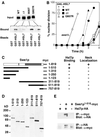
Interaction of Swe1p variants with Hsl7p. (A) Strains containing myc-tagged wild-type or mutant SWE1 expressed from the GAL1 promoter were grown in galactose-containing medium to induce Swe1p expression. Lysates containing Swe1p (Input) were incubated together with beads bound to recombinant bacterially produced GST or GST-Hsl7p as indicated, and the beads were washed and processed to detect bound Swe1p (Bound). Strains were JMY1680 (wild-type), JMY1681 (Δ1), JMY1696 (I806T), and JMY1697 (Q807R). B) cdc24-1 strains containing the indicated SWE1 alleles with or without GAL1-HSL7 were grown at 23°C in galactose-containing medium to induce Hsl7p expression, arrested in G1 with α-factor, and released from the arrest at 37°C to allow cell cycle progression but block bud formation. Aliquots were removed at 30-min intervals, processed to visualize nuclei, and scored to assess the timing of nuclear division. Strains were DLY690 (swe1Δ), JMY1690 (WT−), JMY1718 (WT+), JMY1691 (Δ1−), JMY1719 (Δ1+), JMY1722 (Q807R−), and JMY1756 (Q807R+). (C) Schematic of truncated Swe1p derivatives and summary of Hsl7p binding and localization results. On the diagram of full-length Swe1p, the gray box indicates Swe1p catalytic domain, black bar indicates residues 320–332 identified in the screen as a degradation determinant, and light gray strip at right indicates C-terminal 12myc epitope tag. Hsl7p binding was assessed by coimmunoprecipitation (see part E) and localization was assessed by immunofluorescence. (D) Expression of truncated Swe1p-myc derivatives. Strains DLY4267 (Swe1p1–510), DLY4268 (Swe1p1–310), DLY4269 (Swe1p1–250), DLY4266 (Swe1p1–123), DLY4272 (Swe1p311–819), DLY4270 (Swe1p511–819), and DLY4271 (Swe1p757–819) were induced to express the truncated Swe1p-myc derivatives by growth in galactose-containing medium, and expression was monitored by Western blotting. (E) Coimmunopre-cipitation between Swe1p1–510myc and Hsl7p-HA. Strains DLY4034 (HSL7-HA), DLY4267 (HSL7-HA SWE11–510myc), and DLY3584 (SWE11–510myc) were grown as above and lysed. Immunoprecipitates using α-myc (upper) or anti-HA (lower) antibodies were washed, separated by SDS-PAGE, and immunoblotted for Hsl7p-HA (upper) or Swe1p1–510-myc (lower).
Figure 9
Figure 9
Two-hybrid analysis of Swe1p-Cdc5p interaction. Analysis was performed as described in Figure 3 but with a “prey” plasmid containing C-terminal Cdc5p-encoding sequences.
Figure 10
Figure 10
Sequence comparisons of Swe1p degradation regions among fungal Swe1p homologues. ClustalW alignment of full-length protein sequences was carried out using Macvector software: only the regions of interest are shown. Sequences (from the top) are S. cerevisiae SWE1, A. gossypii SWE1, A. nidulans AnkA, S. pombe wee1, and S. pombe mik1. Numbers indicate first residue shown. Red boxes indicate residues that are identical in three or more homologues. Blue boxes indicate residues that are identical in S. cerevisiae and A. gossypii only. (A) N-terminal Hsl7p-binding region. (B) C-terminal region.

Similar articles

Cited by

References

    1. Bao S, et al. The highly conserved protein methyltransferase, Skb1, is a mediator of hyperosmotic stress response in the fission yeast Schizosaccharomyces pombe. J Biol Chem. 2001;276:14549–14552. - PubMed
    1. Barral Y, Parra M, Bidlingmaier S, Snyder M. Nim1-related kinases coordinate cell cycle progression with the organization of the peripheral cytoskeleton in yeast. Genes Dev. 1999;13:176–187. - PMC - PubMed
    1. Bartholomew CR, Woo SH, Chung YS, Jones C, Hardy CF. Cdc5 interacts with the Wee1 kinase in budding yeast. Mol Cell Biol. 2001;21:4949–4959. - PMC - PubMed
    1. Bi E, Pringle JR. ZDS1 and ZDS2, genes whose products may regulate Cdc42p in Saccharomyces cerevisiae. Mol Cell Biol. 1996;16:5264–5275. - PMC - PubMed
    1. Bishop AC, et al. A chemical switch for inhibitor-sensitive alleles of any protein kinase. Nature. 2000;407:395–401. - PubMed

Publication types

MeSH terms

Substances