. 2002 Oct;13(10):3683-95.
doi: 10.1091/mbc.e02-03-0138.

A novel conserved RNA-binding domain protein, RBD-1, is essential for ribosome biogenesis

Affiliations

A novel conserved RNA-binding domain protein, RBD-1, is essential for ribosome biogenesis

Petra Björk et al. Mol Biol Cell. 2002 Oct.

Abstract

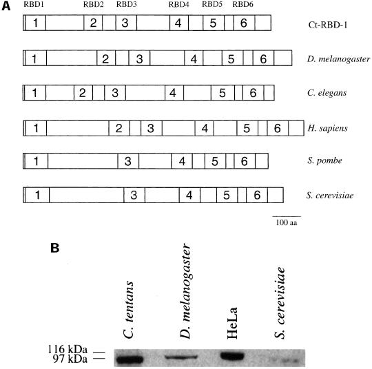
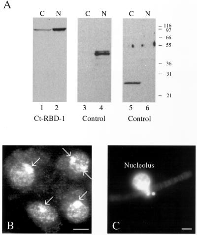
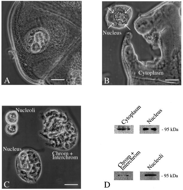
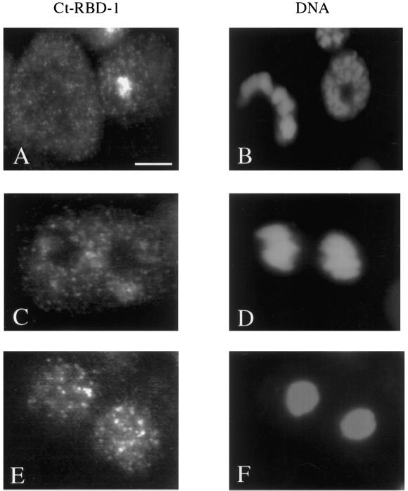
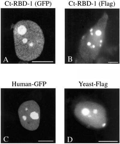
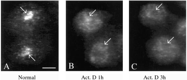
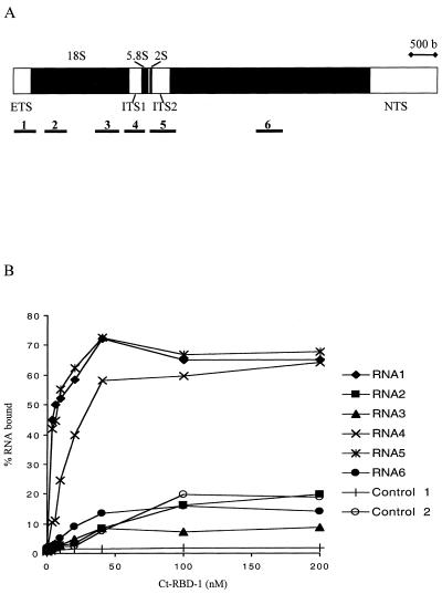
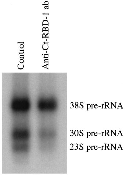
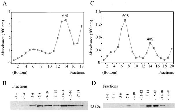
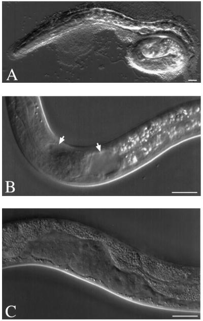
Synthesis of the ribosomal subunits from pre-rRNA requires a large number of trans-acting proteins and small nucleolar ribonucleoprotein particles to execute base modifications, RNA cleavages, and structural rearrangements. We have characterized a novel protein, RNA-binding domain-1 (RBD-1), that is involved in ribosome biogenesis. This protein contains six consensus RNA-binding domains and is conserved as to sequence, domain organization, and cellular location from yeast to human. RBD-1 is essential in Caenorhabditis elegans. In the dipteran Chironomus tentans, RBD-1 (Ct-RBD-1) binds pre-rRNA in vitro and anti-Ct-RBD-1 antibodies repress pre-rRNA processing in vivo. Ct-RBD-1 is mainly located in the nucleolus in an RNA polymerase I transcription-dependent manner, but it is also present in discrete foci in the interchromatin and in the cytoplasm. In cytoplasmic extracts, 20-30% of Ct-RBD-1 is associated with ribosomes and, preferentially, with the 40S ribosomal subunit. Our data suggest that RBD-1 plays a role in structurally coordinating pre-rRNA during ribosome biogenesis and that this function is conserved in all eukaryotes.


Figures

Figure 1
Figure 1
Ct-RBD-1 is conserved in eukaryotes. (A) Comparison of the organization of the RBDs in Ct-RBD-1 (accession no. Q95ZH1) and in the sequence homologues in five different species. The proteins and the RBDs are drawn to scale. The six different RBDs in Ct-RBD-1 are marked by numbers. The proteins in D. melanogaster (accession no. Q9VT19), C. elegans (accession no. Q9XU67), H. sapiens (accession no. Q96E42), S. pombe (accession no. O13620), and S. cerevisiae (accession no. Q06106) have the same domain organization. Sequence comparisons between all the RBDs in the six proteins showed that there is a position-specific conservation of the six RBDs between species, shown by marking homologues RBDs with the same number. In S. pombe and S. cerevisiae, RBD2 is lacking according to the sequence comparisons. (B) Ct-RBD-1–specific antibodies detect similar sized proteins in C. tentans, D. melanogaster, HeLa cells, and S. cerevisiae. Extracts from each species were analyzed by Western blotting and probed with anti-Ct-RBD-1 antibodies.
Figure 2
Figure 2
Ct-RBD-1 is present in nucleoli, nucleoplasm, and cytoplasm. (A) C. tentans diploid tissue culture cells were homogenized and nuclei and cytoplasm were separated by centrifugation. Extracts of the two fractions were analyzed by Western blotting. Ct-RBD-1, migrating at ∼95 kDa, is present both in the cytoplasm and in the nucleus (lane 1 and 2, respectively). Cross-contamination of the fractions was checked using an antibody against the nuclear protein hrp45 (Kiseleva et al., 1994) (lanes 3 and 4) and an antibody recognizing a cytoplasmic protein of unknown function (lanes 5 and 6). (B) Immunocytology of C. tentans diploid tissue culture cells. The cells were centrifuged onto slides, fixed, permeabilized, and stained with the anti-Ct-RBD-1 antibodies and an FITC-conjugated secondary antibody. The nucleoli (marked by arrows) are most heavily stained. In the remaining nucleus and in the cytoplasm, discrete foci are detected against a diffuse overall staining. Bar, 5 μm. (C) Immunostaining of an isolated polytene chromosome III of a C. tentans salivary gland cell. The chromosome was isolated from a fixed salivary gland cell by pipetting and incubated with the anti-Ct-RBD-1 antibodies and a FITC-conjugated secondary antibody. The nucleolus is brightly stained. Bar, 10 μm.
Figure 3
Figure 3
Microdissection analysis of C. tentans salivary gland cells. Salivary glands were isolated, fixed, and prepared for microdissection. The gland and the cellular components were viewed by phase contrast microscopy. (A) Salivary gland cell. The nucleus with the polytene chromosomes and the two nucleoli are seen. Bar, 40 μm. (B) Isolation of the cytoplasm. The nucleus was removed by dissecting well outside the nuclear membrane to avoid nuclear contamination of the cytoplasm. The arrows show the position of the nuclear membrane. Bar, 60 μm. (C) Salivary gland nuclei were carefully dissected to avoid cytoplasmic contamination. Subsequently the two nucleoli and the chromosomes plus interchromatin were isolated. Bar, 30 μm. (D) Western blot analysis of extracted proteins from the cytoplasm (isolated as shown in B), the nucleus, chromosomes plus interchromatin, and nucleoli (isolated as shown in C). Ct-RBD-1 is present in all the analyzed compartments.
Figure 4
Figure 4
Localization of Ct-RBD-1 during mitosis. C. tentans tissue culture cells were fixed, permeabilized, and stained with the anti-Ct-RBD-1 antibodies. The cells were also stained for DNA with DAPI to allow identification of cells in different stages of mitosis. (A) Immunostaining of an interphase cell (right) and a metaphase cell (left). (B) DNA in the same cells as in A, stained with DAPI. (C) Immunostaining of a cell in late anaphase. (D) Same cell as in C, stained with DAPI. (E) Immunostaining of telophase cells. (F) Same cells as in E, stained with DAPI. Bar, 5 μm.
Figure 5
Figure 5
Localization of Ct-RBD-1 and the H. sapiens and S. cerevisiae homologues in HeLa cells. HeLa cells were transfected with cDNA constructs encoding RBD-1 proteins from the different species, tagged with GFP or the FLAG-epitope. Cells expressing GFP-fusion proteins were fixed and directly viewed in the fluorescence microscope, whereas cells expressing the FLAG-tagged proteins were first fixed, permeabilized, and stained with an anti-FLAG antibody. (A) Ct-RBD-1 (GFP) is located throughout the nucleus and highly concentrated in the nucleoli. (B) Ct-RBD-1 (FLAG) has the same location as Ct-RBD-1 (GFP). Many cells also exhibited a weak cytoplasmic staining, and some cells were more strongly stained as shown herein. (C) Hs-RBD-1 (GFP) was located throughout the nucleoplasm but showed the highest concentration in the nucleoli. (D) S. cerevisiae RBD-1 homologue (FLAG) is concentrated in the nucleolus. The nucleoplasm was also stained. Bars, 10 μm.
Figure 6
Figure 6
Nucleolar location of Ct-RBD-1 is dependent on RNA polymerase I transcription. C. tentans tissue culture cells were grown in the presence of actinomycin D (0.05 μg/ml). Cells were fixed, permeabilized, and immunostained with the anti-Ct-RBD-1 antibodies at time 0 (A), after 1 h (B) and after 3 h (C) of actinomycin D treatment. Arrows mark the nucleoli. Bar, 5 μm.
Figure 7
Figure 7
Ct-RBD-1 interacts with pre-rRNA. (A) Schematic representation of the C. tentans rRNA gene. The regions of the pre-rRNA used for binding studies with Ct-RBD-1 are shown below the gene and numbered 1–6. 32P-Labeled RNAs were obtained by in vitro transcription of PCR fragments representing the different regions. The lengths of the RNAs were 438, 442, 477, 412, 519, and 536 nucleotides for RNA 1 to RNA 6, respectively. (B) Filter binding assay with Ct-RBD-1 and RNAs representing different pre-rRNA regions and control RNAs. Then 2–3 fmol of each RNA (in molecules) was incubated with increasing amounts of Ct-RBD-1 and filtered through nitrocellulose filters. The percentage retained RNA was plotted against the concentration of protein. Control RNAs were obtained by transcribing restriction enzyme cleaved pET-15b plasmid DNA (control 2, 430 nucleotides) or a PCR fragment representing a globin gene construct (control 1, 353 nucleotides). A third control RNA (transcribed from the plasmid pBluescript) behaved similarly to control RNA 2 (our unpublished data). The values shown are the averages of two separate measurements. Binding of RNA containing part of the 5′ ETS or ITS 2 (RNA 1 and RNA 5) bound Ct-RBD-1 with similar high affinities (Kd value of ∼5 nM). RNA representing the ITS 1 (RNA 4) bound with slightly less high affinity (Kd value of 10–15 nM). Binding of RNA containing 18S (RNA 2 and 3) or 28S (RNA 6) rRNA sequences was not different compared with the control RNAs.
Figure 8
Figure 8
Anti-Ct-RBD-1 antibodies repress pre-rRNA processing in vivo. Anti-Ct-RBD-1 antibodies were injected into the nuclei of living C. tentans salivary gland cells. RNA was labeled by incubation of the glands in medium containing α-[32P]ATP. The gland cells were fixed and the nucleoli were isolated by microdissection from injected cells and from noninjected cells as controls. The RNA was extracted and separated in agarose gels. The gels were dried and the relative amounts of the 38S, 30S, and 23S pre-rRNA were determined using a PhosphorImager.
Figure 9
Figure 9
Ct-RBD-1 is associated with ribosomes and preferentially with 40S ribosomal subunits. C. tentans tissue culture cells were homogenized and cytoplasmic extracts, treated with deoxycholate and Triton X-100, were centrifuged in sucrose gradients. After fractionation, the fractions were analyzed by measuring the absorbance at 260 nm and by Western blotting. (A) Polysome profile obtained after separation of cytoplasmic extract by using a 15–50% sucrose gradient. (B) Fractions from the gradient shown in A were pooled as indicated and analyzed by Western blotting, by using anti-Ct-RBD-1 antibodies. (C) Profile of ribosomal subunits obtained by EDTA treatment of isolated C. tentans polysomes and subsequent separation in a 10–30% sucrose gradient. (D) Fractions from the gradient shown in C were pooled as indicated and analyzed by Western blotting, with anti-Ct-RBD-1 antibodies.
Figure 10
Figure 10
RNAi phenotype of rbd-1. Double-stranded RNA corresponding to the Ce-RBD-1 mRNA was injected into the gonad of young adult hermaphrodites. The offspring were analyzed 5–30 h after the injection. (A) L1 larva, dying, with detached phenotype, i.e., pseudocoel filled with liquid. (B) L1 larva, vacuoles behind the terminal bulb of pharynx and anterior to it (arrows). (C) L2 larva, abnormal developing gonad with vacuoles. Bars, 10 μm.

Similar articles

Cited by

References

    1. Allain FH-T, Bouvet P, Diecmann T, Feigon J. Molecular basis of sequence specific recognition of pre-ribosomal RNA by nucleolin. EMBO J. 2000;19:6870–6881. - PMC - PubMed
    1. Allain FH-T, Gubser CC, Howe PWA, Nagai K, Neuhaus D, Varani G. Specificity of ribonucleoprotein interaction determined by RNA folding during complex formation. Nature. 1996;380:646–650. - PubMed
    1. Andersen JS, Lyon CE, Fox AH, Leung AKL, Lam YW, Steen H, Mann M, Lamond AI. Directed proteomic analysis of the human nucleolus. Curr Biol. 2002;12:1–11. - PubMed
    1. Baurén G, Jiang W-Q, Bernholm K, Gu F, Wieslander L. Demonstration of a dynamic, transcription-dependent organization of pre-mRNA splicing factors in polytene nuclei. J Cell Biol. 1996;133:929–941. - PMC - PubMed
    1. Biamonti G, Riva S. New insights into the auxiliary domains of eukaryotic RNA binding proteins. FEBS Lett. 1994;340:1–8. - PubMed

Publication types

MeSH terms

LinkOut - more resources