

Beyond 51Cr release: New methods for assessing HIV-1-specific CD8+ T cell responses in peripheral blood and mucosal tissues
- PMID: 12390303
- PMCID: PMC1906532
- DOI: 10.1046/j.1365-2249.2002.01981.x
Beyond 51Cr release: New methods for assessing HIV-1-specific CD8+ T cell responses in peripheral blood and mucosal tissues
Abstract
Much scientific effort has been directed towards elucidating the complexities of cell-mediated immune responses to HIV-1(reviewed in [1,2]). These studies have attempted to explain the immune system's ultimate failure to contain viral replication, leading to development of AIDS disease, and to identify immune responses that will be useful in developing immunomodulatory therapies and novel vaccine strategies. Although many of the complex interactions involved in AIDS pathogenesis remain unsolved, great progress has been made in characterizing the kinetics, specificity and functional dynamics of HIV-1-specific T cell responses. These investigations have come at a time when advances in virology, cellular immunology and molecular biology have converged to provide a variety of methodological approaches not available at the onset of the AIDS pandemic. Application of these tools to other infectious diseases and immunopathological conditions will provide a fertile area of research for future years. This review focuses on recent developments in the assessment of HIV-1-specific T cell responses in peripheral blood and tissues, with a particular emphasis on flow cytometry-based approaches.
Figures


 , bandpass filter.
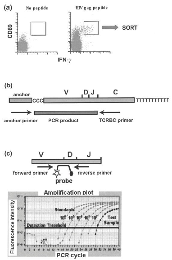
References
-
- Gandhi RT, Walker BD. Immunologic control of HIV-1. Annu Rev Med. 2002;53:149–72. - PubMed
-
- McMichael AJ, Rowland-Jones SL. Cellular immune responses to HIV. Nature. 2001;410:980–7. - PubMed
-
- Appay V, Dunbar PR, Callan M, et al. Memory CD8+ T cells vary in differentiation phenotype in different persistent virus infections. Nat Med. 2002;8:379–85. - PubMed
-
- Appay V, Papagno L, Spina CA, et al. Dynamics of T cell responses in HIV infection. J Immunol. 2002;168:3660–6. - PubMed
Publication types
MeSH terms
Substances
LinkOut - more resources
Full Text Sources
Medical
Research Materials
