. 2002 Oct 21;196(8):1067-78.
doi: 10.1084/jem.20011053.

Nucleolin as activator of human papillomavirus type 18 oncogene transcription in cervical cancer

Affiliations

Nucleolin as activator of human papillomavirus type 18 oncogene transcription in cervical cancer

Edgar Grinstein et al. J Exp Med. .

Abstract

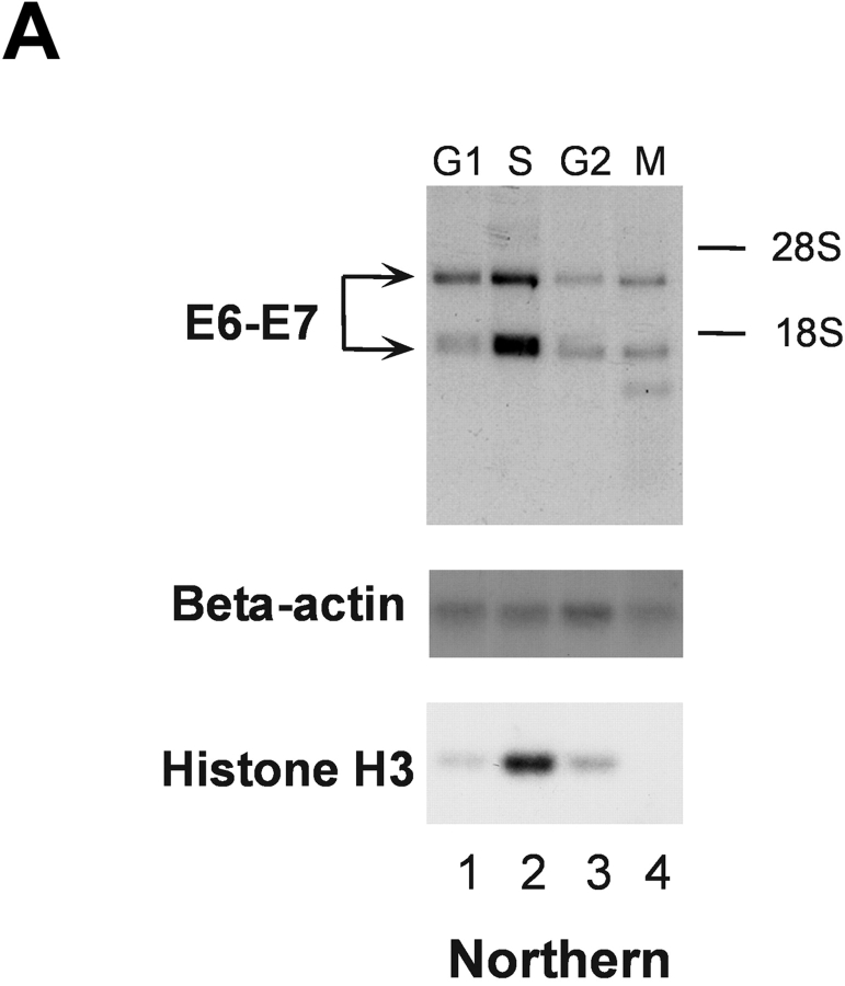
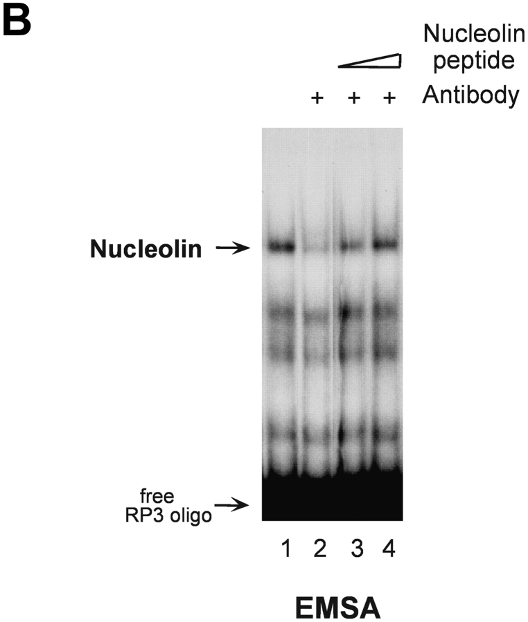
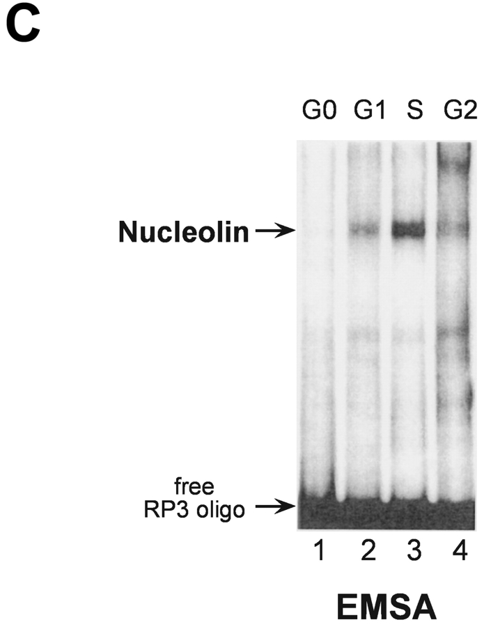
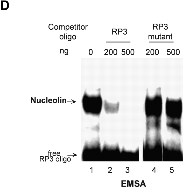
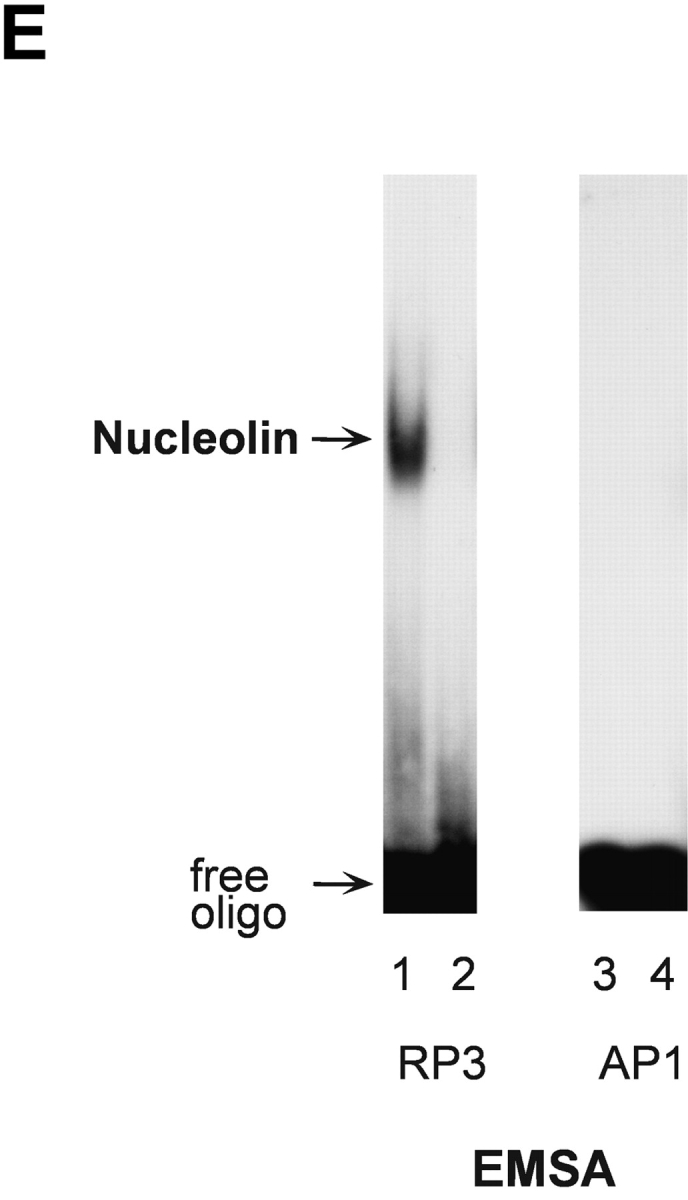
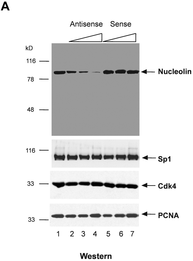
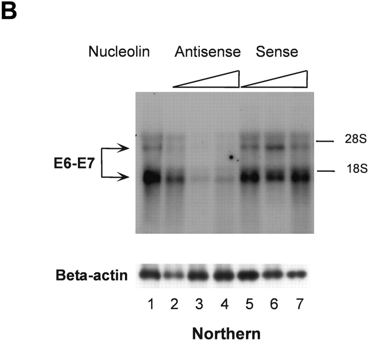
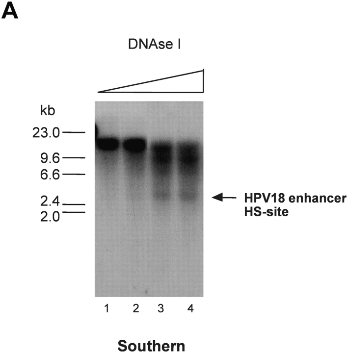
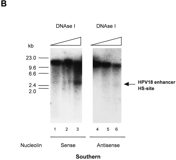
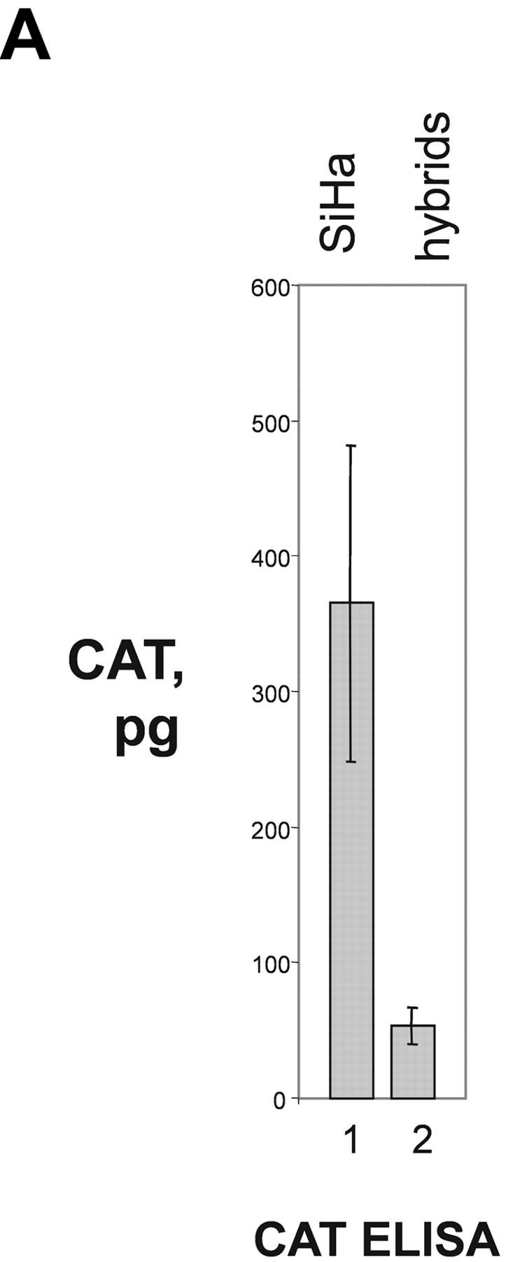
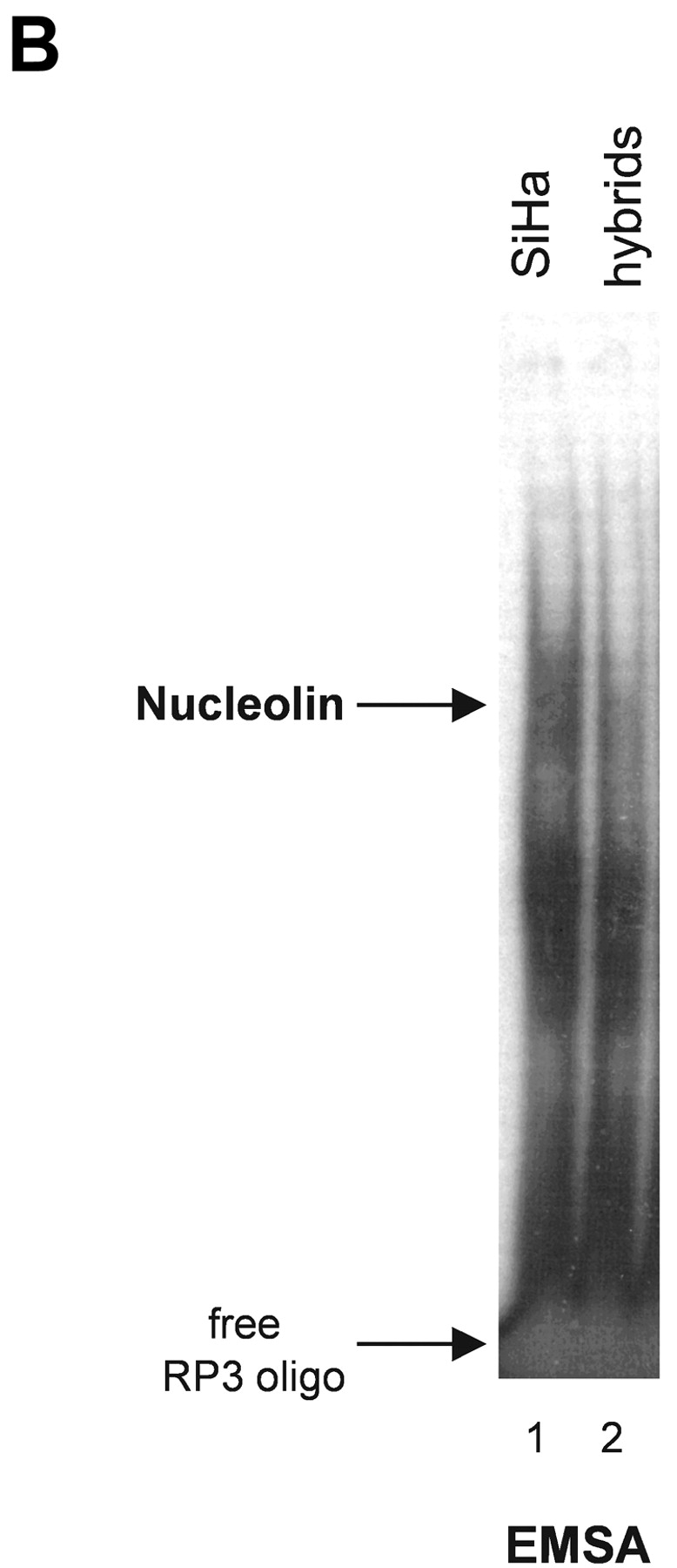
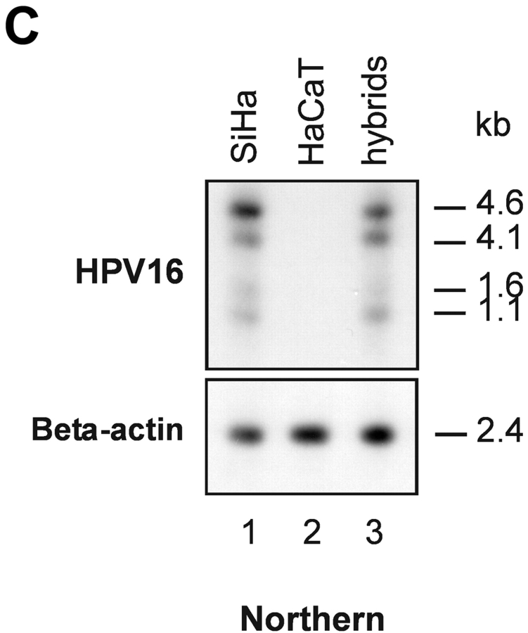
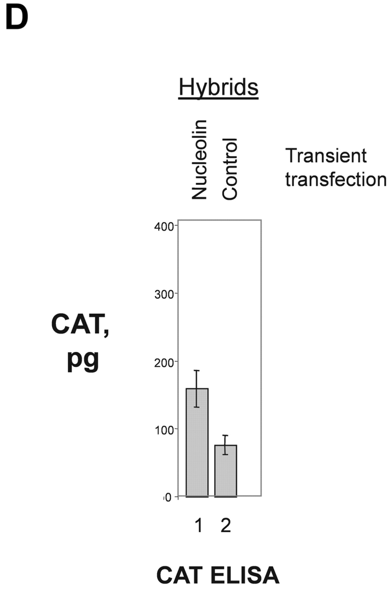
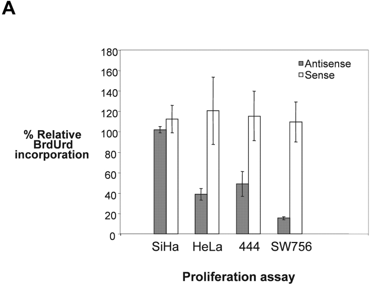
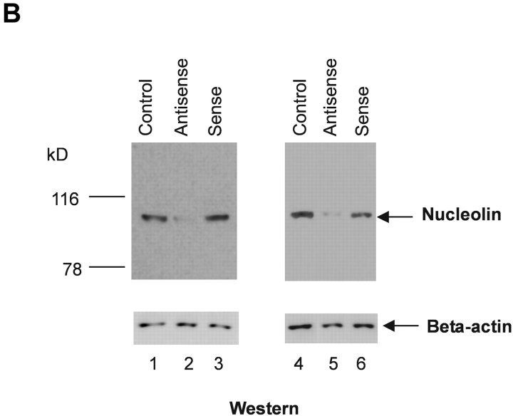
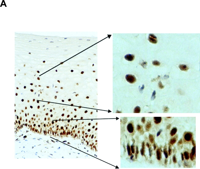
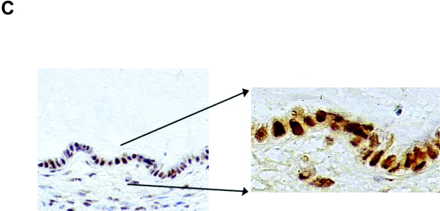
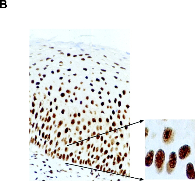
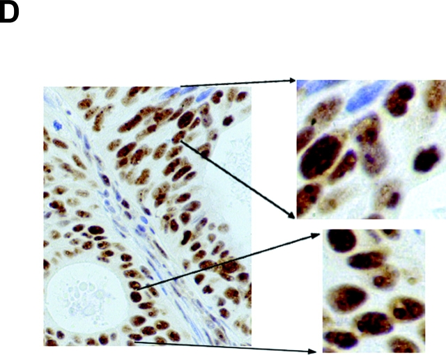
High risk human papillomaviruses (HPVs) are central to the development of cervical cancer and the deregulated expression of high risk HPV oncogenes is a critical event in this process. Here, we find that the cell protein nucleolin binds in a sequence-specific manner to the HPV18 enhancer. The DNA binding activity of nucleolin is primarily S phase specific, much like the transcription of the E6 and E7 oncoproteins of HPV18 in cervical cancer cells. Antisense inactivation of nucleolin blocks E6 and E7 oncogene transcription and selectively decreases HPV18(+) cervical cancer cell growth. Furthermore, nucleolin controls the chromatin structure of the HPV18 enhancer. In contrast, HPV16 oncogene transcription and proliferation rates of HPV16(+) SiHa cervical cancer cells are independent of nucleolin activity. Moreover, nucleolin expression is altered in HPV18(+) precancerous and cancerous tissue from the cervix uteri. Whereas nucleolin was homogeneously distributed in the nuclei of normal epithelial cells, it showed a speckled nuclear phenotype in HPV18(+) carcinomas. Thus, the host cell protein nucleolin is directly linked to HPV18-induced cervical carcinogenesis.


Figures

Figure 1.
Figure 1.
S phase– and sequence-specific binding of nucleolin to the HPV18 enhancer. (A) HPV18 oncogene transcription was examined by Northern blot analysis using nuclear RNA and an E6-E7 cDNA probe (top). S phase synchronization was controlled by hybridization with a histone H3 probe (bottom) and RNA concentrations were verified by hybridization with a β actin probe (middle). The differentially spliced HPV18 early gene transcripts are indicated by arrows (reference 62). (B) Nucleolin binds to the HPV18 enhancer. The labeled HPV18 enhancer oligonucleotide RP3 and a nuclear extract from S phase synchronized cells were used in EMSA. Lane 1, S phase nuclear extract; lane 2, nucleolin Ab was added to the DNA binding reaction; lanes 3 and 4, nucleolin Ab was presaturated with increasing amounts of the immunizing peptide. Electrophoresis was performed for 90 min at 11 V/cm. (C) Cell cycle regulation of nucleolin DNA binding activity. EMSA with nuclear extracts from biochemically synchronized 444 cells and the labeled HPV18 enhancer oligonucleotide RP3. Lane 1, G0 phase; lane 2, G1 phase; lane 3, S phase; lane 4, G2 phase. Electrophoresis was performed for 90 min at 11 V/cm. (D) Sequence specificity of nucleolin binding to the HPV18 enhancer. For EMSA, a partially purified nucleolin protein fraction and labeled RP3 were used. Partially purified nucleolin (all lanes). The binding complexes were competed by the addition of none (lane 1), 200 ng (lane 2), and 500 ng of RP3 (lane 3), and 200 ng (lane 4) and 500 ng (lane 5) of the RP3 mutant. Electrophoresis was performed for 50 min at 11 V/cm. (E) Specific interaction of nucleolin–GST protein with the HPV18 enhancer. Lanes 1 and 3, affinity-purified nucleolin–GST protein; lanes 2 and 4, GST protein; lanes 1 and 2, EMSA with the labeled RP3 HPV18 enhancer oligonucleotide; lanes 3 and 4, EMSA with an unrelated AP1 recognition site. Electrophoresis was performed for 90 min at 11 V/cm.
Figure 1.
Figure 1.
S phase– and sequence-specific binding of nucleolin to the HPV18 enhancer. (A) HPV18 oncogene transcription was examined by Northern blot analysis using nuclear RNA and an E6-E7 cDNA probe (top). S phase synchronization was controlled by hybridization with a histone H3 probe (bottom) and RNA concentrations were verified by hybridization with a β actin probe (middle). The differentially spliced HPV18 early gene transcripts are indicated by arrows (reference 62). (B) Nucleolin binds to the HPV18 enhancer. The labeled HPV18 enhancer oligonucleotide RP3 and a nuclear extract from S phase synchronized cells were used in EMSA. Lane 1, S phase nuclear extract; lane 2, nucleolin Ab was added to the DNA binding reaction; lanes 3 and 4, nucleolin Ab was presaturated with increasing amounts of the immunizing peptide. Electrophoresis was performed for 90 min at 11 V/cm. (C) Cell cycle regulation of nucleolin DNA binding activity. EMSA with nuclear extracts from biochemically synchronized 444 cells and the labeled HPV18 enhancer oligonucleotide RP3. Lane 1, G0 phase; lane 2, G1 phase; lane 3, S phase; lane 4, G2 phase. Electrophoresis was performed for 90 min at 11 V/cm. (D) Sequence specificity of nucleolin binding to the HPV18 enhancer. For EMSA, a partially purified nucleolin protein fraction and labeled RP3 were used. Partially purified nucleolin (all lanes). The binding complexes were competed by the addition of none (lane 1), 200 ng (lane 2), and 500 ng of RP3 (lane 3), and 200 ng (lane 4) and 500 ng (lane 5) of the RP3 mutant. Electrophoresis was performed for 50 min at 11 V/cm. (E) Specific interaction of nucleolin–GST protein with the HPV18 enhancer. Lanes 1 and 3, affinity-purified nucleolin–GST protein; lanes 2 and 4, GST protein; lanes 1 and 2, EMSA with the labeled RP3 HPV18 enhancer oligonucleotide; lanes 3 and 4, EMSA with an unrelated AP1 recognition site. Electrophoresis was performed for 90 min at 11 V/cm.
Figure 2.
Figure 2.
Nucleolin controls HPV18 oncogene transcription. (A) Antisense inhibition of nucleolin expression. 444 cells were treated with phosphorothioate-modified antisense or sense oligonucleotides and then nuclear extracts were prepared. Detection of nucleolin by immunoblotting (top). Lane 1, mock control; lanes 2–4, antisense oligonucleotide: 1 μM, 5 μM, and 9 μM; lanes 5–7, sense oligonucleotide: 1 μM, 5 μM, and 9 μM. The same extracts were analyzed by immunoblotting with an Ab specific for Sp1, Cdk4, and PCNA (bottom). On the side of each immunoblot the positions of detected proteins are indicated by arrows. (B) Nucleolin controls HPV18 oncogene expression. RNA was isolated from aliquots of antisense- or sense-treated cells and analyzed by Northern blotting using an E6-E7 cDNA probe. Lane 1, untreated 444 cells; lanes 2–4, antisense oligonucleotide: 1 μM, 5 μM, and 9 μM; lanes 5–7, sense oligonucleotide: 1 μM, 5 μM, and 9 μM. The differentially spliced HPV18 mRNAs (reference 62) are indicated by arrows. The β actin control is shown at the bottom.
Figure 2.
Figure 2.
Nucleolin controls HPV18 oncogene transcription. (A) Antisense inhibition of nucleolin expression. 444 cells were treated with phosphorothioate-modified antisense or sense oligonucleotides and then nuclear extracts were prepared. Detection of nucleolin by immunoblotting (top). Lane 1, mock control; lanes 2–4, antisense oligonucleotide: 1 μM, 5 μM, and 9 μM; lanes 5–7, sense oligonucleotide: 1 μM, 5 μM, and 9 μM. The same extracts were analyzed by immunoblotting with an Ab specific for Sp1, Cdk4, and PCNA (bottom). On the side of each immunoblot the positions of detected proteins are indicated by arrows. (B) Nucleolin controls HPV18 oncogene expression. RNA was isolated from aliquots of antisense- or sense-treated cells and analyzed by Northern blotting using an E6-E7 cDNA probe. Lane 1, untreated 444 cells; lanes 2–4, antisense oligonucleotide: 1 μM, 5 μM, and 9 μM; lanes 5–7, sense oligonucleotide: 1 μM, 5 μM, and 9 μM. The differentially spliced HPV18 mRNAs (reference 62) are indicated by arrows. The β actin control is shown at the bottom.
Figure 3.
Figure 3.
Regulation of chromatin structure of the HPV18 enhancer by nucleolin. (A) HPV18 enhancer DNase I HS site mapping was performed as previously described (reference 47). Nuclei from S phase cells were digested with DNase I: 0.25 U, 0.5 U, 1.75 U, and 2.5 U (lanes 1–4). Mock-digested nuclei are not shown. Purified nuclear DNA was then digested with EcoR I and analyzed by Southern blotting using an E6-E7 cDNA probe for indirect end labeling of the 3′ end. The position of a DNase I HS site in the HPV18 enhancer is indicated by an arrow. (B) 444 cells were treated with 9 μM nucleolin sense oligonucleotide and isolated nuclei were digested with DNase I: 0.25 U, 1 U, and 1.75 U (lanes 1–3). In parallel, 444 cells were treated with 9 μM antisense oligonucleotide and isolated nuclei were digested with DNase I: 0.25 U, 1 U, and 1.75 U (lanes 4–6). DNA was purified and analyzed by Southern blotting as described in A. The mock-digested controls are not shown. The HPV18 enhancer DNase I HS site is indicated by an arrow.
Figure 3.
Figure 3.
Regulation of chromatin structure of the HPV18 enhancer by nucleolin. (A) HPV18 enhancer DNase I HS site mapping was performed as previously described (reference 47). Nuclei from S phase cells were digested with DNase I: 0.25 U, 0.5 U, 1.75 U, and 2.5 U (lanes 1–4). Mock-digested nuclei are not shown. Purified nuclear DNA was then digested with EcoR I and analyzed by Southern blotting using an E6-E7 cDNA probe for indirect end labeling of the 3′ end. The position of a DNase I HS site in the HPV18 enhancer is indicated by an arrow. (B) 444 cells were treated with 9 μM nucleolin sense oligonucleotide and isolated nuclei were digested with DNase I: 0.25 U, 1 U, and 1.75 U (lanes 1–3). In parallel, 444 cells were treated with 9 μM antisense oligonucleotide and isolated nuclei were digested with DNase I: 0.25 U, 1 U, and 1.75 U (lanes 4–6). DNA was purified and analyzed by Southern blotting as described in A. The mock-digested controls are not shown. The HPV18 enhancer DNase I HS site is indicated by an arrow.
Figure 4.
Figure 4.
Nucleolin as selective activator of HPV18 oncogene transcription. (A) CAT reporter gene assay. HPV16+ SiHa cervical cancer cells containing an integrated contiguous HPV18 URR fused to a CAT reporter gene (HPV18 URR-CAT)-SiHa (lane 1; reference 39). SiHa × HaCaT hybrid cells containing the HPV18 URR-CAT reporter gene (lane 2). HaCaT cells are normal human keratinocytes (reference 53). CAT reporter gene activity was measured by CAT ELISA. The results were normalized to protein concentration. (B) EMSA with nuclear extracts from (HPV18 URR-CAT)-SiHa cells (lane 1) and SiHa × HaCaT hybrid cells where the HPV18 URR-CAT gene is not significantly expressed (lane 2). The position of the retarded RP3–nucleolin complex is indicated by an arrow. Conditions for EMSA are as described in Fig. 1. (C) HPV16 oncogene transcription in (HPV18 URR-CAT)-SiHa cells (lane 1) and SiHa × HaCaT hybrid cells (lane 3). HaCaT cells were used as a HPV16 control (lane 2). For Northern analysis, 0.5 μg of poly A–selected RNA was used per lane. Filters were consecutively hybridized with probes specific for HPV16 and β actin. The sizes of the corresponding mRNAs are indicated. (D) CAT reporter gene assay. SiHa × HaCaT hybrid cells containing the integrated HPV18 URR-CAT construct were transiently transfected with a nucleolin cDNA expression construct (lane 1; reference 46) or the empty vector (lane 2). CAT activity was measured by CAT ELISA. The results were normalized to protein concentration.
Figure 4.
Figure 4.
Nucleolin as selective activator of HPV18 oncogene transcription. (A) CAT reporter gene assay. HPV16+ SiHa cervical cancer cells containing an integrated contiguous HPV18 URR fused to a CAT reporter gene (HPV18 URR-CAT)-SiHa (lane 1; reference 39). SiHa × HaCaT hybrid cells containing the HPV18 URR-CAT reporter gene (lane 2). HaCaT cells are normal human keratinocytes (reference 53). CAT reporter gene activity was measured by CAT ELISA. The results were normalized to protein concentration. (B) EMSA with nuclear extracts from (HPV18 URR-CAT)-SiHa cells (lane 1) and SiHa × HaCaT hybrid cells where the HPV18 URR-CAT gene is not significantly expressed (lane 2). The position of the retarded RP3–nucleolin complex is indicated by an arrow. Conditions for EMSA are as described in Fig. 1. (C) HPV16 oncogene transcription in (HPV18 URR-CAT)-SiHa cells (lane 1) and SiHa × HaCaT hybrid cells (lane 3). HaCaT cells were used as a HPV16 control (lane 2). For Northern analysis, 0.5 μg of poly A–selected RNA was used per lane. Filters were consecutively hybridized with probes specific for HPV16 and β actin. The sizes of the corresponding mRNAs are indicated. (D) CAT reporter gene assay. SiHa × HaCaT hybrid cells containing the integrated HPV18 URR-CAT construct were transiently transfected with a nucleolin cDNA expression construct (lane 1; reference 46) or the empty vector (lane 2). CAT activity was measured by CAT ELISA. The results were normalized to protein concentration.
Figure 5.
Figure 5.
Nucleolin controls proliferation of HPV18+ cervical cancer cells. (A) Proliferation rates of SiHa, HeLa, 444, and SW756 cells were measured by means of BrdUrd incorporation after treatment with phosphorothioate-modified nucleolin antisense or sense oligonucleotides, and are displayed relative to mock-treated samples. (B) Nuclear extracts of HeLa and SiHa cells were analyzed by immunoblotting with an Ab specific for nucleolin after treatment with nucleolin sense and antisense oligonucleotides. Lanes 1–3, HeLa cells; lanes 4–6, SiHa cells; lanes 1 and 3, mock control; lanes 2 and 5, nucleolin antisense oligonucleotide; lanes 3 and 6, nucleolin sense oligonucleotide. The same extracts were analyzed by immunoblotting with an Ab specific for β actin (bottom).
Figure 5.
Figure 5.
Nucleolin controls proliferation of HPV18+ cervical cancer cells. (A) Proliferation rates of SiHa, HeLa, 444, and SW756 cells were measured by means of BrdUrd incorporation after treatment with phosphorothioate-modified nucleolin antisense or sense oligonucleotides, and are displayed relative to mock-treated samples. (B) Nuclear extracts of HeLa and SiHa cells were analyzed by immunoblotting with an Ab specific for nucleolin after treatment with nucleolin sense and antisense oligonucleotides. Lanes 1–3, HeLa cells; lanes 4–6, SiHa cells; lanes 1 and 3, mock control; lanes 2 and 5, nucleolin antisense oligonucleotide; lanes 3 and 6, nucleolin sense oligonucleotide. The same extracts were analyzed by immunoblotting with an Ab specific for β actin (bottom).
Figure 6.
Figure 6.
Nucleolin expression in normal HPV cervical tissue and HPV18+ cervical neoplasias. Paraffin-embedded specimens were processed for immunohistochemistry by standard procedures using an Ab specific for nucleolin. Bound antibodies were visualized by avidin-biotin-peroxidase-diaminobenzidine tetrachloride and are stained brown. As a control, the Ab specific for nucleolin was presaturated with the immunizing nucleolin peptide (not depicted). Specimens were counterstained with hematoxylin. The counterstained nuclei are light blue. (A) Squamous epithelium of the human cervix uteri. High power view of lower epithelial cell layers is shown on the bottom right. High power view of more superficial layers is shown on the top right. (B) HPV18+ HSIL. High power view is shown on the right. (C) Endocervical glandular epithelial cells. High power view is shown on the right. (D) HPV18+ adenocarcinoma of the endocervix. High power views of two areas are shown on the right.
Figure 6.
Figure 6.
Nucleolin expression in normal HPV cervical tissue and HPV18+ cervical neoplasias. Paraffin-embedded specimens were processed for immunohistochemistry by standard procedures using an Ab specific for nucleolin. Bound antibodies were visualized by avidin-biotin-peroxidase-diaminobenzidine tetrachloride and are stained brown. As a control, the Ab specific for nucleolin was presaturated with the immunizing nucleolin peptide (not depicted). Specimens were counterstained with hematoxylin. The counterstained nuclei are light blue. (A) Squamous epithelium of the human cervix uteri. High power view of lower epithelial cell layers is shown on the bottom right. High power view of more superficial layers is shown on the top right. (B) HPV18+ HSIL. High power view is shown on the right. (C) Endocervical glandular epithelial cells. High power view is shown on the right. (D) HPV18+ adenocarcinoma of the endocervix. High power views of two areas are shown on the right.

Similar articles

Cited by

References

    1. zur Hausen, H., and E.-M. de Villiers. 1994. Human papillomaviruses. Annu. Rev. Microbiol. 48:427–447. - PubMed
    1. Shah, K.V. 1997. Human papillomaviruses and anogenital cancers. N. Engl. J. Med. 337:1386–1388. - PubMed
    1. Walboomers, J.M., M.V. Jacobs, M.M. Manos, F.X. Bosch, J.A. Kummler, K.V. Shah, P.J. Snijders, J. Peto, C.J. Meijer, and N. Munoz. 1999. Human papillomavirus is a necessary cause of invasive cervical cancer worldwide. J. Pathol. 189:12–19. - PubMed
    1. Bosch, F.X., M.M. Manos, N. Munoz, M. Sherman, A.M. Jansen, J. Peto, M.H. Shiffman, V. Moreno, R. Kurman, and K.V. Shah. 1995. Prevalence of human papillomavirus in cervical cancer: a worldwide perspective. International biological study on cervical cancer (IBSCC) study group. J. Natl. Cancer Inst. 88:1361–1365. - PubMed
    1. Barnes, W., G. Delgado, R.J. Kurman, E.S. Petrilli, D.M. Smith, S. Ahmed, A.T. Lorincz, G.F. Temple, A.B. Jenson, and W.D. Lancaster. 1988. Possible prognostic signficance of human papillomavirus type in cervical cancer. Gynecol. Oncol. 29:267–273. - PubMed

Publication types