. 2002 Oct 28;159(2):245-54.
doi: 10.1083/jcb.200203089. Epub 2002 Oct 21.

Distinct cell cycle-dependent roles for dynactin and dynein at centrosomes

Affiliations

Distinct cell cycle-dependent roles for dynactin and dynein at centrosomes

Nicholas J Quintyne et al. J Cell Biol. .

Abstract

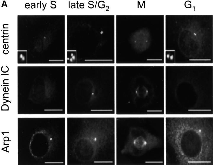
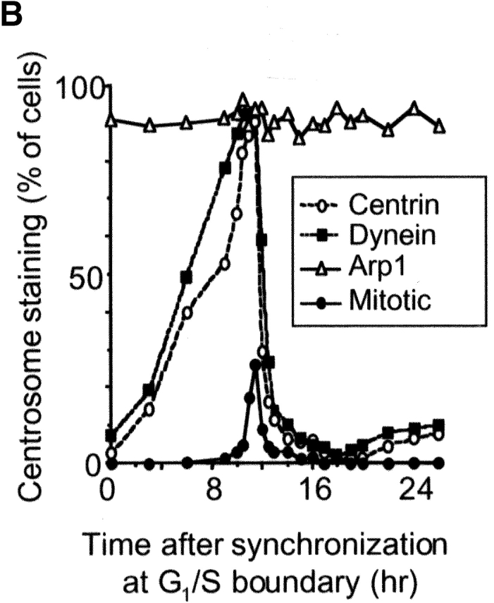
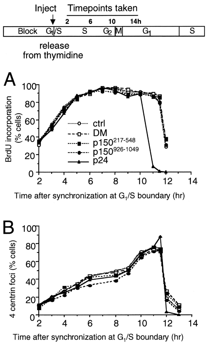
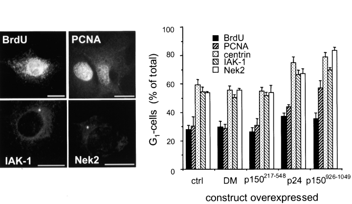
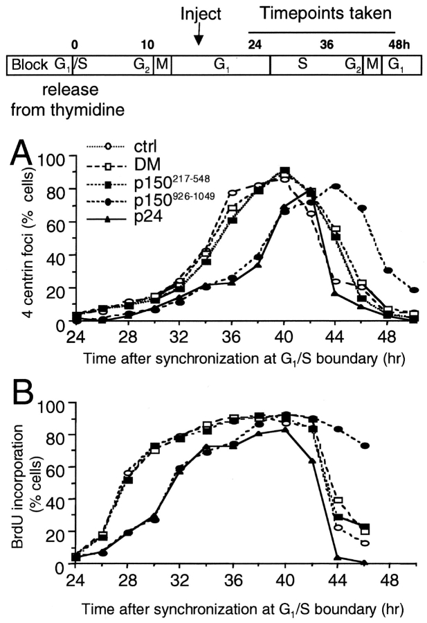
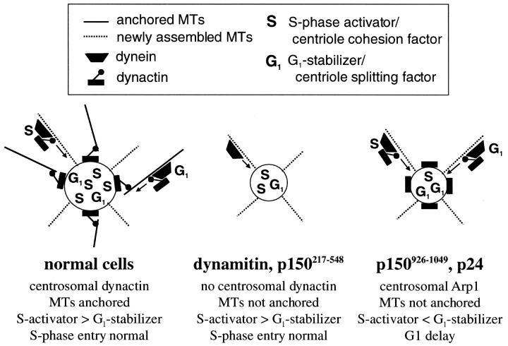
Centrosomal dynactin is required for normal microtubule anchoring and/or focusing independently of dynein. Dynactin is present at centrosomes throughout interphase, but dynein accumulates only during S and G2 phases. Blocking dynein-based motility prevents recruitment of dynactin and dynein to centrosomes and destabilizes both centrosomes and the microtubule array, interfering with cell cycle progression during mitosis. Destabilization of the centrosomal pool of dynactin does not inhibit dynein-based motility or dynein recruitment to centrosomes, but instead causes abnormal G1 centriole separation and delayed entry into S phase. The correct balance of centrosome-associated dynactin subunits is apparently important for satisfaction of the cell cycle mechanism that monitors centrosome integrity before centrosome duplication and ultimately governs the G1 to S transition. Our results suggest that, in addition to functioning as a microtubule anchor, dynactin contributes to the recruitment of important cell cycle regulators to centrosomes.


Figures

Figure 1.
Figure 1.
Cell cycle localization of dynein and dynactin to the centrosome. (A) Cells were synchronized using a double thymidine block and then released for increasing intervals before being fixed and labeled with Abs to the centriole marker, centrin, dynein (IC), or Arp1. Bars, 10 μm. (B) Cell populations were scored for either a centrosomal focus of dynein IC or dynactin subunit (p150Glued or Arp1) and for four centrin foci. At each time point, the number of mitotic cells was also determined on the basis of the characteristic mitotic patterns of centrin and dynein–dynactin staining (as in A). At least 400 cells were scored per time point in two independent experiments.
Figure 1.
Figure 1.
Cell cycle localization of dynein and dynactin to the centrosome. (A) Cells were synchronized using a double thymidine block and then released for increasing intervals before being fixed and labeled with Abs to the centriole marker, centrin, dynein (IC), or Arp1. Bars, 10 μm. (B) Cell populations were scored for either a centrosomal focus of dynein IC or dynactin subunit (p150Glued or Arp1) and for four centrin foci. At each time point, the number of mitotic cells was also determined on the basis of the characteristic mitotic patterns of centrin and dynein–dynactin staining (as in A). At least 400 cells were scored per time point in two independent experiments.
Figure 2.
Figure 2.
Localization of dynactin at centrioles. (Large panels) Cells were double labeled with Abs to p150Glued or Arp1 (green) and centrin or ɛ-tubulin (red); fiduciary markers were used to verify that the offset seen here is real. Insets show the boxed centrosome enlarged 5×. (Bottom) Deconvolution microscopy of the centrosome boxed in the panels above. Right panels, merge. Bars, 10 μm.
Figure 3.
Figure 3.
Effects of dynactin subunit overexpression on accumulation of centrosomal dynein and dynactin during microtubule regrowth. Microtubules in transiently transfected cells were depolymerized by nocodazole/cold treatment and then allowed to regrow (see Materials and methods for details). The appearance of a centrosomal focus of dynein intermediate chain (DIC; similar results were obtained using Abs to dynein light IC) or dynactin (Arp1 or p150Glued) was analyzed by immunofluorescence. Overexpressing cells were identified by GFP (dynamitin, p24, and p150926–1049) or DsRed (p150217–548) fluorescence. (A) Nonexpressing cells in the transfected population. (B) Cells overexpressing dynamitin or p150217–548. (C) Cells overexpressing p24 or p150 926–1049. DM, dynamitin.
Figure 4.
Figure 4.
Effects of dynactin subunit overexpression on S phase events. Cells were synchronized using a double thymidine block, microinjected with cDNAs encoding dynactin subunits, and then released from the block and incubated at 37°C for increasing lengths of time (the experimental scheme is cartooned above the graphs). Some cells were fixed and labeled with Abs to centrin or treated with BrdU to label DNA before fixation and BrdU Ab labeling. The percent of cells in the population showing BrdU incorporation (A) or four centrioles (B) was determined. Overexpressing cells were identified by GFP or DsRed fluorescence. Ctrl, noninjected control cells on the coverslip; DM, dynamitin.
Figure 5.
Figure 5.
Localization and prevalence of S phase markers in cells overexpressing dynactin subunits. (Left) Typical S phase distribution of BrdU (nuclear staining), PCNA (nuclear accumulation), or the kinases IAK-1 and Nek2 (centrosomal accumulation). Bars, 10 μm. (Right) Unsynchronized cells were transfected with dynactin shoulder/sidearm expression vectors and scored for expression of S phase markers. Overexpressing cells were identified by GFP or DsRed fluorescence. Ctrl, nonexpressing control cells in the transfected population; DM, dynamitin.
Figure 6.
Figure 6.
Effects of dynactin subunit overexpression on cell cycle progression. Cells were synchronized using a double thymidine block, released, and allowed to proceed through mitosis, and then microinjected with cDNAs encoding dynactin subunits. At different times after microinjection, cells were fixed and stained for centrin (A) or labeled with BrdU and fixed and stained with BrdU Abs (B). Ctrl, noninjected control cells on the coverslip; DM, dynamitin.
Figure 7.
Figure 7.
Model figure summarizing the effects of dynactin subunit overexpression on microtubule anchoring and centrosomal protein targeting on G1 centrosomes. For simplicity, the centrosome is depicted as a single object and mother and daughter centrioles are not shown. In dynamitin and p150217–548 overexpressers, dynein-based delivery of proteins to centrosomes is blocked, but an acceptable balance of S phase activators and G1 stabilizers is maintained to allow cells to enter S phase at the normal time. In p24 and p150926–1049 overexpressers, dynein-based transport brings the normal complement of cell cycle regulators to the centrosome, but G1 stabilizing activities predominate due directly or indirectly to the localized perturbation of dynactin integrity. S phase entry is delayed.

References

    1. Allan, V. 2000. Dynactin. Curr. Biol. 10:R432. - PubMed
    1. Bornens, M. 2002. Centrosome composition and microtubule anchoring mechanisms. Curr. Opin. Cell Biol. 14:25–34. - PubMed
    1. Bouckson-Castaing, V., M. Moudjou, D.J. Ferguson, S. Mucklow, Y. Belkaid, G. Milon, and P.R. Crocker. 1996. Molecular characterisation of ninein, a new coiled-coil protein of the centrosome. J. Cell Sci. 109:179–190. - PubMed
    1. Bravo, R., and J.E. Celis. 1985. Changes in the nuclear distribution of cyclin (PCNA) during S-phase are not triggered by post-translational modifications that are expected to moderately affect its charge. FEBS Lett. 182:435–440. - PubMed
    1. Burkhardt, J.K., C.J. Echeverri, T. Nilsson, and R.B. Vallee. 1997. Overexpression of the dynamitin (p50) subunit of the dynactin complex disrupts dynein-dependent maintenance of membrane organelle distribution. J. Cell Biol. 139:469–484. - PMC - PubMed

Publication types