. 2002 Nov;22(22):7731-43.
doi: 10.1128/MCB.22.22.7731-7743.2002.

Alternative splicing controls the mechanisms of FAK autophosphorylation

Affiliations

Alternative splicing controls the mechanisms of FAK autophosphorylation

Madeleine Toutant et al. Mol Cell Biol. 2002 Nov.

Abstract

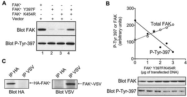
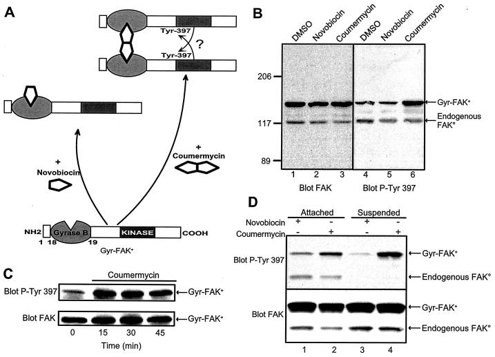
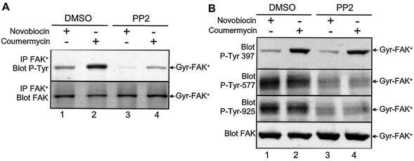
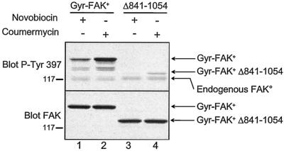
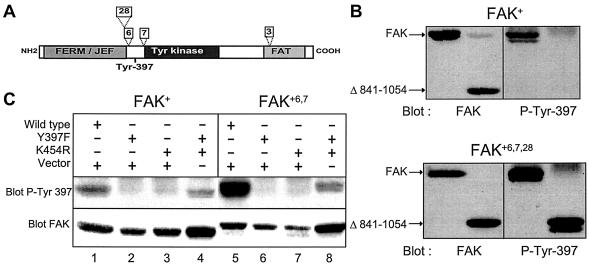
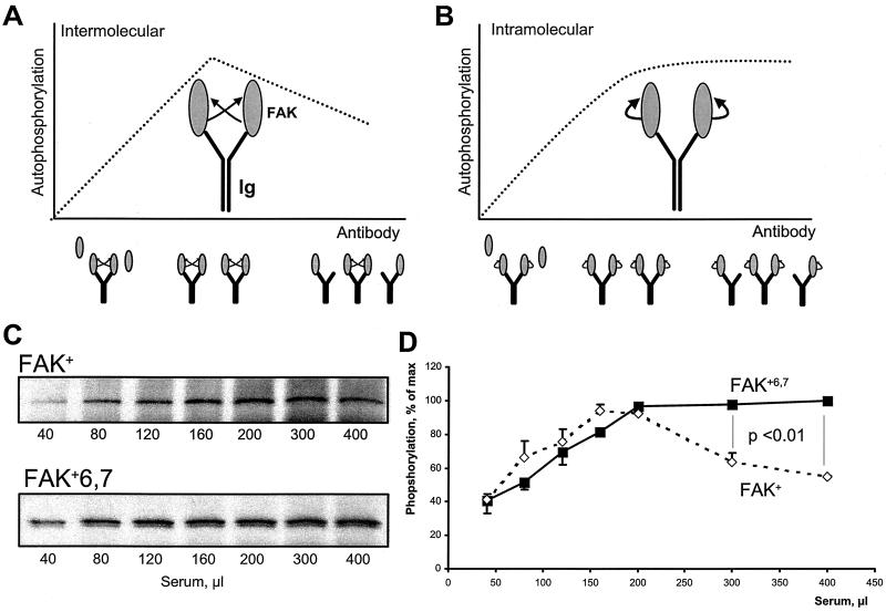
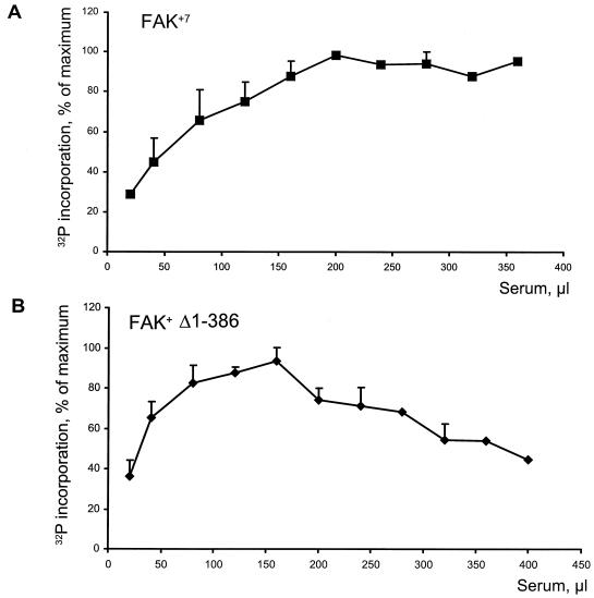
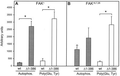
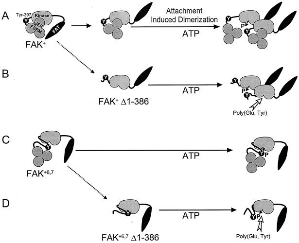
Focal adhesion kinase (FAK) is activated following integrin engagement or stimulation of transmembrane receptors. Autophosphorylation of FAK on Tyr-397 is a critical event, allowing binding of Src family kinases and activation of signal transduction pathways. Tissue-specific alternative splicing generates several isoforms of FAK with different autophosphorylation rates. Despite its importance, the mechanisms of FAK autophosphorylation and the basis for differences between isoforms are not known. We addressed these questions using isoforms of FAK expressed in brain. Autophosphorylation of FAK(+), which is identical to that of "standard" FAK, was intermolecular in transfected cells, although it did not involve the formation of stable multimeric complexes. Coumermycin-induced dimerization of gyrase B-FAK(+) chimeras triggered autophosphorylation of Tyr-397. This was independent of cell adhesion but required the C terminus of the protein. In contrast, the elevated autophosphorylation of FAK(+6,7), the major neuronal splice isoform, was not accounted for by transphosphorylation. Specifically designed immune precipitate kinase assays confirmed that autophosphorylation of FAK(+) was intermolecular, whereas autophosphorylation of FAK(+6,7) or FAK(+7) was predominantly intramolecular and insensitive to the inhibitory effects of the N-terminal domain. Our results clarify the mechanisms of FAK activation and show how alternative splicing can dramatically alter the mechanism of autophosphorylation of a protein kinase.


Figures

FIG. 1.
FIG. 1.
Transphosphorylation of FAK+ Tyr-397 in COS-7 cells. (A) COS-7 cells were transfected with 4 μg of plasmids coding for FAK+, FAK+ Y397F, FAK+ K454R (kinase dead), or empty pBK-CMV2 vector (vector), as indicated. Immunoblotting of cell lysates was carried out with antibodies recognizing specifically FAK phosphorylated on Tyr-397 (SL625857, blot P-Tyr 397) or independently of its phosphorylation state (A-17, blot FAK). Results are representative of three independent experiments. (B) COS-7 cells were transfected with 2 μg of plasmid coding for wild-type FAK+ and increasing amounts of the FAK+ K454R/Y397F double mutant. The total amount of DNA was kept constant by the addition of vector. The phospho-Tyr 397 immunoreactivity (blot P-Tyr-397) of transfected FAK+ and the total immunoreactivity of FAK+ and FAK+ K454R/Y397F (blot FAK) were quantified from immunoblots by densitometry scanning of autoradiograms and analysis with NIH Image software. Plotted data are the means of two independent experiments. (C) To determine whether stable complexes comprising several molecules of FAK were formed in cells, COS-7 cells were cotransfected with HA-FAK+ and FAK+-VSV. After 48 h, cells were lysed, and the lysates were subjected to immunoprecipitation with anti-HA (IP HA) or anti-VSV (IP VSV) antibodies. The samples were immunoblotted with anti-HA (blot HA) or anti-VSV (blot VSV) antibodies.
FIG. 2.
FIG. 2.
Coumermycin-induced dimerization enhances phosphorylation of gyrase B-FAK+ fusion proteins (Gyr-FAK+) on Tyr-397. (A) Schematic representation of the gyrase B-inducible dimerization system (17) applied to FAK+. The positions of the dimerization domain of the B subunit of bacterial gyrase (gyrase B), the central catalytic domain of FAK+ (kinase), and Tyr-397 are indicated. Addition of coumermycin, a divalent ligand of gyrase B, leads to the formation of Gyr-FAK+ homodimers. Novobiocin, a monovalent molecule also able to bind to gyrase B, is unable to induce its dimerization and was used as a negative control. (B) COS-7 cells transfected with Gyr-FAK+ were treated for 15 min with dimethyl sulfoxide (DMSO, vehicle), 10 μM novobiocin, or 10 μM coumermycin. Cell lysates were analyzed directly by immunoblotting with an antibody reacting with all isoforms of FAK (A-17, blot FAK). The apparent molecular weights of endogenous FAK° and Gyr-FAK+ were approximately 125,000 and 150,000, respectively. Phosphorylation of Tyr-397 was assessed with specific antibodies (SL625857, blot P-Tyr 397). (C) Time course of coumermycin-induced phosphorylation of Tyr-397. COS-7 cells transfected with Gyr-FAK+ were incubated in the presence of 10 μM coumermycin for the indicated times. Gyr-FAK+ immune precipitates were subjected to immunoblotting with anti-phospho-Tyr-397 (SL625857, blot P-Tyr 397) or anti-FAK (A17, blot FAK). Results are representative of three independent experiments. (D) The effects of coumermycin treatment on Gyr-FAK+ were examined in attached or suspended transfected COS-7 cells. After incubation in the presence of 10 μM novobiocin or coumermycin, the cells were lysed in RIPA buffer. An aliquot of the lysate was used for immunoblotting with antibodies specific for phospho-Tyr 397 (Biosource, blot P-Tyr 397), and the total amount of Gyr-FAK+ was determined by immunoblotting with an antibody reacting with FAK independently of its level of phosphorylation (A-17, blot FAK). Gyr-FAK+ Tyr-397 phosphorylation was: novobiocin/attached, 7 ± 1; coumermycin/attached, 75 ± 10; novobiocin/suspended, 3 ± 1; coumermycin/suspended, 94 ± 5 (arbitrary units, mean ± standard error of the mean of three independent experiments) (analysis of variance: P < 0.05).
FIG. 3.
FIG. 3.
Dimerization-induced phosphorylation of Tyr-397 in Gyr-FAK+ is independent of Src family kinases. COS-7 cells transfected with Gyr-FAK+ were treated for 15 min with 10 μM novobiocin or 10 μM coumermycin. A specific Src family kinase inhibitor, PP2 (10 μM), or vehicle (dimethyl sulfoxide, DMSO) was added 20 min prior to coumermycin or novobiocin. (A) Gyr-FAK+ was immunoprecipitated with antibodies specific for FAK+, and its total phosphorylation was assessed with anti-phospho-Tyr antibodies (IP-FAK+ blot P-Tyr). and with antibodies reacting with FAK independently of its state of phosphorylation (IP-FAK+ blot FAK). (B) Total lysates from the same cells were immunoblotted with antibodies specific for phospho-Tyr-397, phospho-Tyr-577, phospho-Tyr-925, or total FAK. Results are representative of at least three experiments.
FIG. 4.
FIG. 4.
Dimerization-induced autophosphorylation of Gyr-FAK+ requires its C-terminal region. COS-7 cells were transfected with Gyr-FAK+ or Gyr-FAK+Δ841-1054 and incubated for 15 min in the presence of novobiocin or coumermycin. Cells were lysed, and phosphorylation of Tyr-397 and the total amount of FAK were determined by immunoblotting with specific antibodies. Note that endogenous FAK was not apparent in total FAK immunoblotting at the short exposure time used to allow comparison of Gyr-FAK+ and Gyr-FAK+Δ841-1054 levels. Results are representative of at least three experiments.
FIG. 5.
FIG. 5.
Differences in autophosphorylation between FAK splice isoforms in transfected cells. (A) The positions of the N-terminal FERM/JEF domain, the catalytic domain (Tyr kinase), the focal adhesion targeting (FAT) domain, and the peptides (boxes 28, 6, 7, and 3) coded by alternatively spliced exons are indicated. Tyr-397 is the autophosphorylation site. (B) Deletion of the C-terminal region (Δ841-1054) abolished autophosphorylation of FAK+ in transfected COS-7 cells, whereas it did not alter the autophosphorylation of FAK+6,7,28. The level of phosphorylation of Tyr-397 in intact cells was measured by immunoblotting with specific antibodies for phospho-Tyr-397 (blot P-Tyr-397). The total amount of FAK was measured with antibodies reacting with FAK independently of its state of phosphorylation (blot FAK). (C) Transphosphorylation in intact cells did not fully restore the increased autophosphorylation of FAK+6,7. COS-7 cells were transfected with 4 μg of plasmids coding for the wild-type, Y397F, or K454R forms of FAK+ or FAK+6,7, as indicated. Empty pBK-CMV2 vector was added when necessary to keep the total amount of transfected DNA constant. Cells were lysed on the culture dish. Immunoblotting was carried out with antibodies recognizing FAK phosphorylated on Tyr-397 (SL625857, blot P-Tyr 397) or independently of its phosphorylation state (A-17, blot FAK). Results are representative of three independent experiments which gave similar results.
FIG. 6.
FIG. 6.
Different mechanisms of autophosphorylation of FAK+ and FAK+6,7. (A and B) Principle of the CITIK assay. Equal amounts of lysates from COS-7 cells transfected with FAK are immunoprecipitated in the presence of increasing amounts of antiserum and constant amounts of protein A-Sepharose. Autophosphorylation assays are carried out in the immune precipitates, and the results are expected to be affected by the reaction mechanism as follows. For small amounts of antiserum, the quantity of specific antibodies against FAK is limiting, and all the available binding sites are expected to be occupied, as schematically indicated by two molecules of FAK per immunoglobulin (Ig). In these conditions, autophosphorylation increases with the amount of antiserum, reflecting the increasing amount of immunoprecipitated FAK, regardless of the cis or trans mechanism (ascending part of the curve). When the quantity of serum is further increased, the number of FAK-specific binding sites becomes greater than the number of FAK molecules, and an increasing proportion of immunoglobulin is expected to bind only one molecule of FAK or none. In these conditions, the trans and cis mechanisms of autophosphorylation can be clearly distinguished: intermolecular autophosphorylation will diminish with increasing the amount of serum, since the probability that two FAK molecules will be bound to the same immunoglobulin will decrease (A); intramolecular autophosphorylation is independent of immunoglobulin G-mediated interactions and will reach a plateau corresponding to the total amount of FAK (B). CITIK assays were carried out for FAK+ and FAK+6,7 as described in the text. Reaction products were separated by SDS-PAGE, transferred to nitrocellulose, and analyzed by immunoblotting (C), and incorporated 32P was quantified with an Instant Imager (D). The total amount of immunoprecipitated FAK, estimated by immunoblotting with FAK antibodies, was maximal with approximately 200 μl of antiserum and remained constant with higher amounts of antiserum (C). The amount of 32P incorporated was plotted as a function of the amount of antiserum for the two FAK isoforms (D). Results are expressed as percent of maximum to correct for the higher activity of FAK+6,7 compared to FAK+. Results are means ± standard error of the mean of three independent experiments. Statistical analysis was done with two-way analysis of variance. The results show a significant difference between FAK+6,7 and FAK+ for the relevant portion of the curve, supporting the hypothesis that phosphorylation is intramolecular in the case of FAK+6,7 and intermolecular in the case of FAK+.
FIG. 7.
FIG. 7.
Autophosphorylation mechanism of FAK is altered by the presence of box 7 but not by N-terminal truncation. (A) The presence of box 7 is sufficient for intramolecular phosphorylation of Tyr-397. The CITIK assay was carried out with FAK+7 as described in the legend to Fig. 6. The results indicate that phosphorylation was intramolecular. (B) Deletion of the N-terminal FERM/JEF domain does not alter the autophosphorylation mechanism of FAK+. The CITIK assay was carried out with a truncated form of FAK+ (Δ1-386). The results indicate that autophosphorylation is intermolecular. In both cases, data are means ± standard error of the mean of three independent experiments.
FIG. 8.
FIG. 8.
Effects of deletion of the N-terminal FERM/JEF domain on autophosphorylation of FAK isoforms and their ability to phosphorylate an exogenous substrate. Wild-type (wt) FAK+, its N-terminally truncated form (FAK+Δ1-386) (left panel), and wild-type (wt) FAK+6,7,28 and its truncated form (FAK+6,7,28Δ1-386) (right panel) were expressed in COS-7 cells, dephosphorylated in vitro, and immunoprecipitated, and their autophosphorylation was assayed in the presence of [γ-32P]ATP (Autophos). In the same conditions, the ability of each immunoprecipitated isoform of FAK to phosphorylate an exogenous substrate, poly(Glu, Tyr), was examined. The amount of radioactivity incorporated into FAK or poly(Glu, Tyr) was measured with an Instant Imager after SDS-PAGE, and the counts per minute were normalized to the total amount of immunoprecipitated FAK, measured by immunoblotting. The autophosphorylation of FAK+6,7,28 was higher than that of FAK+ (○; P < 0.01, two-tailed Student's t test). N-terminal truncation increased the ability of FAK+ to autophosphorylate (∗; P < 0.005, two-tailed Student's t test), whereas it did not significantly alter the autophosphorylation of FAK+6,7,28 (left panel). In contrast, N-terminal truncation dramatically increased the ability of both isoforms to phosphorylate poly(Glu, Tyr) (∗, P < 0.05, two-tailed Student's t test). Values are means ± standard error of the mean for four (autophosphorylation) or three [phosphorylation of poly(Glu, Tyr)] experiments.
FIG. 9.
FIG. 9.
Schematic representation of FAK autophosphorylation mechanisms and differences between isoforms, based on the results of the present study. (A) FAK+ (as well as FAK°, which has the same low autophosphorylation) is in a poorly active state in basal conditions, presumably due to inhibition by the N-terminal domain. The FERM/JEF domain is indicated here as a trilobate structure, on the basis of the recently determined structure of several ERM domains (24, 41). The C-terminal region, including the FAT domain, is necessary for phosphorylation in intact cells. Cell attachment or artificial dimerization (as shown in the present study) promotes intermolecular phosphorylation of Tyr-397. (B) Deletion of the FERM/JEF domain of FAK+ increases intermolecular autophosphorylation in vitro by removing the inhibitory effect of the N-terminal FERM/JEF and facilitating the access of substrates to the active site [arrow, poly(Glu, Tyr)]. (C) The neuronal isoform, FAK+6,7 (as well as FAK+6,7,28, which has the same high autophosphorylation), has an increased autophosphorylation because of its ability to undergo intramolecular phosphorylation. The presence of box 7 is sufficient to allow intramolecular phosphorylation, presumably by allowing access of Tyr-397 to the active site located on the same peptide chain. However, in FAK+6,7, the N-terminal FERM/JEF domain still hampers the access of external substrates. (D) The deletion of the FERM/JEF domain has little effect on FAK+6,7 autophosphorylation but increases the accessibility of substrates to the active site [arrow, poly(Glu, Tyr)]. Whereas the C-terminal FAT domain is essential for the attachment- or artificial dimerization-induced autophosphorylation of FAK+, this does not appear to be the case for FAK+6,7. This is symbolized by the ordered open position of the C terminus in A and B and its loosely closed position in C and D.

Similar articles

Cited by

References

    1. Baron, V., V. Calléja, P. Ferrari, F. Alengrin, and E. Van Obberghen. 1998. p125Fak focal adhesion kinase is a substrate for the insulin and insulin-like growth factor-I tyrosine kinase receptors. J. Biol. Chem. 273:7162-7168. - PubMed
    1. Burgaya, F., and J. A. Girault. 1996. Cloning of focal adhesion kinase from rat stiatum reveals multiple transcripts. Mol. Brain Res. 37:63-73. - PubMed
    1. Burgaya, F., M. Toutant, J. M. Studler, A. Costa, M. Le Bert, M. Gelman, and J. A. Girault. 1997. Alternatively spliced focal adhesion kinase in rat brain with increased autophosphorylation activity. J. Biol. Chem. 272:28720-28725. - PubMed
    1. Calalb, M. B., T. R. Polte, and S. K. Hanks. 1995. Tyrosine phosphorylation of focal adhesion kinase at sites in the catalytic domain regulates kinase activity: A role for Src family kinases. Mol. Cell. Biol. 15:954-963. - PMC - PubMed
    1. Calalb, M. B., X. E. Zhang, T. R. Polte, and S. K. Hanks. 1996. Focal adhesion kinase tyrosine-861 is a major site of phosphorylation by Src. Biochem. Biophys. Res. Commun. 228:662-668. - PubMed

Publication types

MeSH terms